Multiple Genitourinary Malformations Associated with Malformations of Other Organs – As a Rarity of Case Report
Article Information
Fjolla Hyseni1* , Guri Hyseni2, Valon Vokshi3, Loran Rakovica4, Ali Guy 5, Breta Kotorri6,
Ina Kola7, Masum Rahman8, Anisa Cobo9, Juna Musa10
1Department of Urology, NYU Langone Health, New York, USA
2Department of Pediatric Surgery, Hospital and University Clinical Service of Kosova, Prishtina, Kosova
3Department of Anesthesiology, Hospital and University Clinical Service of Kosova
4Department of Gastroenterology, Hospital and University Clinical Service of Kosova, Prishtina, Kosova
5Clinical Assistant Professor, Department of Physical Medicine and Rehabilitation, New York University School of Medicine, NYU Medical Center, New York, USA
6Medical Doctor, University of Prishtina, Faculty of Medicine, Kosovo
7Plastic Surgery Resident,Department of “Burns and Plastic Surgery”, University Hospital Center “Mother Theresa”, Tirana, Albania
8Department of Neurologic Surgery, Mayo Clinic, Rochester, MN, USA
9University of Medicine, Tirana, Albania
10Department of Surgery Physiology and Biomedical Engineering Mayo Clinic, Rochester, Minnesota, USA
*Corresponding Author: Fjolla Hyseni Vokshi, Department of Urology, NYU Langone Health, New York, USA
Received: 25 January 2021; Accepted: 06 February 2021; Published: 31 March 2021
Citation: Fjolla Hyseni, Guri Hyseni, Valon Vokshi, Loran Rakovica, Ali Guy, Breta Kotorri, Ina Kola, Masum Rahman, Anisa Cobo, Juna Musa. Multiple Genitourinary Malformations Associated with Malformations of Other Organs – As a Rarity of Case Report. Journal of Surgery and Research 4 (2021): 172-178.
View / Download Pdf Share at FacebookAbstract
Congenital anomalies are not uncommon. Urogenital disorders can be isolated or associated with other malformations, but they are usually misdiagnosed at birth and found in physical examination or as urologic signs at adolescence. This case report presents a rare case of a 3-year-old female patient with multiple malformations including ectopic anus, crossed fused ectopic kidney, vaginal duplication, bladder duplication, and multiple organ malformations. Prenatal diagnosis, a detailed physical examination and early surgical treatment are particularly important for prevention of further complications. In this case report, the surgical approach was performed using the cutback technique for rectovaginal separation among other surgical procedures in order to achieve functionality.
Keywords
Malformations; Ectopic; Surgical; Urogenital
Article Details
1. Introduction
Congenital anomalies are not uncommon disorders. In 2010 the World Health Organization (WHO) estimated about 7% neonatal deaths as being caused by congenital anomalies [1].However, urogenital disorders are not usual findings in newborns and at the same time are usually misdiagnosed [2]. Female congenital urogenital anomalies incidence varies from 2-4% and is usually diagnosed in reproductive women [3]. Urogenital malformations can be isolated or associated with urinary tract malformations, skeletal malformations and gynecologic and obstetrical problems later in life [4]. Some of these anomalies are diagnosed by physical exam and others are discovered at puberty or menarche, or as a result of urologic signs and symptoms [5].
2. Embryology
The development of the urogenital systems occurs from the intermediate mesothelium of the peritoneal cavity and the endoderm of the urogenital sinus [2]. Paramesonephric ducts (Mullerian) develops: Fallopian tubes, uterus and cervix. Embryogenesis disorders of the vagina and uterus occurs at approximately 9 weeks of gestation involving a complete or partial failure at the moment of the fusion of the Mullerian ducts [6, 7]. The development of the vagina has its origin at the caudal part of the Wolffian and Mullerian ducts [8]. There is not a system to classify the urogenital abnormalities. The most common anomalies are problems with uterine fusion with an incidence of 90% of the cases for septate uteri, bicornate uteri 5% and double cervices with two separate vaginas known as didelphic uterus 5% 9. Another deformity is the double uteri with a single vagina called uterus duplex bicollis, or the two uteri fused with a single cervix and a single vagina (uterus duplex unicollis or bicornuate uterus).
The fetal kidneys start producing urine at 10 weeks of pregnancy. They can be observed on ultrasound as early as [9] weeks of gestation, for they later are the main contributors to the amniotic fluid [10]. The association between congenital malformations of the vagina and anomalies of the urinary systems is very common, with approximately one third of these patients having renal abnormalities [11, 12].
3. Diagnosis
Ultrasonic imaging of the fetus may play an important role due to the possibility to visualize the fetal urogenital tract antenatally [13]. Treatment modalities are wide according to the time of diagnosis. In developed countries there is a multidisciplinary team including perinatology, neonatology, pediatric urology and nursing. The broad spectrum of abnormalities that can be found in the urinary and genital tract lead to various general health and obstetrical conditions and implications, hence the importance of a timely diagnosis and early treatment [14].
4. Case Presentation
We have recently observed a 3-year-old, female patient with multiple genitourinary malformations and malformations of other organs. At the time of delivery, the neonate was discovered to have an ectopic anus. Anus was located in the vaginal vestibulum and was presented with very hard feces and trouble to defecate. When she was 1-month old, multiple congenital anomalies were diagnosed and she was hospitalized. Because of the coprostasis (the impaction of feces in the intestine), the perineal dissection of the ectopic anus by the cutback procedure was performed according to the A. Pena schedule, bougie dilation technique of anus using “Hegar Dilatators”.
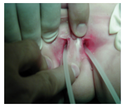
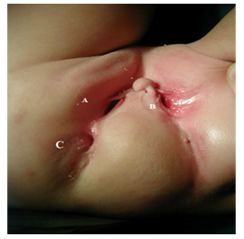
At the age of 3, the child was admitted in our clinic. During physical examination, we found that she had two uteri and two cervices with two separate vaginas. She passed urine from both urethras inside vaginal openings. Cystoscopic examination under anesthesia revealed a normal vagina, urethra, ureteric orifice, and a small bladder on the left side. On the other hand, on the right side the vagina was normal and urethral opening was at the fornices of the vagina. On the left side normal vaginal orifice was found, while on the right side, the orificium vaginae was deep in the wall of vaginal fornix and it was incompetent (Figures 1, 2).
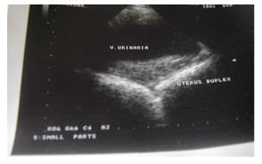
Pelvic ultrasound examination revealed bicornuate uterus (Figure 3).
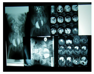
Ultrasonic and urography examinations showed a complete bladder duplication, crossed fused ectopic kidney (ectopic left kidney that was crossed and fused with the right kidney), two ureters deriving from the fused kidney and terminated separately into the duplicated bladder. The kidneys were functioning properly. In addition, CT showed two fused spleens (Figure 4) and several skeletal abnormalities (spina bifida, hemivertebrae).
The diagnosis was confirmed by ultrasound, intravenous urography, retrograde pyelography, miction cystography, CT, scintigraphy and radioisotope renography (Figure 4). At age 3, both bladders were exposed and opened via a single transverse incision. The complete separating wall was excised to the bladder base, the remaining mucosa of the right urethra was stripped out, the mucosal defect was closed, and the resulting single bladder was drained. Three months after surgery the urinary functions appeared normal for her age. The girl is still object of surgical treatment and follow-up observation.
5. Discussion
Congenital kidney abnormalities are often associated with the urinary tract malformation, and there is a wide range of anomalies resulting from disorders in the development process. Developmental anomalies of the urinary tract affect 10 percent of the population, accounting for one third of all congenital malformations [6]. Over two thirds of those found to have renal anomalies have anomalies in other organ systems [6, 7].
Renal and extrarenal anomalies [8] are very common in renal ectopy. A 55% incidence of associated anomalies is reported in series of 378 cases of crossed fused renal ectopy [9]. In our case left kidney was crossed and fused with the right kidney. Because there is an increased incidence of renal anomalies (unilateral renal agenesis, pelvic or horseshoe kidneys, or irregularities in the collecting system),additional radiologic evaluation should be pursued in the setting of a congenital Müllerian anomaly. Disorders of sex development comprise a heterogeneous group of congenital conditions associated with atypical development of internal and external genitalia. Congenital uterine anomalies arise owing to abnormal development of the Mullerian ducts during embryogenesis and have been associated with reduced fertility, miscarriage, preterm birth, and other adverse fetal outcomes. (M. Prior et al. 2018).
The most common anomalies of the uterus result from either: Incomplete fusion of the paramesonephric ducts, Incomplete dissolution of the midline fusion of those ducts and formation failures. Most common Anomalies are: Uterus didelphys represent two separate uterine bodies, each with its own cervix and attached fallopian tube and vagina. A bicornuate uterus with a rudimentary horn represents a fusion failure. bicornuate uterus with or without double cervices. Bicornuate and unicornuate uteri are associated with second-trimester pregnancy loss, malpresentation, and preterm labor and delivery. Septate uterus represents incomplete dissolution of the midline fusion of the paramesonephrica, 25% of women with uterine septa may suffer from recurrent first-trimester pregnancy loss.Unicornuate uterus represent failure of formation with normal karyotypic and phenotypic females + anomalies of the urinary system such as a horseshoe or pelvic kidney. Complex urogenital anomalies associated with imperforate anus, uterus didelphys represent two separate uterine bodies, each with its own cervix and attached fallopian tube and vagina (M. Prior et al. 2018).As to our knowledge, there have been no report published for this syndrome in Balkan`s region, and in literature. We consider that it would be of interest to report our case.
Knowledge of the correct genitourinary embryology is essential for the understanding, study, diagnosis and subsequent treatment of genital and other malformations, especially complex ones and those that lead to gynecological and urinary malfunctions. Adequate management of any associated bladder dysfunction is essential to preserving renal function, minimizing risk of urinary tract infection, and potentially avoiding need for future reconstructive surgery. Vaginal duplication along with renal anomalies need multiple studies prior to surgical intervention because different anatomical versions might require different surgical procedures. Multidisciplinary approach involving the urologist, pediatric surgeon, neonatologists, endocrinologists, perinatologists, radiologist and the gynecologist with expertise in this field.
6. Conclusion
Errors in development can occur, from minor, clinically insignificant disorders to severe abnormalities that are devastating to the child and parents. In our unique case congenital abnormalities affected the external genitalia in combination with internal genital anomalies involving other organ systems. At the time of delivery, child was required immediate intervention due to the clinical obstructive anomalies, and clinically significant symptoms. In this complicated case, additional information can be obtained by further examination under anesthesia, (vaginoscopy, hysteroscopy and laparoscopy, and maybe another surgical intervention unless the child has reached reproductive age.
References
- World Health Organization.Executive summary (2009).
- Lloyd DA. Principles of pediatric surgery. 2nd Edn. O'Neill JA Jr, Grosfeld JL, Fonkalsrud EW, Coran AG, Caldamone AC (eds). 220 × 283 mm. Pp. 895. Illustrated. 2004. Mosby (Elsevier): New York, British Journal of Surgery 92 (2005): 123-123.
- Keith Edmonds D, Gillian L Rose. Outflow tract disorders of the female genital tract. The Obstetrician and Gynaecologist 15 (2013): 11-17.
- Carlos Simón, Luis Martinez, Francisco Pardo, et al. Müllerian defects in women with normal reproductive outcome', Fertility and Sterility 56 (1991): 1192-1193.
- Edmonds DK. Congenital malformations of the genital tract', Obstetrics and Gynecology Clinics of North America 27 (2000): 49-62.
- Kevin A Burbige,Terry W Hensle. Uterus Didelphys and Vaginal Duplication with Unilateral Obstruction Presenting as a Newborn Abdominal Mass. Journal of Urology 132 (1984): 1195-1198.
- Lloyd DA. Principles of pediatric surgery. 2nd Edn. O'Neill Jr JA, Grosfeld JL, Fonkalsrud EW, Coran AG, Caldamone C (eds). 220 × 283 mm. Pp. 895. Illustrated. 2004. Mosby (Elsevier): New York. British Journal of Surgery 92 (2005): 123-123.
- Dewhurst CJ. Congenital Malformations of the Genital Tract In Childhood, Bjog: An International Journal of Obstetrics and Gynaecology 75 (1968): 377-391.
- Kruepunga N, Hikspoors JPJM, Mekonen HK, et al. The development of the cloaca in the human embryo. J. Anat 233 (2018): 724-739.
- Goossens WJ, de Blaauw I, Wijnen MH, et al. Urogenital anomalies in anorectal malformations in the Netherlands: effects of screening all patients on long- term outcome. Pediatr. Surg. Int. (2011).
- Daoub TM Drake. Congenital abnormalities of the urogenital tract: the clue is in the cord? Case Reports 2014 (2014): bcr2014208172-bcr2014208172.
- Mileto A, Itani M, Katz D, et al. Fetal Urinary Tract Anomalies: Review of Pathophysiology, Imaging, and Management. Imaging od Fetal Urinary Tract Anomalies. AJR: 210 (2018).
- Gerard S Letterie. Combined Congenital Absence of the Vagina and Cervix. Gynecologic and Obstetric Investigation 46 (1998): 65-67.
- Gough MH. Anorectal Agenesis with Persistence of Cloaca. Proceedings of the Royal Society of Medicine 52 (1959): 886-889.


