Chylothorax: An Unsuspected Presentation of Lymphoproliferative Disease
Article Information
Áurea Lima1-3*, Diana Dias4, Alcinda Reis5, Joana Malheiro4, Joana Rodrigues4
5Radiology Service, Centro Hospitalar de Entre o Douro e Vouga, EPE, São Sebastião Hospital, Portugal
*Corresponding Author: Áurea Lima, Hospital de São Sebastião. Serviço de Oncologia Médica. R. Dr. Cândido
Received: 18 January 2021; Accepted: 02 February 2021; Published: 01 March 2021
Citation: Áurea Lima, Diana Dias, Alcinda Reis, Joana Malheiro, Joana Rodrigues. Chylothorax: An Unsuspected Presentation of Lymphoproliferative Disease. Archives of Clinical and Medical Case Reports 5 (2021): 221-225.
View / Download Pdf Share at FacebookKeywords
Chylothorax; Hodgkin lymphoma; Lymphoproliferative disease; PET/CT
Chylothorax articles Chylothorax Research articles Chylothorax review articles Chylothorax PubMed articles Chylothorax PubMed Central articles Chylothorax 2023 articles Chylothorax 2024 articles Chylothorax Scopus articles Chylothorax impact factor journals Chylothorax Scopus journals Chylothorax PubMed journals Chylothorax medical journals Chylothorax free journals Chylothorax best journals Chylothorax top journals Chylothorax free medical journals Chylothorax famous journals Chylothorax Google Scholar indexed journals Hodgkin lymphoma articles Hodgkin lymphoma Research articles Hodgkin lymphoma review articles Hodgkin lymphoma PubMed articles Hodgkin lymphoma PubMed Central articles Hodgkin lymphoma 2023 articles Hodgkin lymphoma 2024 articles Hodgkin lymphoma Scopus articles Hodgkin lymphoma impact factor journals Hodgkin lymphoma Scopus journals Hodgkin lymphoma PubMed journals Hodgkin lymphoma medical journals Hodgkin lymphoma free journals Hodgkin lymphoma best journals Hodgkin lymphoma top journals Hodgkin lymphoma free medical journals Hodgkin lymphoma famous journals Hodgkin lymphoma Google Scholar indexed journals Lymphoproliferative disease articles Lymphoproliferative disease Research articles Lymphoproliferative disease review articles Lymphoproliferative disease PubMed articles Lymphoproliferative disease PubMed Central articles Lymphoproliferative disease 2023 articles Lymphoproliferative disease 2024 articles Lymphoproliferative disease Scopus articles Lymphoproliferative disease impact factor journals Lymphoproliferative disease Scopus journals Lymphoproliferative disease PubMed journals Lymphoproliferative disease medical journals Lymphoproliferative disease free journals Lymphoproliferative disease best journals Lymphoproliferative disease top journals Lymphoproliferative disease free medical journals Lymphoproliferative disease famous journals Lymphoproliferative disease Google Scholar indexed journals PET/CT articles PET/CT Research articles PET/CT review articles PET/CT PubMed articles PET/CT PubMed Central articles PET/CT 2023 articles PET/CT 2024 articles PET/CT Scopus articles PET/CT impact factor journals PET/CT Scopus journals PET/CT PubMed journals PET/CT medical journals PET/CT free journals PET/CT best journals PET/CT top journals PET/CT free medical journals PET/CT famous journals PET/CT Google Scholar indexed journals CIED articles CIED Research articles CIED review articles CIED PubMed articles CIED PubMed Central articles CIED 2023 articles CIED 2024 articles CIED Scopus articles CIED impact factor journals CIED Scopus journals CIED PubMed journals CIED medical journals CIED free journals CIED best journals CIED top journals CIED free medical journals CIED famous journals CIED Google Scholar indexed journals treatment articles treatment Research articles treatment review articles treatment PubMed articles treatment PubMed Central articles treatment 2023 articles treatment 2024 articles treatment Scopus articles treatment impact factor journals treatment Scopus journals treatment PubMed journals treatment medical journals treatment free journals treatment best journals treatment top journals treatment free medical journals treatment famous journals treatment Google Scholar indexed journals CT articles CT Research articles CT review articles CT PubMed articles CT PubMed Central articles CT 2023 articles CT 2024 articles CT Scopus articles CT impact factor journals CT Scopus journals CT PubMed journals CT medical journals CT free journals CT best journals CT top journals CT free medical journals CT famous journals CT Google Scholar indexed journals surgery articles surgery Research articles surgery review articles surgery PubMed articles surgery PubMed Central articles surgery 2023 articles surgery 2024 articles surgery Scopus articles surgery impact factor journals surgery Scopus journals surgery PubMed journals surgery medical journals surgery free journals surgery best journals surgery top journals surgery free medical journals surgery famous journals surgery Google Scholar indexed journals Pathogenesis articles Pathogenesis Research articles Pathogenesis review articles Pathogenesis PubMed articles Pathogenesis PubMed Central articles Pathogenesis 2023 articles Pathogenesis 2024 articles Pathogenesis Scopus articles Pathogenesis impact factor journals Pathogenesis Scopus journals Pathogenesis PubMed journals Pathogenesis medical journals Pathogenesis free journals Pathogenesis best journals Pathogenesis top journals Pathogenesis free medical journals Pathogenesis famous journals Pathogenesis Google Scholar indexed journals ABVD articles ABVD Research articles ABVD review articles ABVD PubMed articles ABVD PubMed Central articles ABVD 2023 articles ABVD 2024 articles ABVD Scopus articles ABVD impact factor journals ABVD Scopus journals ABVD PubMed journals ABVD medical journals ABVD free journals ABVD best journals ABVD top journals ABVD free medical journals ABVD famous journals ABVD Google Scholar indexed journals
Article Details
Abbreviations:
ABVD: Adriamycin-Bleomycin-Vinblastine-Dacarbazine; CT: computed tomography; LDH: lactate dehydrogenase; PET/CT: positron emission computed tomography; 18F-FDG: 18-F-fluoro-2-deoxyglucose
1. Clinical Images of Interest
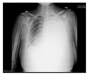
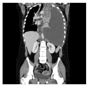
A healthy 20-year-old woman intended to the emergency department because of a 2-months clinical course of progressive asthenia and dyspnea, associated with left neck pain that is unresponsive to the instituted analgesia. No other positive findings were issued in the anamnesis. At physical examination it was worth mentioning sinus tachycardia, tachypnea and auscultatory silence throughout the left hemithorax, without respiratory failure. Chest radiography (Figure 1) revealed opacity of the entire left pulmonary hemithorax, with significant contralateral mediastinal deviation. Analytical studies revealed: hypochromic microcytic anemia, increased lactate dehydrogenase (LDH) and increased C-reactive protein. Thoraco-abdomino-pelvic computed tomography (CT) (Figure 2) showed a solid mass (78 × 94 × 121 mm) apparently centered in the anterior mediastinum and with supra-clavicular extension, with lobulated contours and without cleavage plans with the pericardium, the aortic cross or the thoracic operative, as well as, a total left lung atelectasis and pleural effusion in the entire left hemithorax, with signs of compression and contralateral mediastinal deviation. Thoracentesis was performed with drainage of 1000 mL of milky-looking pleural fluid whose analysis was compatible with chylothorax (amicrobial, pH 7.5, 935 leukocytes/µL with 29% polymorphonuclear cells and a clear predominance of lymphocytes, 5.3 g/dL proteins, 684 mg/dL triglycerides, 123 mg/dL cholesterol, normal glucose and LDH).
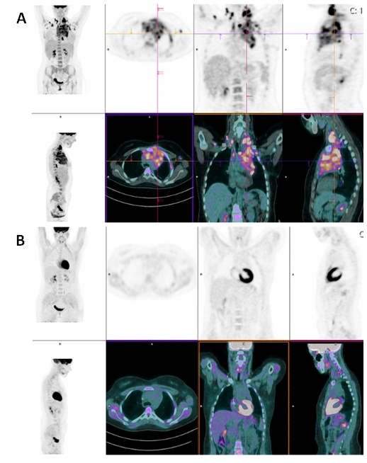
Patient was admitted at a cancer service and the diagnosis of classic Hodgkin's lymphoma of the nodular sclerosis subtype (stage II-B) was established. The staging 18-F-fluoro-2-deoxyglucose (18F-FDG) positron emission computed tomography (PET/CT) (Figure 3A) showed abnormal 18F-FDG uptake in the anterior mediastinum and in the left superior and posterior cervical, supra-clavicular and axillary ganglia bilaterally, and in the upper trachea, as well as in the left pleura. First line chemotherapy with ABVD (Adriamycin-Bleomycin-Vinblastine-Dacarbazine) was performed without relevant adverse drug events. After the 2nd cycle of chemotherapy, the PET/CT revealed a complete remission (Figure 3B).
Chylothorax is a rare but serious condition caused by obstruction or disruption of the lymphatic branches draining the lower body and gastrointestinal tract. Chylothorax is characterized by the presence of lymphatic fluid with triglycerides and chylomicrons in the pleural cavity [1]. Etiologically, it can be divided into traumatic or non-traumatic, and this separation is of pathophysiological importance, since the treatment should be individualized aiming at the underlying cause of the condition. Traumatic chylothoraxes’ represent the majority of cases, with most arising as postoperative complications of surgery; the most common cause of non-traumatic chylothoraxes’ is cancer [1]. The images presented aim to exemplify one possible presentation of Hodgkin's lymphoma: indolent, without serious clinical repercussions, even though the extensive pleural effusion and the large associated mediastinal mass. It also reinforces the importance of the differential diagnosis of pleural effusion, recalling the hypothesis of chylothorax as an underlying cause. Furthermore, it highlights the role of 18F-FDG-PET/CT as a functional imaging modality that has become a standard tool in the management of Hodgkin lymphoma [2].

Figure 1: Chest radiography demonstrates opacity of the entire left pulmonary hemithorax, with significant contralateral deviation of the mediastinum.

Figure 2: Coronal reformatted contrast-enhanced thoraco-abdomino-pelvic CT image shows: an anterior mediastinic solid mass (78 × 94 × 121mm), with heterogeneous enhancement, lobulated contours, without fat or calcifications, encircling the aortic arch and abutting the pericardium; left lung atelectasis and massive pleural effusion, occupying the entire left hemithorax with contralateral mediastinal shift.

Figure 3: PET/CT scan before starting chemotherapy (A) shows an heterogeneous increased uptake of 18F-FDG in a large mass of the anterior mediastinum, in left superior and posterior cervical lymphadenopathies, in right paratraqueal and in supraclavicular and axillary bilaterally lymph nodes, as well as in the left pleura. After 2 cycles of ABVD chemotherapy (B) hypermetabolic changes of 18F-FDG are not apparent suggesting complete remission of the disease.
Conflicts of Interest
I declare on behalf of all authors that we do not have financial interests related to the work described in this paper.
Communication with the Media and Embargo
We do not have plans for publicizing this paper.
References
- Hvass M, Fransen JL, Bruun JM. Chylothorax. Ugeskr Laeger 179 (2017): pii: V05170429.
- Andrew M Evens, Lale Kostakoglu. The role of FDG-PET in defining prognosis of Hodgkin lymphoma for early-stage disease. Blood 124 (2014): 3356-3364.
