Bilateral Optic Disc Pit, Bilateral Iris Chorioretinal Coloboma and Unilateral Congenital Ptosis in a Person with Persistent Patent Ductus Arterious: A Rare Case with Review of Literatures
Article Information
Raba Thapa MD, PhD*
Vitreoretina Specialist, Tilganga Institute of Ophthalmology, Kathmandu, Nepal
*Corresponding Author: Dr. Raba Thapa MD, PhD, Associate Professor, National Academy of Medical Sciences, Vitreo-retinal Service, Tilganga Institute of Ophthalmology, Kathmandu, Nepal
Received: 05 September 2019; Accepted: 02 October 2019; Published: 10 April 2020
Citation: Raba Thapa. Bilateral Optic Disc Pit, Bilateral Iris Chorioretinal Coloboma and Unilateral Congenital Ptosis in a Person with Persistent Patent Ductus Arterious: A Rare Case with Review of Literatures. Archives of Clinical and Medical Case Reports 4 (2020): 302-306.
View / Download Pdf Share at FacebookAbstract
We report a 24 year old female presented with drooping of left eye upper lid since birth. Examination revealed left eye upper lid congenital ptosis, both eyes iris, chorioretinal coloboma and both eyes optic disc pit. Her medical records showed ligation of patent ductus arteriosus in her childhood. The ductus arteriosus closes soon after birth, but sometimes it fails to close leading to persistent patent ductus arteriosus. Optic disc pit, iris chorioretinal coloboma and congenital ptosis are among the rare congenital anomalies of eye. We report this case because of its rarity with review of literatures.
Keywords
Congenital ptosis; Chorioretinal coloboma; Iris coloboma; Optic disc pit; Patent ductus arteriosus
Congenital ptosis articles, Chorioretinal coloboma articles, Iris coloboma articles, Optic disc pit articles, Patent ductus arteriosus articles
Congenital ptosis articles Congenital ptosis Research articles Congenital ptosis review articles Congenital ptosis PubMed articles Congenital ptosis PubMed Central articles Congenital ptosis 2023 articles Congenital ptosis 2024 articles Congenital ptosis Scopus articles Congenital ptosis impact factor journals Congenital ptosis Scopus journals Congenital ptosis PubMed journals Congenital ptosis medical journals Congenital ptosis free journals Congenital ptosis best journals Congenital ptosis top journals Congenital ptosis free medical journals Congenital ptosis famous journals Congenital ptosis Google Scholar indexed journals ptosis articles ptosis Research articles ptosis review articles ptosis PubMed articles ptosis PubMed Central articles ptosis 2023 articles ptosis 2024 articles ptosis Scopus articles ptosis impact factor journals ptosis Scopus journals ptosis PubMed journals ptosis medical journals ptosis free journals ptosis best journals ptosis top journals ptosis free medical journals ptosis famous journals ptosis Google Scholar indexed journals Chorioretinal coloboma articles Chorioretinal coloboma Research articles Chorioretinal coloboma review articles Chorioretinal coloboma PubMed articles Chorioretinal coloboma PubMed Central articles Chorioretinal coloboma 2023 articles Chorioretinal coloboma 2024 articles Chorioretinal coloboma Scopus articles Chorioretinal coloboma impact factor journals Chorioretinal coloboma Scopus journals Chorioretinal coloboma PubMed journals Chorioretinal coloboma medical journals Chorioretinal coloboma free journals Chorioretinal coloboma best journals Chorioretinal coloboma top journals Chorioretinal coloboma free medical journals Chorioretinal coloboma famous journals Chorioretinal coloboma Google Scholar indexed journals coloboma articles coloboma Research articles coloboma review articles coloboma PubMed articles coloboma PubMed Central articles coloboma 2023 articles coloboma 2024 articles coloboma Scopus articles coloboma impact factor journals coloboma Scopus journals coloboma PubMed journals coloboma medical journals coloboma free journals coloboma best journals coloboma top journals coloboma free medical journals coloboma famous journals coloboma Google Scholar indexed journals Iris coloboma articles Iris coloboma Research articles Iris coloboma review articles Iris coloboma PubMed articles Iris coloboma PubMed Central articles Iris coloboma 2023 articles Iris coloboma 2024 articles Iris coloboma Scopus articles Iris coloboma impact factor journals Iris coloboma Scopus journals Iris coloboma PubMed journals Iris coloboma medical journals Iris coloboma free journals Iris coloboma best journals Iris coloboma top journals Iris coloboma free medical journals Iris coloboma famous journals Iris coloboma Google Scholar indexed journals treatment articles treatment Research articles treatment review articles treatment PubMed articles treatment PubMed Central articles treatment 2023 articles treatment 2024 articles treatment Scopus articles treatment impact factor journals treatment Scopus journals treatment PubMed journals treatment medical journals treatment free journals treatment best journals treatment top journals treatment free medical journals treatment famous journals treatment Google Scholar indexed journals Optic disc pit articles Optic disc pit Research articles Optic disc pit review articles Optic disc pit PubMed articles Optic disc pit PubMed Central articles Optic disc pit 2023 articles Optic disc pit 2024 articles Optic disc pit Scopus articles Optic disc pit impact factor journals Optic disc pit Scopus journals Optic disc pit PubMed journals Optic disc pit medical journals Optic disc pit free journals Optic disc pit best journals Optic disc pit top journals Optic disc pit free medical journals Optic disc pit famous journals Optic disc pit Google Scholar indexed journals Patent ductus arteriosus articles Patent ductus arteriosus Research articles Patent ductus arteriosus review articles Patent ductus arteriosus PubMed articles Patent ductus arteriosus PubMed Central articles Patent ductus arteriosus 2023 articles Patent ductus arteriosus 2024 articles Patent ductus arteriosus Scopus articles Patent ductus arteriosus impact factor journals Patent ductus arteriosus Scopus journals Patent ductus arteriosus PubMed journals Patent ductus arteriosus medical journals Patent ductus arteriosus free journals Patent ductus arteriosus best journals Patent ductus arteriosus top journals Patent ductus arteriosus free medical journals Patent ductus arteriosus famous journals Patent ductus arteriosus Google Scholar indexed journals arteriosus articles arteriosus Research articles arteriosus review articles arteriosus PubMed articles arteriosus PubMed Central articles arteriosus 2023 articles arteriosus 2024 articles arteriosus Scopus articles arteriosus impact factor journals arteriosus Scopus journals arteriosus PubMed journals arteriosus medical journals arteriosus free journals arteriosus best journals arteriosus top journals arteriosus free medical journals arteriosus famous journals arteriosus Google Scholar indexed journals Surgery articles Surgery Research articles Surgery review articles Surgery PubMed articles Surgery PubMed Central articles Surgery 2023 articles Surgery 2024 articles Surgery Scopus articles Surgery impact factor journals Surgery Scopus journals Surgery PubMed journals Surgery medical journals Surgery free journals Surgery best journals Surgery top journals Surgery free medical journals Surgery famous journals Surgery Google Scholar indexed journals
Article Details
1. Introduction
During fetal life, most of the blood from pulmonary artery passes through the ductus arteriosus in to the aorta just below the origin of left subclavian artey. Normally, the ductus closes soon after birth. Persistence of patent ductus arteriosus (PDA) may be associated with other abnormalities and is much commoner in females. Small shunts are asymptomatic but large shunts may lead to retardation of growth and development. It can ultimately lead to cardiac failure. Most of the persistent ductus can be closed by percutaneous occlusion with the Rashkind umbrella or similar devices. Treatment with surgical division is also safe [1]. Iris and chorioretinal colobomas of the eye are caused by failure of the embryonic fissure of the optic cup to close during development. Typically, colobomas are located at the lower part of iris, chorioretina and optic disc. Chorioretinal coloboma can involve the part of optic disc and macula. Parents bring child for the poor vision, deviated eyes, small eyes or marked diminution of vision due to retinal detachment [2, 3]. Congenital ptosis may be simple or complicated associated with other ocular anomalies. Ptosis covering the visual axis could lead to amblyopia if not corrected on time [4].
We report a case of PDA associated with multiple congenital anomalies of eye; unilateral congenital ptosis, both eyes iris, chorioretinal coloboma and optic disc pit for its rarity. This case can help the ophthalmologist and cardiologist for referral and cross-referral of such cases for prompt treatment to avoid the sight and life threatening complications.
2. Case Report
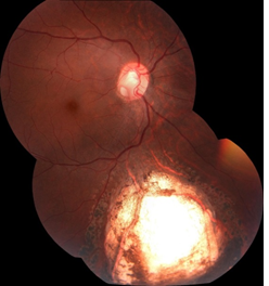
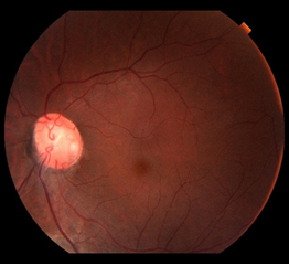
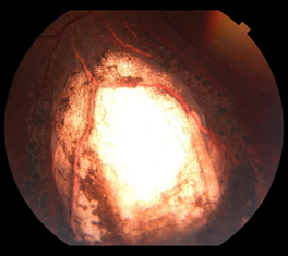
Twenty four years female was presented at Tilganga Institute of Ophthalmology with the chief complains of drooping of left eye lid since birth. She had no history of visual problems and other ocular problems. She had past history of Patent Ductus Arteriosus ligation 16 years back at the age of 8 years. On ocular examination, her best corrected visual acuity (BCVA) was 6/6 in both eyes (right eye: -0.25/-0.5 *90 degree, left eye: -0.25/-0.75*90 degree). There was congenital ptosis of left upper eye lid. The levator palpebral superioris (LPS) function was 7 mm, margin reflex distance (MRD1) was +1, and interpalpebral height was 8 mm in left eye. On anterior segment examination, she had bilateral iris coloboma located in the inferior part in both iris. Lens was normal. Fundus examination revealed bilateral optic disc pit and inferior chorioretinal coloboma not involving the optic disc and macula (Figure 1-3). Macula was normal. The rest of the retina was also flat. Eye lid revealed normal finding in right eye. Her intraocular pressure was 18 mmHg in both eyes on applanation tonometry. She was diagnosed as left eye upper lid congenital ptosis with bilateral iris and chorio-retinal coloboma with bilateral optic disc pit with refractive error. She was treated with barrage laser in both eyes for chorioretinal coloboma in 2015. She is in regular follow up since then and at her last follow up on July 2019, her BCVA was 6/6 and stable anterior and posterior segment findings. She was again advised for regular use of glass and follow up at six months interval or earlier as needed.

Figure 1: Right eye fundus photo showing chorioretinal coloboma and optic disc pit.

Figure 2: Left eye fundus photo showing optic disc pit.

Figure 3: Left eye fundus photo showing chorioretinal coloboma.
3. Discussion
Congenital anomalies can involve any systems of the body. Careful suspicion and thorough evaluation is required to assess for its multisystem involvement. Here we report a case of multiple ocular congenital anomalies with congenital defect in heart. To the best of our knowledge, there are no typical reported cases of congenital ptosis, optic disc pit and iris, chorioretinal coloboma in a single person with PDA. Congenital ptosis can be simple or complicated. It may lead to occlusion amblyopia in severe cases [4]. Mild ptosis may be associated with Char syndrome, a rare genetic multiple congenital anomalies characterized by triad of PDA, facial dysmorphism and hand anomalies [3]. Although there was ptosis and PDA in our cases, there were absence of other abnormalities suggestive of Char syndrome. Optic disc pit may lead to serous detachment of macula. If not treated on time, it can lead to pigmentary changes in macula and macular scar leading to irreversible visual impairment. Treatment of such cases are also challenging [5, 6]. Iris and chorioretinal coloboma often coexist together [3]. Our cases was consistent with this findings. Chorioretinal coloboma may lead to retinal detachment. Hussain et al. reported bilateral chorioretinal coloboma in 65.5% of his cases series and retinal detachment was found in 29.5% of the cases [7]. Presence of bilateral chorioretinal coloboma was consistent with the majority of their cases series. Uhumwangho and Jalali reported presence of retinal detachment in 17.6% and 87.2% of these cases had other ocular anomalies. The prevalence of retinal detachment among those who received prophylactic laser was 2.9% [8]. Absence of retinal detachment in the follow up of our patient could be because of the prior prophylactic laser in chorioretinal coloboma. Nakaura reported both presence of iris and chorioretinal coloboma in 24% of their cases series like in our case [9]. Ming et al reported colobomas and other ophthalmic anomalies in Kabuki Syndrome [10]. However, our cases had no other features suggestive of this syndrome. Our patient presented for the first time for cosmetic reason due to ptosis. Iris, chorioretinal coloboma and optic disc pit were diagnosed during her routine evaluation of anterior and posterior segment of eye. Furthermore, despite the presence of PDA diagnosis and its management at childhood, patient was not consulted for ophthalmic anomalies until the adolescent. So timely referral and systemic evaluation could help in timely detection of such congenital anomalies to save the sight and life.
4. Conclusion
Persistent ductus Arteriosus could be associated with multiple congenital anomalies of eye like congenital ptosis, iris, chorioretinal coloboma and optic disc pit. This case report highlights the importance of timely ocular evaluation for possible other congenital anomalies of eye in cases with PDA to save the sight. Cross referral has to be strengthened between ophthalmologist and cardiologist for timely diagnosis and treatment of life and sight threatening complications from these congenital disorders.
Funding
None
Conflict of Interest
None
References
- Edwards CRW, Bouchier IAD, Haslett C. Davidson’s Principles and Practice of Medicine. ELBS, 17th Edition (1995).
- Sun M, Wei D, lin H, et al. Coloboma of Iris and Choroid: Two Case Reports. HSOA J Ophthalmic Clin Res (2017): 034.
- Satoda M, Pierpont ME, Diaz GA, et al. Char Syndrome, an Inherited Disorder With Patent Ductus Arteriosus, Maps to Chromosome 6p12-p21. Circulation (1999): 3036-3042.
- Thapa R. Refractive error, Strabismus and Amblyopia in Congenital Ptosis. J Nepal Med Assoc (2010): 43-46.
- Chatziralli I, Theodossiadis P, Theodossiadis GP. Optic disc pit maculopathy: current management strategies. Clinical Ophthalmol (2018): 1417-1422.
- Moisseiev E, Joseph Moisseiev J, Anat Loewenstein A. Optic disc pit maculopathy: when and how to treat? A review of the pathogenesis and treatment options. Int J Retin Vitr (2015): 13.
- Hussain RM, Abbey AM, Shah AR, et al. Chorioretinal Coloboma Complications: Retinal Detachment and Choroidal Neovascular Membrane. J Ophthalmic Vis Res (2017): 3-10.
- Uhumwangho OM, Jalali S. Chorioretinal coloboma in a paediatric population. Eye (2014): 728-733.
- Nakamura KM, Diehl NN, Mohney BG. Incidence, Ocular Findings and Systemic Associations of Ocular Coloboma: A Population-Based Study. Arch Ophthalmol (2011): 69-74.
- Ming JE, Russell KL, Bason L, et al. Coloboma and other ophthalmologic anomalies in Kabuki syndrome: distinction from charge association. Am J Med Genet A (2003): 249-252.
