Aberrant Left Subclavian Artery with Kommerell’s Diverticulum And Right Sided Aortic Arch: Hybrid Approach
Article Information
Juliette Strella1, Quentin Langouet1, Etienne Marchand1, Julien Pucheux2, Antoine Legras1*, Robert Martinez1
1Department of Thoracic and Cardio-Vascular Surgery, Tours University Hospital, Tours, France
2Department of Radiology, Tours University Hospital, Tours, France
*Corresponding Author: Antoine Legras, Department of Thoracic and Cardio-Vascular Surgery, Tours University Hospital, Tours, France
Received: 25 May 2022; Accepted: 31 May 2022; Published: 22 June 2022
Citation: Juliette Strella, Quentin Langouet, Etienne Marchand, Julien Pucheux, Antoine Legras, Robert Martinez. Aberrant Left Subclavian Artery with Kommerell’s Diverticulum And Right Sided Aortic Arch: Hybrid Approach. Journal of Surgery and Research 5 (2022): 361-364.
View / Download Pdf Share at FacebookAbstract
A 58 years-old woman presented a rare right-sided aortic arch with an aberrant left subclavian retro-esophageal artery, originates from Kommerell’s diverticulum. After left subclavian to carotid transposition, we implanted a thoracic endoprosthesis under ventricular fibrillation. Type IA symptomatic proximal endoleak was treated with a second endograft a week later. We shared here technical aspects and challenges of endovascular management, including precise preoperative imaging (CT angiography, lymphangio-MRI), the need of a hybrid operative room, conformable endoprosthesis and right ventricle overstimulation.
Keywords
Subclavian artery, Endovascular technique, Minimally invasive technique, Aortic aneurysm, Congenital vascular abnormality
Article Details
1. Introduction
Combination of right sided aorta and aberrant left subclavian retro-esophageal artery originates from a Kommerell’s diverticulum is a rare entity and its treatment remains complex. Here we reported a case of a 58 years-old woman presenting dysphagia to share technical aspects and challenges of endovascular management.
2. Case report
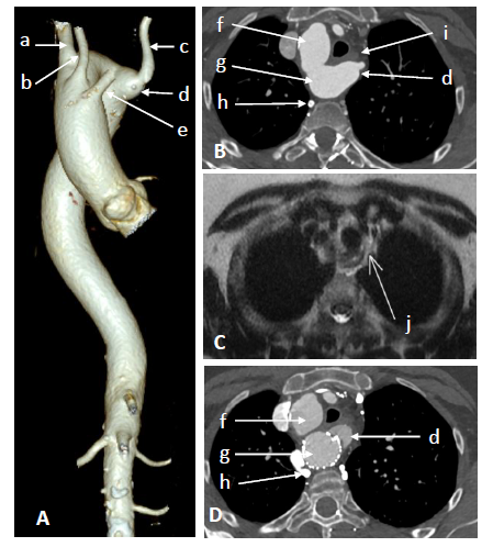
A 58 years-old woman was admitted for dysphagia for about one year and a half and permanent retro-sternal pain associated with a weight loss of 25kg. A chest computerized tomography angiography (CTA) showed a right-sided 25 mm aortic arch with an aberrant left subclavian retro-esophageal artery, originates from a 17 mm (orificial diameter) Kommerell’s diverticulum (Figure 1A, B) and hypoplasia of left vertebral artery. Distal aorta was normal. Lymphangio-MRI showed that the thoracic duct was at the left side of esophagus (Figure 1C). Consent from the patient was obtained.
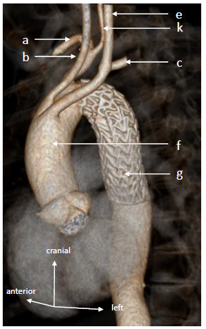
A left subclavian to carotid transposition was first performed. Despite proximal left subclavian artery transection and ligature, persistent dysphagia led us to implant 3 months later, in a second step, a 28-28-10 thoracic endoprosthesis GORE (DEL, USA), under ventricular fibrillation [1] in a hybrid operative room, leading to exclusion of the diverticulum. Endograft was post-plastied with an aortic balloon after placement. At day one, back pain occurred. A chest CTA showed that endoprosthesis had moved backward and diverticulum was still permeable with type IA endoleak (Figure 1D). A new 28-28-10 endoprosthesis was implanted upstream the first, excluding the diverticulum. Symptoms disappeared. At 3 months, dysphagia disappeared, and CTA showed a complete retraction of the diverticulum (Figure 2).
Figure 1: A: 3-Dimensional reconstruction of CT angiography, front view, showing : a : right subclavian artery, b : right carotid artery, c : left subclavian artery, d : kommerell’s diverticulum, e : left carotid artery. B: Axial section of CT angiography showing : f : ascending aorta, g : descending right sided aorta, h : azygos vein, i : esophagus, d : kommerell’s diverticulum. C: Axial section of lymphangio-MRI showing the thoracic duct (j). D: Axial section of CT angiography showing the diverticulum after the first endoprosthesis (d). CT, chest tomography; MRI, magnetic resonance imaging.
3. Discussion
During aortic arch development, the persistence of the right aortic arch, instead of the left one, lead to definitive right sided aortic arch [2], classified by Edwards [3]. Kommerell’s diverticulum was first described in 1936, as a diverticulum of the descending thoracic aorta, giving birth to an aberrant right subclavian artery. Position and anatomy of lymph pathways are unknown in case of such complex vascular variation. We proposed preoperative thoracic duct imaging using lymphangio-MRI, to localize thoracic duct and limit the risk of postoperative lymphatic leak. Atherosclerotic changes of the vessels may lead to rupture of compression of surrounding structures, responsible for dysphagia, dyspnea, cough, chest pain, recurrent pneumonia [4].
Mini-invasive hybrid approaches were developed to treat aortic arch lesions, associated with supra aortic debranching [5]. However, it was exceptionally performed in case of Kommerell’s diverticulum associated to right-sided aortic arch, due to the rarity of this condition [6]. Technical aspects and challenges of endovascular management should be shared, knowing that we experienced backward movement of the endoprosthesis, as previously described [6].
1) Debranching alone could sometimes eradicate symptoms [7]. 2) Hybrid operative room with 3-dimensional reconstruction offers accurate visualization of the diverticulum. 3) In case of right aortic arch with narrow angle, endoprosthesis should be conformable with orientable proximal segment, for a better congruence. 4) For precise endoprosthesis landing, we recommend right ventricle overstimulation [1]. 5) Be aware of symptoms recurrence that could reveal a leak due to endoprosthesis migration and may require another endovascular treatment.
4. Conclusion
Planning hybrid treatment of dysphagia due to Kommerell’s diverticulum should include CTA and evoke the interest of lymphangio-MRI. Complex aortic disease endovascular treatment should be considered in a hybrid operative room, under ventricular fibrillation with appropriate endoprosthesis.
Disclosure statement
Authors have no conflicts of interest to declare.
References
- Lhommet P, Espitalier F, Merlini T, et al. Tolerance of rapid right ventricular pacing during thoracic endovascular aortic repair. Ann Vasc Surg 29 (2015): 578-585.
- Cinà CS, Althani H, Pasenau J, et al. Kommerell’s diverticulum and right-sided aortic arch: a cohort study and review of the literature. J Vasc Surg 39 (2004): 131-139.
- Edwards JE. Anomalies of the derivatives of the aortic arch system. Med Clin North Am 32 (1948): 925-949.
- Austin EH, Wolfe WG. Aneurysm of aberrant subclavian artery with a review of the literature. J Vasc Surg 2 (1985): 571-577.
- Canaud L, Hireche K, Berthet JP, et al. Endovascular repair of aortic arch lesions in high-risk patients or after previous aortic surgery: midterm results. J Thorac Cardiovasc Surg 140 (2010): 52-58.
- Raymond SL, Gray SE, Peters KR, et al. Right-sided aortic arch with aberrant left subclavian artery and Kommerell diverticulum. J Vasc Surg Cases Innov Tech 5 (2019): 259-260.
- Thoreau B, Bigot A, Diallo BD, et al. A digital ischemia of unusual cause. Rev Med Interne 38 (2017): 218-219.


