Use of Locking Compression Plate with or without Bone Grafting in Intra Articular and/or Highly Comminuted Distal End Radius Fractures: Our Experience in Adult Working Population in a Semi Urban Setup
Article Information
Gautam A Taralekar1*, Girish Shinde1, Sasha Martyres1, Sachin G Shetti2, Lokesh Singh3
1Department of Orthopaedics, Bharati Vidyapeeth (Deemed to be University) Medical College and Hospital, Sangli, Maharashtra, India
2Department of Physiotherapy, Bharati Vidyapeeth (Deemed to be University) Medical College and Hospital, Sangli, Maharashtra, India
3Department of Radiodiagnosis, ESIC PGIMSR, New Delhi, India
*Corresponding Author: Gautam Taralekar, Department of Orthopaedics, Bharati Vidyapeeth (Deemed to be University) Medical College and Hospital, Sangli, Maharashtra, India, E-mail: dr.gautambvu@gmail.com
Received: 13 December 2020; Accepted: 23 December 2020; Published: 11 January 2021
Citation: Mohan Sai Krishna, Gautam A Taralekar, Vaidyanath Rahate, Aditya Deshmukh, Lokesh Singh. Use of Locking Compression Plate with or without Bone Grafting in Intra Articular and/or Highly Comminuted Distal End Radius Fractures: Our Experience in Adult Working Population in a Semi Urban Setup. Archives of Clinical and Biomedical Research 5 (2021): 16-26.
View / Download Pdf Share at FacebookAbstract
Background and Objective: Incidence of fractures of distal radius are increasing due to more geriatric population and road traffic accidents and at the same time surgical treatment options for the same are modified continuously. The fundamental goal of treating distal radial fractures is restoration of normal or near normal alignment and articular congruity. Internal fixation using different plates can rigidly stabilize cancellous fragmented bone along with augmentation with bone graft which may not be amenable to k-wire or External fixation. Thus, this study was carried out in adult working population to study the functional outcome and effectiveness of operative management and study the complications of the same in a semi urban population.
Method: This was a prospective observational study conducted in orthopaedic department of a tertiary center.30 patients with fractures of distal radius were included in the study on the basis of predefined inclusion and exclusion criteria. Patients were treated with open reduction and internal fixation with Locking Compression Plate through a volar approach and were followed up at 6 weeks, 3 months and with final assessment done at the end of 6 months post operatively and assessed clinically and radiologically.
Result: The study comprised of 18 male and 12 female patients aged from 19 to 74 years with mean age of 47.1 years and more of left side involvement. Intra articular fractures (frykman type VIII and III) are most common. 29 (97%) cases had union within 3-4 months. Using the demerit scoring system of Gartland and Werley we had 50% excellent, 40% good, 10% fair and 0 poor results.
Interpretation and Conclusion: In carefully selected patients even in the face of osteoporosis, fixation of fractures of distal end of radius with Locking Compression Plate along with bone grafting especially has satisfac
Keywords
Distal radius; Comminuted; Intra-articular & extra-articular; Open reduction internal fixation; Locking Compression Plate; Osteoporosis
Distal radius articles; Comminuted articles; Intra-articular & extra-articular articles; Open reduction internal fixation articles; Locking Compression Plate articles; Osteoporosis articles
Article Details
1. Introduction
Fractures of distal end of radius continue to pose a therapeutic challenge. Intraarticular and extra articular malalignment can lead to various complications like post traumatic osteoarthrosis, decreased grip strength and endurance, as well as limited motion and carpal instability [1]. Open reduction and internal fixation is indicated to address the unstable distal end radius fractures with articular incongruity and extra articular fractures with severe comminution in the metaphyseal bone with significant loss of radial length, that cannot be anatomically reduced and maintained through external manipulation and ligamentotaxis, provided sufficient bone stock is present to permit early range of motion [2]. Types of plates routinely used for fixing fractures of distal radius (a) Conventional Buttress plates and (b) Fixed angle locking compression plates. Conventional Buttress plates are used when there is good bone stock and where stability is achieved by compression of plate to bone by bicortical screws fixation [3, 4]. With severely comminuted fractures with intraarticular extension or in osteoporotic extra articular fractures with loss of radial length LCP provides support to the subchondral bone with the help of locking screws forming scaffold under the articular surface and allowing early post-operative rehabilitation [5, 6]. With Buttress plate or Locking Compression Plate, reduction can be maintained overtime so that secondary displacement is no longer a problem [7].
Primary stability achieved with screw and plate prevents secondary displacement irrespective of the bone quality enabling good results in osteoporotic bones and young patients. And with the addition of bone graft or cement in severely comminuted fractures due to osteoporosis or high energy injuries where there is difficulty in restoration of length due to collapse of soft metaphyseal subchondral bone, bone grafting augments the stability and length of the radius [8]. The development of this fixation technique theoretically improves the stability to maintain the reduction of fractures in osteoporotic bones and fractures that are considered to be unstable [9, 10]. The purpose of this study was to evaluate functional, clinical and radiological outcome of patients with distal radius fractures treated with volar plates with or without bone grafting to assess the effectiveness of the operative management and also study the complications associated.
2. Material and Methods
This was a prospective observational study conducted in orthopaedic department of a tertiary care center. 30 patients with fractures of distal radius were included in the study on the basis of predefined inclusion and exclusion criteria. Patients were treated with open reduction and internal fixation with Locking Compression Plate through a volar approach and were followed up at 6 weeks, 3 months and with final assessment done at the end of 6 months post operatively and assessed clinically and radiologically.
2.1 Inclusion Criteria
- Adults - Working population (aged between 19 to 74 years), both male and female with unstable, comminuted extra or intra articular fractures of distal end radius.
- Patients willing for treatment and given-informed written consent.
- Radiological findings confirming intra and extra articular fracture with combination of distal end Radius.
2.2 Exclusion Criteria
- Patients aged below 19 years.
- Patients medically unfit for surgery.
- Pathological fractures
- Patients not willing for surgery.
2.3 Pre-Operative Evaluation
2.3.1 Immediate Management: Following admission to the hospital, a careful history was elicited from the patients and / or attendants to reveal the mechanism of injury and the severity of trauma. Primary treatment in the form of slab was followed by filling up of the patient proforma which included the demographics, clinical examination and radiological findings.
2.4 Radiographic Examination
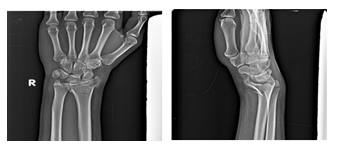
Standard radiographs in PA and lateral views were taken for confirmation of the diagnosis and also to know the type of fracture. Oblique views or CT scan were also taken in a few patients who had complex comminuted fractures. The fracture fragments were analysed and involvement of radiocarpal and distal radioulnar joints were assessed and classified according to the Frykman’s classification. Frykman’s classification is one of the most popular classifications established in 1967, was used to classify the fractures, to delineate the fracture geometry and plan surgery.
2.4.1 Pre-operative planning: Consent for surgery was taken and patients were operated after a pre-anaesthetic checkup and obtaining a pre-operative fitness for surgery after carrying out the required investigations as per the case.
2.5 Surgical Procedure
2.5.1 Anaesthesia: The operations were performed under interscalene or supra clavicular block and in those who needed bone grafting general anaesthesia was the choice.
2.5.2 Position: The patient was placed supine on the operating table with the affected limb placedon a side arm board. Forearm, hand and iliac crest (for those cases where bone grafting is planned) were thoroughly scrubbed, painted with betadine and spirit and draped.
2.5.3 Procedure
All the 30 were cases fixed with Locking Compression plate using a volar Henry approach.
2.5.4 Instruments And Implants Used
- 5 system BP/LC plates of varying length
- 7 mm drill bit and sleeve system
- Power drill
- Tap for 3.5 mm cortical screws and depth guage
- Hexagonal screw driver for 3.5 mm cortical screws and locking scew driver
- General instruments like retractors, periosteal elevators, reduction clamps bone levers, osteotomes, hammer for bone grafting etc.
2.5.5 Technique
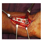
Henry's approach was used. An incision was made between the flexor carpi radialis (FCR)tendon and the radial artery. This interval was developed, revealing the flexor pollicis longus (FPL) muscle at the proximal extent of the wound and the pronator quadratus muscle more distally. The radial artery was carefully retracted laterally, and the tendons of the FCR and FPL are retracted medially. The pronator quadratus was divided at its most radial aspect, leaving a small cuffof muscle for later reattachment. Any elevation of the muscle of the FPL was to be performed at its most radial aspect, as it receives its innervation from the anterior interosseous nerve on its ulnar side. After the pronator quadratus has been divided and elevated, the fracture is readily visualized, and reduction maneuvers were accomplished under direct vision. After exposure and debridement of the fracture site, the fracture is reduced and provisionally fixed under fluoroscopy with K-wires, reduction forceps or suture fixation or temporary distractor. Reduction aids when placed should not interfere with placement of the plate.
In cases where the bone is osteoporotic or more loss of radial length due to gross collapse of subchondral bone bone graft is taken to compensate the lost radial length and to augment the stability and fixation. The optimal placement of the distal screws is important: they must be inserted at the radial styloid, beneath the lunate facet, and near the sigmoid notch. All the screws should have purchase of both cortices when using BP or monocortical or bicotical when using LCP system. More volar tilt can be achieved during distal screw placement when the wrist is volarly flexed as much as possible by an assistant. Moreover, radial length can be further improved by pushing the whole plating system distally while using the oval plate hole and screw as a glide and by augmenting with bone graft. The final position of the plate was confirmed using fluoroscopy. Pronator quadratus muscle was used at the time of closure, to cover, in part, the implant that were applied to the anterior surface of the radius.
Once stable fixation was achieved and hemostasis secured, the wound was closed in layers and sterile compression dressing was applied. The tourniquet was removed and capillary refilling was checked in the fingers. The operated limb was supported with a below elbow POP slab with the wrist in neutral position.
Post Operative Evaluation was carried out by radiographic assessment. Acceptable radiographic parameters considered for healed radius fracture in an active healthy patient are given in Table 1.
2.6 Criteria to assess results
Clinical assessment was done based on Demerit point system of Gartland and Werley taking into consideration residual deformity (range 0-3), subjective evaluation (range 0-6), objective evaluation (range 0-5) and complications (range 0-5). The final results are given in Table 2.
|
Radial length |
>8mm |
|
Radial angulation |
< 5 degree loss |
|
Palmar tilt |
Neutral (0 degree) |
|
Ulnar variance |
<50 loss |
|
Intra articular step off |
<+2 mm |
Table 1: Acceptable radiographic parameters considered for healed radius fracture in an active healthy patient.
|
Final result |
Range of points |
|
Excellent |
0-2 |
|
Good |
3-8 |
|
Fair |
9-20 |
|
Poor |
21 & above |
Table 2: Final results.
Objective evaluation is based on range of motion. The minimum required for normal function:
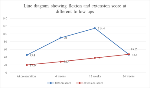
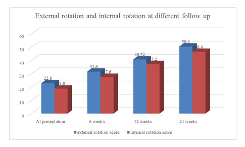
Dorsiflexion 45°, Palmar flexion 30°, Radial deviation 15°, Ulnar deviation 15°, Pronation 50°, Supination 50°
2.7 Outcome measures
The patient was discharged on 4th post-operative day. Sutures were removed on 10th post operative day. Then they were followed up at 6 weeks, 3 months, 6 months. During the follow up visits, patients were evaluated clinically and radiologically. The clinical outcome was evaluated using DEMERIT SCORING SYSTEM OF GARTLAND AND WERLEY. The fracture was to be considered as united if three or more than three cortices are continuous on two radiographic views. Complications during this period were entered in the proforma.
2.8 Statistical Analysis
The relationship of the functional outcome was analysed and will be related to fracture pattern, time gap between injury and surgery and quality of reduction. The data was analysed using SPSS version 20.0. Summary statistics (mean, SD, Skewness, Kurtosisetc) was used to describe the clinical characteristics and functional and radiological outcome. Bivariate association was studied using Pearson chi – square test. The strength of association was carried out using Karl Pearson’s or Spearman’s rank correlation coefficient.Various comparisons were made either using t-test or analysis of variance (ANOVA).
3. Results
The study comprised of thirty cases of distal radial fractures in adults. All patients were treated with open reduction and internal fixation either with Locking Compression plate. The average age was 47.1 years with the fracture being more common in the 4th to 6th decades. The follow-up ranged from 6-9 months. Male preponderance with left wrist affection more than right. All fractures were either due to road traffic accident or fall on the outstretched hand, with RTA being more common of the two. Most of the fractures were of Frykman Type III and Type VIII. Surgery was delayed till the fourth day in 3 (10%) cases, to make the patient medically fit for surgery. All (100%) the patients had their range of motion within the normal functional range. None of the patients had wrist stiffness. Complications were minimal. There was 1 (3%) case of extensor pollicis longus tendon irritation which was because of long volar to dorsal screw. There was 1 (3%) case with a intra articular fracture developed grade I radiocarpal arthritis doing well with physiotherapy. Though we had 22 (73.4%) cases of intra articular fracture, the complication of post traumatic arthritis was in only 1 (3%) case. Long term follows up is needed to assess the arthritic changes. Using the Demerit score system of Gartland and Werley, we had 15 (50%) excellent results, 12 (40%) good results, 3 (10%) fair results and 0 poor result.
3.1 Evaluation of Results
The assessment of results was made using the demerit score system of Gartland and Werley based on objective and subjective criteria, residual deformity and complications. Using the Demerit score system of Gartland and Werley, we had 15 (50%) excellent results, 12 (40%) good results, 3 (10%) fair results and no poor results.
|
Time of Union |
No.of Cases |
Percentage |
|
2-3 months |
24 |
80 |
|
3-4 months |
5 |
17 |
|
> 4 months |
1 |
3 |
Table 3: Study comprising of 30 cases.
4. Discussion
Distal radius fractures are the most frequently seen upper extremity fractures. The main objective of its treatment is the re-establishment of anatomic integrity and function. In unstable intra-articular fractures, re-establishment of intra-articular integrity and in extra-articular fractures with more subchondra l/ metaphyseal bone loss, maintaining the radial length is often not possible with closed methods. In such cases, where an open positioning is required, various surgical methods and fixation materials can be used. A better understanding of wrist anatomy and functioning through the studies conducted in the recent years, as well as the increasing expectations of patients have expanded the borders of surgical treatment. Besides, improvements in fixation materials have provided new opportunities. Due to their extra-articular nature with loss of radial length and intra articular unstable nature, distal radius fractures are treated surgically. Today, open positioning and plate fixation are the widely recognized surgical methods. Locking Compression plates are facilitating the positioning. These plates with screw have more biomechanical strength against forces applied on the fracture surfaces. Bone grafting is necessary in osteoporotic bones with trivial trauma or high energy injuries where there is severe loss of radial length due to comminution of metaphyseal bone with extra or intra articulars. However, there is no consensus either about how to approach the distal radius or the positioning of the plate. During the recent years, volar approach has become more popular.
Though many cases of distal radius fractures come to our OPD, only 30 cases were undertaken which have radio carpal extension or distal radio ulnar joint step or gap deformities greater than 1-2 mm, gross radio ulnar joint instability or those with extensive metaphyseal comminution making them particularly unstable after closed reduction. Hence operative management with open reduction and internal fixation of distal radial fractures using Locking Compression plates with bone graft was done. We evaluated our results and compared them with those obtained by various other studies utilizing different modalities of treatment.
5. Conclusion
The present study was undertaken to assess the functional outcome of operative management of distal radial fractures in adults by open reduction internal fixation and the following conclusions were drawn. Many of the literature denotes fracture of the distal radius are common in older individuals 4th to 6th decade, as our clinical trial was to study the effectiveness of the open reduction and internal fixation of the distal radius fractures, we included the cases, requiring surgery which have radio carpal extension or distal radio ulnar joint step or gap deformities greater than1-2 mm or gross radio ulnar joint instability or those with extensive metaphyseal comminution leading them particularly unstable after closed reduction or extra articulars with loss of radial length. LCP plates used provided successful results as they are effective in anatomic realignment, allows early joint motion, owing to its fixation strength. Close placement to joint interface and screwing capability are its biomechanical superiorities.
Locking compression plates are more beneficial especially in intra articular fractures and severely osteoporotic bone as they combine the advantage of locking plate and screws that creates a construct that resists angular collapse and also provide a scaffold for osteoporotic bone, which may not be possible with the conventional buttress plates as they require good bone stock for purchase of screw to bone, which necessarily not required in Locking Compression plate. In elderly patients with osteoporosis with trivial trauma and in young patients with high energy injuries causing intra or extra articular fractures there is significant bone loss in the metaphyseal/ subchondral area with loss of normal radiological parameters, which cannot be addressed by pop casting, CRIF with K wires and External Fixator. Bone grafting can resolve this problem as it increases the rate of union, provide short term mechanical support, prevent loss of fixation and restore the acceptable radiological parameters. Volar approach provides both access with minimal surgical trauma on distal radius and fixation with a better adaptation to surrounding tissues. In the subjects of our study, a successful anatomic alignment was acquired with volar approach, regardless the direction of fracture angulation and the plate used. The patients who were old in majority, went back to their daily activities with 90% recovery.
We encountered 2 complications (6%) in our study. One being extensor tendon irritation, which was because of long screws projecting dorsally, other complication being arthritis in one patient which was because of improper reduction and articular step. These complications can be prevented once the surgeon gets adapted to the procedure. By using Locking Compression plates in distal radius fractures, augmentation of bone loss with bone graft provide good to excellent results and are effective in the correction and maintenance of distal radius anatomy. By using these plates, joint motions and daily functioning is recovered in a shorter time. All said and done, a multicentric study, may be a comparitive study with a larger sample size would be needed to dogmatically conclude about its effectiveness. A single centric study with 30 cases is too small a study to conclude anything.
Financial Support
Nil
Conflict of Interest
There is no conflict of interests.
References
- Fitoussi F and Chow SPF. Treatment of displaced Intra articular fractures of the distal end of Radius with Plates. J Bone Joint Surg 79-A (1997): 1303-1311.
- Gerostathopoulos N, Kalliakmanis A, Fandridis E, et al. Trimed Fixation System for Displaced Fractures of the Distal Radius. The Journal of Trauma: Injury, Infection, and Critical Care 62 (2007): 913-918.
- Ruch David S. Fractures of the distal Radius and Ulna, Chapter 26 in Rockwood and Green’s Fractures in Adults, Philadelphia: Lippincott Williams &Wikins 6th (2006): 909-964.
- Crenshaw Andrew H. Jr. Fractures of shoulder, arm, and forearm. Chapter-54 In: Campbell’s operative orthopaedics, Philadelphia: Mosby Inc., 11th, 3 (2008): 3447-3449.
- Cognet J, Geanah A, Marsal C, et al. Ostéosynthèse des fractures du radius distal par plaque à visbloquée. Revue de ChirurgieOrthopédique et Réparatrice de l'AppareilMoteur 92 (2006): 663-672.
- Adani R, Tarallo L, Amorico M, et al. The Treatment Of Distal Radius Articular Fractures Through Lcp System. Hand Surg 13 (2008): 61-72.
- Pichon H, Chergaoui A, Jager S, et al. Volar fixed angle plate LCP 3.5 for dorsally distal radius fracture. About 24 cases. Rev Chir Orthop Reparatrice Appar Mot 94 (2008): 152-159.
- Leung F Palmar. Plate Fixation Of Ao Type C2 Fracture Of Distal Radius Using A Locking Compression Plate – A Biomechanical Study In A Cadaveric Model. The Journal of Hand Surgery: Journal of the British Society for Surgery of the Hand 28 (2003): 263-266.
- Chen N. Management of Distal Radial Fractures. The Journal of Bone and Joint Surgery (American) 89 (2007): 2051.
- Cooney WP III, Dobyns JH, Linscheid RL. Complications of colles fractures. J Bone joint surg 62-A (1980): 613.

