UPLC-MS analysis of characterization of bioactive compounds produced by endophytic Bacillus tequilensis ALR-2 from Aloe vera plant
Article Information
Akinsanya Mushafau Adewale1,2*, Goh Joo Kheng2, Ting Adeline Su Yien2
1Department of Medical Biochemistry, Faculty of Basic Medical Sciences, College of Medicine, Lagos State University, Lagos, Nigeria
2School of Science, Monash University Malaysia, 47500 Bandar Sunway, Selangor, Malaysia
*Corresponding Author: Akinsanya Mushafau Adewale, Department of Medical Biochemistry, Faculty of Basic Medical Sciences, College of Medicine, Lagos State University, Lagos, Nigeria
Received: 14 July 2022; Accepted: 19 July 2022; Published: 01 October 2022
Citation: Akinsanya Mushafau Adewale, Goh Joo Kheng, Ting Adeline Su Yien. UPLC-MS analysis of characterization of bioactive compounds produced by endophytic Bacillus tequilensis ALR-2 from Aloe vera plant. Journal of Analytical Techniques and Research 4 (2022): 130-138.
View / Download Pdf Share at FacebookAbstract
Endophytic Bacillus tequilensis ALR-2 isolated from Aloe vera plant, was found to have antimicrobial activities attributed to a series of lipopeptide antibiotics and biosurfactants produced. Partial-purification of ethylacetate extracts by column chromatography revealed that fraction SF5 (5mg mL-1) effectively inhibited Bacillus cereus and Staphylococcus aureus (28.6±0.5 and 30.3±0.5 mm zone of inhibition, respectively), and was significantly more effective than the standard antibiotics Ciprofloxacin (5μg) (23.6 ±0.5 and 18.6 ±0.5 mm, respectively) and Kanamycin (30μg) (20.3 ±0.5 and 13.6 ±0.5 mm, respectively). UPLC-MS analysis of SF5 showed three series of ion peaks. The quadrupole-orthogonal ionization time-of-flight mass spectrometer revealed [M+H]+ of 678.4601; 679.4690 and 680.5076 which corresponded to the oligopeptide antibiotic monamycin (mol. wt. 677.84). The [M+H]+ of 1119.8870; 1120.8992 and 1121.9119 corresponded to antibiotic complex 61-26 (mol. wt. 1120.348), a lipopeptide. The third peak of [M+H]+ of 1356.0151 and 1358.0664 could not be classified to existing known compounds in the literatures.
Keywords
Aloe vera, Biosurfactants, Microbes, Drug discovery, Monamycin, UPLC-MS
Aloe vera articles; Biosurfactants articles; Microbes articles; Drug discovery articles; Monamycin articles; UPLC-MS articles
Aloe vera articles Aloe vera Research articles Aloe vera review articles Aloe vera PubMed articles Aloe vera PubMed Central articles Aloe vera 2023 articles Aloe vera 2024 articles Aloe vera Scopus articles Aloe vera impact factor journals Aloe vera Scopus journals Aloe vera PubMed journals Aloe vera medical journals Aloe vera free journals Aloe vera best journals Aloe vera top journals Aloe vera free medical journals Aloe vera famous journals Aloe vera Google Scholar indexed journals Biosurfactants articles Biosurfactants Research articles Biosurfactants review articles Biosurfactants PubMed articles Biosurfactants PubMed Central articles Biosurfactants 2023 articles Biosurfactants 2024 articles Biosurfactants Scopus articles Biosurfactants impact factor journals Biosurfactants Scopus journals Biosurfactants PubMed journals Biosurfactants medical journals Biosurfactants free journals Biosurfactants best journals Biosurfactants top journals Biosurfactants free medical journals Biosurfactants famous journals Biosurfactants Google Scholar indexed journals Microbes articles Microbes Research articles Microbes review articles Microbes PubMed articles Microbes PubMed Central articles Microbes 2023 articles Microbes 2024 articles Microbes Scopus articles Microbes impact factor journals Microbes Scopus journals Microbes PubMed journals Microbes medical journals Microbes free journals Microbes best journals Microbes top journals Microbes free medical journals Microbes famous journals Microbes Google Scholar indexed journals Drug discovery articles Drug discovery Research articles Drug discovery review articles Drug discovery PubMed articles Drug discovery PubMed Central articles Drug discovery 2023 articles Drug discovery 2024 articles Drug discovery Scopus articles Drug discovery impact factor journals Drug discovery Scopus journals Drug discovery PubMed journals Drug discovery medical journals Drug discovery free journals Drug discovery best journals Drug discovery top journals Drug discovery free medical journals Drug discovery famous journals Drug discovery Google Scholar indexed journals Monamycin articles Monamycin Research articles Monamycin review articles Monamycin PubMed articles Monamycin PubMed Central articles Monamycin 2023 articles Monamycin 2024 articles Monamycin Scopus articles Monamycin impact factor journals Monamycin Scopus journals Monamycin PubMed journals Monamycin medical journals Monamycin free journals Monamycin best journals Monamycin top journals Monamycin free medical journals Monamycin famous journals Monamycin Google Scholar indexed journals UPLC-MS articles UPLC-MS Research articles UPLC-MS review articles UPLC-MS PubMed articles UPLC-MS PubMed Central articles UPLC-MS 2023 articles UPLC-MS 2024 articles UPLC-MS Scopus articles UPLC-MS impact factor journals UPLC-MS Scopus journals UPLC-MS PubMed journals UPLC-MS medical journals UPLC-MS free journals UPLC-MS best journals UPLC-MS top journals UPLC-MS free medical journals UPLC-MS famous journals UPLC-MS Google Scholar indexed journals lipopeptide antibiotics articles lipopeptide antibiotics Research articles lipopeptide antibiotics review articles lipopeptide antibiotics PubMed articles lipopeptide antibiotics PubMed Central articles lipopeptide antibiotics 2023 articles lipopeptide antibiotics 2024 articles lipopeptide antibiotics Scopus articles lipopeptide antibiotics impact factor journals lipopeptide antibiotics Scopus journals lipopeptide antibiotics PubMed journals lipopeptide antibiotics medical journals lipopeptide antibiotics free journals lipopeptide antibiotics best journals lipopeptide antibiotics top journals lipopeptide antibiotics free medical journals lipopeptide antibiotics famous journals lipopeptide antibiotics Google Scholar indexed journals ethylacetate articles ethylacetate Research articles ethylacetate review articles ethylacetate PubMed articles ethylacetate PubMed Central articles ethylacetate 2023 articles ethylacetate 2024 articles ethylacetate Scopus articles ethylacetate impact factor journals ethylacetate Scopus journals ethylacetate PubMed journals ethylacetate medical journals ethylacetate free journals ethylacetate best journals ethylacetate top journals ethylacetate free medical journals ethylacetate famous journals ethylacetate Google Scholar indexed journals chromatography articles chromatography Research articles chromatography review articles chromatography PubMed articles chromatography PubMed Central articles chromatography 2023 articles chromatography 2024 articles chromatography Scopus articles chromatography impact factor journals chromatography Scopus journals chromatography PubMed journals chromatography medical journals chromatography free journals chromatography best journals chromatography top journals chromatography free medical journals chromatography famous journals chromatography Google Scholar indexed journals quadrupole-orthogonal ionization articles quadrupole-orthogonal ionization Research articles quadrupole-orthogonal ionization review articles quadrupole-orthogonal ionization PubMed articles quadrupole-orthogonal ionization PubMed Central articles quadrupole-orthogonal ionization 2023 articles quadrupole-orthogonal ionization 2024 articles quadrupole-orthogonal ionization Scopus articles quadrupole-orthogonal ionization impact factor journals quadrupole-orthogonal ionization Scopus journals quadrupole-orthogonal ionization PubMed journals quadrupole-orthogonal ionization medical journals quadrupole-orthogonal ionization free journals quadrupole-orthogonal ionization best journals quadrupole-orthogonal ionization top journals quadrupole-orthogonal ionization free medical journals quadrupole-orthogonal ionization famous journals quadrupole-orthogonal ionization Google Scholar indexed journals
Article Details
Introduction
Endophytes are bacteria or fungi, which reside in the tissues of higher plants without causing any symptoms or apparent harm to the host. These microbes are not pathogenic to the host, neither are they providing benefit to other microbial residents [1]. They have been found to have a wide range of antimicrobial activities, hence are important sources of antimicrobial substances [2]. In recent years, endophytes have been found to produce novel metabolites exhibiting a variety of biological activities against different pathogens [3,4]. Thus, endophytes have become increasingly popular for isolation and characterization of bioactive compounds [5]. Some endophytes have been found to produce bioactive compounds that may be involved in a symbiotic association with a host plant [6]. In recent studies, it has been recognized that endophytic bacteria play an important role in resistance to diseases and mediate some beneficial role between the endophyte and its host [5,7]. Similarly, various studies have been conducted on the plant growth-promoting abilities of endophytes and this has indicated their ability to increase plant growth through the improvement of nutrient/mineral recycling, and their ability to inhibit invading phytopathogens [8]. Bacillus tequilensis ALR-2 was isolated from Aloe vera, along with other endophytic Proteobacteria, Firmicutes and Bacteriodetes, and was found to produce the most potent antimicrobial bioactive compounds to inhibit gram-positive and gram-negative pathogens [9]. Although studies on Bacillus sp. is quite common, there are not many on Bacillus tequilensis, particularly on their antimicrobial potential [10]. As such, this study characterized the bioactive compounds from B. tequilensis ALR-2 using Ultra-Performance Liquid Chromatography (UPLC) and Mass Spectrometer, which essentially determines accurate mass of compounds. This method is superior due to its efficiency and high resolution of molecular ions compared to other mass-to-charge ratio (m/z) analyzers [11]. This study reports on the antimicrobial activities of crude extracts of endophytic Bacillus tequilensis ALR-2, and the characterization of the bioactive compounds produced.
Experimental procedures
Culture establishment
The Bacillus tequilensis ALR-2 strain (KJ689792) was isolated from A. vera and identified as described in our previous study [9]. For this study, cultures were prepared on nutrient agar (Sigma-Aldrich) and incubated at incubation at 30 ± 2 ºC for 18-24 h.
Extraction of bioactive compounds
Bacillus tequilensis ALR-2 colony was inoculated into sterile Erlenmeyer flask (1L) containing 500 mL nutrient broth (Sigma-Aldrich) and incubated at 35 ± 2ºC (150 rpm, 36 - 40 h) in Rotary Flask Shaker. The culture was centrifuged at 8,000 × g for 5 min to obtain the cell free supernatant. The extracellular metabolites in the cell free supernatant were exhaustively extracted with ethylacetate, diethylether and n-hexane separately in the ratio of 1:1 (supernatant:solvent). The organic phase was concentrated in rotatory evaporator (Rotavap) at 37ºC, freeze-dried (LABCONCO freeZone 4.5-105ºC) and weighed. The percentage yield of each extracts (% w/v) was calculated from the following equation:
% extract yield = weight of dry extract (mg)/volume taken for extraction (1L) x 100
Antimicrobial activity screening of the extracts via TLC-bioautography
The ethylacetate extract was fractionated on aluminium-backed thin layer chromatography (TLC) plates (Merck, silica gel 60 F254). The plates were developed under saturated conditions with the eluent systems, developed in our laboratory. The mobile phase used for separation of the bioactive metabolites was Chloroform:Methanol (4:1 v/v). The separations were detected under ultraviolet light (254 nm) and the Rf values were calculated. TLC-bioautography assay was carried out as described in our earlier publication [12] using immersion method. To develop the chromatograms, 1µL (20 mg mL-1) of the extract was loaded onto TLC plates in a narrow band and eluted using the mobile solvent system in a covered chromatography jar. The developed plates were dried under the laminar flow for 6 h to remove traces of solvent on the TLC plates. One mL of 0.5 McFarland standard bacteria pathogens were mixed with molten Mueller Hinton agar in sterile petri dish. The prepared chromatograms were immersed in the molten Mueller Hinton agar and removed to solidify. This process was carried out in a laminar flow cabinet (Labotec, SA). The plates were then incubated in petri dish overnight at 37 ± 2ºC and 100% relative humidity in the dark. The chromatograms were then sprayed with a 5 mg mL-1 solution of tetrazolium compound (TTC) and incubated overnight. Formation of white bands indicated bacterial growth suppression due to the presence of compounds that inhibited the growth of tested organisms that prevented the reduction of TTC to formazan (reddish coloration) [13].
Extraction and purification of bioactive compounds
The crude ethylacetate extract of ALR-2 was purified via column chromatography using a LiChroprep RP-18 (15-25 µm) silica gel column (Merck, EMD Millipore Corporation, USA) and chloroform:methanol (ratio 4:1) as eluent. A total of 10 (5 mL each) fractions were collected and were lyophilized to obtain powder forms for the antimicrobial assay. Three fractions with good antimicrobial activities were subsequently combined and purified with acetonitrile:methanol as eluent to obtain a further six subfractions. The purity of each subfraction was analysed by reversed phase HPLC (COSMOSIL Packed C18 column, 4.6 ID × 150 mm, Agilent Technologies 1260 infinity). The following mobile phase was used; methanol:water with gradient elution method of 95% deionized water and 5% methanol for (0 – 20) min for solvent-A, and 5% deionized water and 95% methanol for (20 – 30) min for solvent-B.
Antimicrobial assay
The antimicrobial activities of the six subfractions were assayed using the Kirby-Bauer disc diffusion method [14, 15], against bacteria and yeast pathogens, which include-Pseudomonas aeruginosa ATCC 10145, Enterococcus faecalis ATCC 29212, Staphylococcus aureus ATCC 33591, Bacillus cereus ATCC 14579, Salmonella Typhimurium ATCC 14028, Proteus vulgaris-ATCC 8427, Klebsiella pneumoniae ATCC 10031, Escherichia coli ATCC 25922, Streptococcus pyogenes ATCC and Candida albicans ATCC 90028. All pathogens were obtained from the Microbiology Laboratory of Monash University Malaysia. The bacteria pathogens were first pre-cultured overnight in Mueller Hinton broth (Difco, USA.) at 35 ± 2ºC. Then, 5 mL of the culture were pipetted and centrifuged at 6,000 × g for 5 min. The pellets were re-suspended in sterile distilled water and the cell density was subsequently adjusted to 0.5 McFarland standard. The bacterial suspension was then seeded onto Mueller Hinton agar (Difco, USA.) plates for antimicrobial testing. The subfractions (10 µL of 10 mg mL-1) were impregnated onto sterile discs and placed on seeded agar plates. The plates were incubated at 35 ± 2ºC for 48 h and the zone of inhibition was determined by measuring the diameter of annular clear zone. The experiment was performed in triplicates. Ciprofloxacin (5 µg) and Kanamycin (30 μg) standard antimicrobial discs were used as positive control.
Minimum inhibitory concentration (MIC) and Minimum bactericidal concentration (MBC)
Microdilution method was used as described by Andrews [14]. The dilution range of the antimicrobial compound tested was modified to 0.1 mg mL-1 to 10 mg mL-1 concentrations. The bacterial inoculum was prepared and adjusted to 108 cfu mL-1 (0.5 McFarland standards). Each antimicrobial extract (75 µL) was added into two rows of sterile 96-wells. This was followed with 75 µL of tested bacterial inoculum which was added into one row and 75 µL of control sterile broth was added into the second row of wells. In addition, inoculated and uninoculated wells of antimicrobial free broth were included to determine adequacy of the broth to support growth and to check for sterility of the broth. The microplate was covered with lid and incubated at 37 ± 2ºC for 18 h. The absorbance of the culture turbidity was then read at 625 nm using the microplate reader (Infinite 200 TECAN). To assess MBC, wells with absence of growth (non-turbid wells) detected at the lowest concentration was sub-cultured onto Mueller Hinton agar (Difco, USA.) plates to determine the MBC. The concentration with no growth of tested organism is the MBC for the antimicrobial agent against the tested organism.
Ultra-Performance Liquid Chromatography-Mass Spectrometry (UPLC-MS) analysis of antimicrobial compound
Ultra-Performance Liquid Chromatography-Mass Spectrometry (UPLC-MS) analysis was performed on the partially-purified antimicrobial substance using AcquityTM Waters Ultra Performance Liquid Chromatography (UPLC) (Waters Corporation, USA). The electrospray source was operated at a capillary voltage of 2.7kV. Column specification used was ACQUITY UPLC BEH C18 1.7 µm, 2.1 x 50 mm. Mass spectrometer used was Synapt high definition mass spectrometer quadrupole-orthogonal acceleration with time of flight detector (Waters Corporation, USA). Mobile phase used was: milliQ water + 0.1% formic acid (solvent A) and methanol + 0.1% formic acid (Solvent B). Flow rate was 0.5 mL/min. Gradient program was pre-determined as follows: 95-95% (solvent A) 0-2.8 min, 5-5% (solvent B) 0-2.8 min, 95-5% (solvent A) 2.8-3.5 min, 5-95% (solvent B), 5-95% (solvent A) 3.5-4.0 min 95-5% 3.5-4.0 min, 95-95% (solvent A) 4.0-5.0 min 5-5% (solvent B) 4.0-5.0 min.
Statistical analysis
Results were expressed as mean ± Standard deviation (SD). One-way ANOVA was used to analyse all data obtained. The analysis was carried out using the Statistical Package for Social Science (SPSS) version 20.0 and means were compared using Tukey’s Studentized Range Test (HSD (0.05)), with differences considered statistically significant when p<0.05.
Data availability
All data are contained in the manuscript. Accession number of organisms used are also indicated in the manuscript.
Results and Discussion
Bacillus tequilensis ALR-2 is a Gram-positive, endospore-forming, motile and rod-shape bacterium. As reported in our earlier study, the nucleotide sequence of this isolate (16S rDNA) showed the closest similarity to that of Bacillus tequilensis (with a homology of 97%) [9]. This strain is reported as an endophyte from Aloe vera plant for the first time, with the GenBank accession number KJ689792 assigned [9]. This isolate has excellent antimicrobial activity towards Staphylococcus aureus, Bacillus cereus, Proteus vulgaris, Klebsiella pneumoniae, Escherichia coli, Streptococcus pyogenes and Candida albicans as documented in our earlier studies [9].
Antimicrobial activities screening of extracts via TLC-bioautography
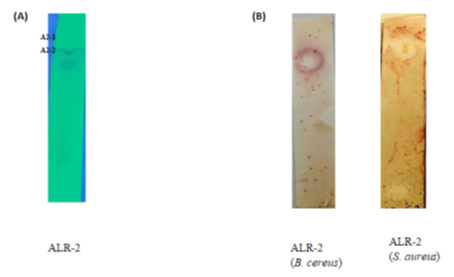
The results indicated that only ethylacetate-derived extracts showed bioactivities in the TLC-bioautography (Fig. 1). This suggested the bioactive components are present in the more polar solvent (ethylacetate with polarity index 4.4) than the less and non-polar solvents such as diethylether and n-hexane, respectively. The ethylacetate extract, when subjected to TLC, indicated 2 separate fractions in the chromatogram: A2-1 and A2-2 with Rf values 0.98 and 0.86, respectively (Figure 1). The two fractions showed antimicrobial activities against S. aureus and B. cereus (Fig. 1) as the zone of inhibition was wider and appeared to map the two fractions with Rf values (0.98 and 0.86). Hence, the TLC-bioautography technique has enabled us to rapidly identify the fractions with bioactive compounds of interest and the right separating mixture for the isolation of the bioactive compounds [16]. Therefore, B. tequilensis (ALR-2) may be said to possess very promising antimicrobial compounds as revealed by the prominent zone of inhibition seen on the chromatogram.

Figure 1: (A) Bioautography plates. Chromatograms plate of ethylacetate extracts of ALR-2 showing fractions (A2-1, A2-2). (B) Clear areas showing inhibition zones of chromatograms when immersed in molten agar seeded with bacterial pathogens, incubated at 37 ± 2ºC for 18 h then sprayed with tetrazolium compound and then incubated at 37 ± 2ºC for 24 h. (Images scale x2)
Extraction and purification of bioactive compound
The ethyl acetate extraction of Bacillus tequilensis ALR-2 yielded 3.4 mg of lyophilized powder forms (from 1 L broth culture), the highest yield compared to diethylether (2.02 mg) and n-hexane (0.69 mg). Upon fractionation via the LiChroprep RP-18 (15-25 µm) silica gel column (Merck, EMD Millipore Corporation, USA), 10 fractions were obtained (see supplementary Table 1). Fractions F6 and F7 have the highest yield with 6443 and 6283 mg mL-1, respectively, while fractions F2 and F3 produce the lowest yield of 110 and 156 mg mL-1, respectively. Since ALR-2 was extracted with polar solvent, it is expected to contain more polar compounds than non-polar compounds. The combined three fractions of F7, F8 and F9 gave six subfractions (see supplementary Table 1).
|
Fraction (20 mg mL-1) |
E. faecalis |
B. cereus |
S. pyogenes |
S. aureus |
K. pneumoniae |
C. albicans |
|
F1-F3 |
- |
- |
- |
- |
- |
- |
|
F4 |
13.6±0.5abc |
16.3±0.5bc |
- |
18.3±0.5b |
- |
- |
|
F5 |
13.6±0.5abc |
17.6±0.5c |
- |
19.3±0.5bc |
- |
- |
|
F6 |
13.0±0.5ab |
16.3±0.5bc |
- |
20.3±0.5c |
- |
- |
|
F7 |
15.6±0.5d |
15.3±0.5b |
14.3±0.5b |
19.3±0.5bc |
- |
- |
|
F8 |
14.6±0.5cd |
15.3±0.5b |
14.3±0.5b |
20.0±0.5c |
- |
- |
|
F9 |
14.3±0.5bcd |
16.0±0.5bc |
15.0±0.5b |
20.6±0.5c |
- |
- |
|
F10 |
12.3±0.5a |
12.6±0.5a |
12.3±0.5a |
16.3±0.5a |
- |
12.0±0.5 |
Table 1: Antimicrobial activities of fractions (F1-F10) against bacteria and C. albicans pathogens
Data are mean ±SD values. One-way ANOVA was used to analyse all data and means were compared using Tukey’s Studentized Range Test (HSD (0.05)), with differences considered statistically significant when p<0.05 represented by alphabets (a-d). F1-F10 represent the fractions from first fractionation. Pathogens-Pseudomonas aeruginosa ATCC 10145, Salmonella Typhimurium ATCC 14028, Proteus vulgaris ATCC 8427 and Escherichia coli ATCC 25922 were resistance to all the fractions. (-) represent no inhibition zone. One of these compounds was detected in SF3 and SF6 at retention time 22.9 min with 25.6% and 2.0%, respectively. It was also observed that SF3 was equally active against S. aureus, B. cereus and K. pneumonia, with zone of inhibitions 9.3 ±0.5, 13.6 ±0.5 and 10.6 ±0.5 mm, respectively (Table 2). We also observed SF6 was active against S. aureus and B. cereus, and this may be attributed to the compound detected at peak 24.4-minute retention time of 26%. This compound was also detected in SF3 (8%), SF4 (7%) and SF5 (2%) retention time of 24.4 min. The results agreed with statements of researchers’ bioprospecting for new antibiotics from either soil microbes or endophytes, that these microbes produce several antimicrobial and antifungal compounds in their culture, and some exist in isoforms [17-19]. Hence, the most potent of the compound was targeted for identification which we believed can be found in SF5.
|
Subfraction (10 mg mL-1) |
S. aureus |
B. cereus |
S. pyogenes |
E. faecalis |
K. pneumoniae |
|
SF1-SF2 |
- |
- |
- |
- |
- |
|
SF3 |
9.3±0.5a |
13.6±0.5a |
- |
- |
10.6±0.5a |
|
SF4 |
13.6±0.5b |
13.3±0.5a |
- |
- |
16.6±0.5b |
|
SF5 |
30.3±0.5e |
28.6±0.5e |
15.6±0.5a |
8.6±0.5a |
18.6±0.5c |
|
SF6 |
21.3±0.5d |
17.6±0.5b |
- |
- |
- |
|
Ciprofloxacin (5µg) |
18.6±0.5c |
23.6±0.5d |
25.0±0.5b |
26.0±0.5c |
34.6±0.5d |
|
Kanamycin (30 µg) |
13.6±0.5b |
20.3±0.5c |
24.6±0.5b |
18.0±0.5b |
16.6±0.5b |
Table 2: Antimicrobial activities of subfractions (SF1-SF6) against bacteria pathogens
Data are mean ±SD values. One-way ANOVA was used to analyse all data and means were compared using Tukey’s Studentized Range Test (HSD (0.05)), with differences considered statistically significant when p<0.05 represented by alphabets (a-e). SF1-SF6 represent the sub-fractions from the second fractionation. Pathogens-Pseudomonas aeruginosa ATCC 10145, Salmonella Typhimurium ATCC 14028, Proteus vulgaris ATCC 8427, Escherichia coli ATCC 25922 and Candida albicans ATCC 90028 were resistance to all the fractions. (-) represent no inhibition zone.
Antimicrobial activity
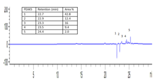
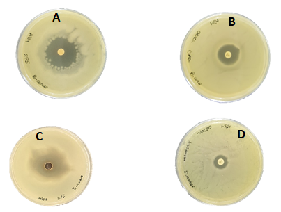
Seven fractions (F4 – F10) inhibited three to five pathogens tested with zone of inhibition from 12.3 to 20.6 ±0.5 mm, while the first three fractions (F1-F3) appeared to have no inhibition against all the pathogens tested (Table 1). Gram-positive bacteria: S. aureus, B. cereus, E. faecalis and S. pyogenes were more susceptible to fractions F7-F10 while C. albicans was only inhibited by F10 with inhibition zone of 12.0 ±0.5 mm. Other studies have also reported that the bioactive compounds produced by Bacillus species is active against gram-positive bacteria pathogens [20, 21]. However, it appeared fractions-F7, F8 and F9 relatively inhibited the pathogens more than fractions F4, F5, F6 and F10 (Table 1). Hence, they were considered to have high bioactivity and were selected for further purification analysis. The antimicrobial activities of the subfractions (SF1-SF6) revealed subfraction SF5 to have the highest antimicrobial activity against all the Gram-positive bacteria (with zone of inhibition from 8.6 to 30.3±0.5 mm) (Table 2). It is interesting to note that antimicrobial activities of SF5 against S. aureus and B. cereus were significantly higher than the standard antibiotics used; Ciprofloxacin (5 µg) and Kanamycin (30 μg) as positive control (Table 2, Figure 3). Subfractions SF3 and SF4 also showed antimicrobial activities against three of the five pathogens tested, with zone of inhibition from 9.3 to 16.6 ±0.5 mm. Only subfraction SF6 inhibited two pathogens with zone of inhibition from 10.6 to 21.3 ±0.5 mm. The inhibitory activities can be seen to increase as the major bioactive compound(s) increases in the subfractions. Consequently, this result confirmed the presence of the most active bioactive compound in the subfraction SF5 which has also been revealed by the result of the quantification analysis done with HPLC (Figure 2).

Figure 2: HPLC chromatogram of subfraction SF5 at 293 nm showing five major peaks (1) = 42.8% area at retention time 22.7 min., (2) = 12.4% at retention time 22.9 min., (3) = 16% area at retention time 23.3 min., (4) = 9.4% area at retention time 23.5 min. (5) = 2%; 24.4 min.

Figure 3: Zone of inhibitions shown by SF5 (10 mg mL-1), Ciprofloxacin (5 µg) and Kanamycin (30 µg) standard antibiotics against B. cereus ATCC 14579 and S. aureus ATCC 33591. (A) Zone of inhibition of SF5 on B. cereus, (B) Zone of inhibition of Ciprofloxacin on B. cereus, (C) Zone of inhibition of SF5 on S. aureus and (D) Zone of inhibition of Kanamycin on S. aureus.
Minimum inhibitory concentration (MIC) and minimum bactericidal concentration (MBC) was evaluated to ascertain the potency of the subfractions. The MIC and MBC values of the subfractions against the bacteria pathogens were 0.1 to 5 mg ml-1 (Table 3). The most susceptible pathogen to subfraction SF5 is S. aureus. The results indicated that SF5, with concentrations between 0.1 and 1 mg mL-1, could inhibit (bacteriostatic) and kill all the five pathogens (bactericidal) effectively. The three subfractions SF3, SF4 and SF6, with concentrations of 1 to 5 mg mL-1, were only able to inhibit (bacteriostatic) and kill (bactericidal) B. cereus and K. pneumoniae. In addition, 5 mg mL-1 of SF6 could only inhibit K. pneumoniae (bacteriostatic). Consequently, it is highly suggestive that SF5 comprise of the most active compounds against the five pathogens (Table 3).
|
Subfraction |
SF3 (mg mL-1) |
SF4 (mg mL-1) |
SF5 (mg mL-1) |
SF6 (mg mL-1) |
||||
|
Organisms |
MIC |
MBC |
MIC |
MBC |
MIC |
MBC |
MIC |
MBC |
|
S. aureus |
1 |
10 |
0.5 |
1 |
0.1 |
0.1 |
0.5 |
1 |
|
B. cereus |
1 |
5 |
0.1 |
1 |
0.1 |
0.5 |
1 |
5 |
|
S. pyogenes |
5 |
- |
5 |
- |
0.1 |
0.5 |
5 |
- |
|
E. faecalis |
5 |
- |
5 |
- |
0.1 |
0.5 |
5 |
- |
|
K. pneumoniae |
1 |
5 |
1 |
5 |
0.1 |
1 |
5 |
- |
Table 3: Minimum inhibitory concentration (MIC) and minimum bactericidal concentration (MBC) of Subfractions SF1-SF6 against five bacteria pathogens
SF1-SF6 represents the sub-fractions from the second fractionation. Pathogens-Pseudomonas aeruginosa ATCC 10145, Salmonella Typhimurium ATCC 14028, Proteus vulgaris ATCC 8427, Escherichia coli ATCC 25922 and Candida albicans ATCC 90028 were resistance to all the fractions.
Figure 4: (A) Chromatogram of UPLC-MS of Subfraction SF5 for the time of flight (TOF) of 5 mins., showing the three series of ion peaks at [M+H]+ m/z = 197.1575; 169.0823, at [M+H]+ m/z = 678.4408, at [M+H]+ m/z = 244.1696; 270.1950; 270.2010 and (B) Magnified spectra of precursor ions of m/z = 1356.0151; 1358.0664 and 1105.8010; 1119.8870; 1120.8992; 1121.9119 (Lipopeptides homologues).
Ultra-Performance Liquid Chromatography-Mass Spectrometry analysis (UPLC-MS)
The chromatogram of UPLC/MS analysis of the subfraction SF5 showed three series of ion peaks at [M+H]+ m/z = 197.1575 and 169.0823, at [M+H]+ m/z = 678.4408, at [M+H]+ m/z = 244.1696; 270.1950 and 270.2010 (Fig. 4A). In the first series, a set of mass peaks with an interval of 28 was observed and it has been reported that for the lipopeptides (depending on the type) a set of 14 or 28 molecular weight is often observed with different numbers of methylene (?CH2?) or ethylene (?CH2?CH2?) in fatty acyl chains [22-24]. The second series of peaks corresponds to a class of oligopeptide antibiotics such as monamycin (mol. wt. 677.84) and Alphostatin (mol. wt 668.637) produced by B. megaterium [25]. The third series of peaks with an interval of 26 molecular weight, cannot be immediately classified to compounds reported in the literature, though the mass data shows that it could be an ionic product of larger compounds. The first and second series of peaks in this study can be said to represent different fengycin and surfactin homologues by comparing their mass data with product ions obtained in previous studies [23,26,27]. The ion peak results showed product ions of [M+H]+ m/z = 965.6798; 966.7001 and 967.7208, which corresponds to the collision ionization dissociation (CID) spectra of precursor ions of [M+H]+ m/z = 1356.0151 and 1358.0664 (Fig. 4B). The product ion fragments of [M+H]+ m/z = 965.6798; 966.7001 and 967.7208 can be said to be losses of fatty acid-Glu (amino acid residue) from the N-terminus segment of a lipopeptide antibiotic (fengycin B) [23,27]. Similarly, the product ions of [M+H]+ m/z = 678.4601; 679.4690 and 680.5076 can be explained as ionized product of oligopeptide antibiotics, i.e. Monamycin (mol. wt. 677.84), as its accurate mass correspond to the [M+H]+ 678.4601, which has also been reported as one of the products of Bacillus sp. [17,28]. The third series of peaks [M+H]+ m/z = 120.0921; 233.1971; 277.2599; 361.2691; 460.3424; 544.3967; 545.4211; 554.4237 and 560.4314 see Figure 4B, can also be explained to be product ions from the precursor ions of [M+H]+ m/z =1105.8010; 1119.8870; 1120.8992 and 1121.9119 that belonged to fengycin isoforms [23]. The precursor ion [M+H]+ m/z = 1119.8870 observed, corresponded to Antibiotic complex 61-26 (mol. wt. 1120.348) produced by Bacillus sp. isolated by Wakisaka and Koizumi [29] and Shoji, Sakazaki [17]. Bacillus strains have been reported to produce several lipopeptide antibiotics and lipopeptide biosurfactants that have the capacity to inhibit Gram-positive bacteria and phytopathogenic fungi. They are so diverse that different strains of Bacillus produce different types of these substances [27,30]. It is evident that endophytic Bacillus tequilensis ALR-2, has equal potential to produce series of lipopeptides antibiotics and fengycin isoforms which are responsible for its antimicrobial activities. This study is also the first to report on the production of Antibiotic complex 61-26 (mol. wt. 1120.348) and Monamycin (mol. wt. 677.84) by the endophytic Bacillus tequilensis ALR-2. Conclusively, the UPLC-MS analysis has suggested that the precursor ions are lipopeptides (fengycins) and surfactins. Fengycins and surfactins are natural lipopeptides that show antimicrobial, antiviral and antitumor activities [23] and they are said to be safer in the environment when compared with other chemical agents. Fengycin are known to inhibit filamentous fungi but it does not inhibit yeast. In this study Bacillus tequilensis ALR-2 was found to be a co-producer of fengycins and surfactin as its antimicrobial activities and UPLC-MS have suggested. These natural lipopeptide compounds can be of use in the food preservation and control of plant diseases. Moreover, we suggest the determination of its amino acids and fatty acids residues to be able to elucidate its structure which may be useful in predicting it mechanism of action.
Acknowledgements
This work was supported by scholarship of Higher Degree for Research (HDR) School of Science, Monash University Malaysia.
Conflict of interest
The authors report no conflict of interest and are responsible for the content and writing of the manuscript.
References
- Kobayashi D, Palumbo J. Bacterial endophytes and their effects on plants and uses in agriculture. Microbial endophytes 12 (2000): 199-233.
- Strobel GA. Endophytes as sources of bioactive products. Microbes and infection 5 (2003): 535-44.
- Sun L, Lu Z. Advance on antibiotics produced by endophytes. Food Ferment Ind (China) 31 (2005): 39-41.
- Yu H, Zhang L, Li L, et al. Recent developments and future prospects of antimicrobial metabolites produced by endophytes. Microbiological research 165 (2010): 437-449.
- Cho SJ, Park SR, Kim MK, et al. Endophytic Bacillus sp. isolated from the interior of balloon flower root. Bioscience, biotechnology, and biochemistry 66 (2002): 1270-1275.
- Yang X, Strobel G, Stierle A, et al. Fungal endophyte-tree relationship: Phoma sp. in Taxus wallachiana. Plant Science 102 (1994): 1-9.
- Huang JS. Ultrastructure of bacterial penetration in plants. Annual review of phytopathology 24 (1986): 141-57.
- Guo B, Dai JR, Ng S, et al. Cytonic acids A and B: novel tridepside inhibitors of hCMV protease from the endophytic fungus Cytonaema species. Journal of Natural Products 63 (2000): 602-604.
- Akinsanya MA, Goh JK, Lim SP, et al. Diversity, antimicrobial and antioxidant activities of culturable bacterial endophyte communities in Aloe vera. FEMS microbiology letters 362 (2015): 12-18.
- Gatson JW, Benz BF, Chandrasekaran C, et al. Bacillus tequilensis sp. nov., isolated from a 2000-year-old Mexican shaft-tomb, is closely related to Bacillus subtilis. International journal of systematic and evolutionary microbiology 56 (2006): 1475-1484.
- Dawson J, Guilhaus M. Orthogonal?acceleration time?of?flight mass spectrometer. Rapid Communications in Mass Spectrometry 3 (1989): 155-159.
- Akinsanya MA, Ting ASY, Sanusi MO. Extraction methods and TLC-bioautography for evaluation of antimicrobial activities of endophytic bacteria from medicinal plants. LASU Journal of Medical Sciences 2 (2017): 1-19.
- Begue WJ, Kline RM. The use of tetrazolium salts in bioauthographic procedures. Journal of Chromatography A 64 (1972): 182-184.
- Bauer A, Kirby W, Sherris JC, et al. Antibiotic susceptibility testing by a standardized single disk method. American journal of clinical pathology 45 (1966): 493.
- Andrews JM. Determination of minimum inhibitory concentrations. Journal of antimicrobial Chemotherapy 48 (2001): 5-16.
- Suleiman MM, McGaw L, Naidoo V, et al. Detection of antimicrobial compounds by bioautography of different extracts of leaves of selected South African tree species. African Journal of Traditional, Complementary and Alternative Medicines 7 (2010).
- Shoji JI, Sakazaki R, Wakisaka Y, et al. Studies on antibiotics from the geneus Bacillus. V. Isolation of a new peptide antibiotic complex 61-26. The Journal of antibiotics 28 (1975): 129-131.
- Ruma K, Kumar S, Prakash H. Antioxidant, Anti-Inflammatory, Antimicrobial and Cytotoxic Properties of Fungal Endophytes from Garcinia Species. International Journal of Pharmacy and Pharmaceutical Sciences 5 (2013): 889-97.
- Sun L, Lu Z, Bie X, et al. Isolation and characterization of a co-producer of fengycins and surfactins, endophytic Bacillus amyloliquefaciens ES-2, from Scutellaria baicalensis Georgi. World Journal of Microbiology and Biotechnology 22 (2006): 1259-66.
- Joseph B, Priya RM. Bioactive compounds from endophytes and their potential in pharmaceutical effect: a review. Am J Biochem Mol Biol 1 (2011): 291-309.
- Roongsawang N, Thaniyavarn J, Thaniyavarn S, et al. Isolation and characterization of a halotolerant Bacillus subtilis BBK-1 which produces three kinds of lipopeptides: bacillomycin L, plipastatin, and surfactin. Extremophiles 6 (2002): 499-506.
- Thaniyavarn J, Roongsawang N, Kameyama T, et al. Production and characterization of biosurfactants from Bacillus licheniformis F2. 2. Bioscience, biotechnology, and biochemistry 67 (2003): 1239-1244.
- Kowall M, Vater J, Kluge B, et al. Separation and Characterization of Surfactin Isoforms Produced byBacillus subtilisOKB 105. Journal of colloid and interface science 204 (1998): 1-8.
- Morikawa M, Hirata Y, Imanaka T. A study on the structure- function relationship of lipopeptide biosurfactants. Biochimica et Biophysica Acta (BBA)-Molecular and Cell Biology of Lipids 1488 (2000): 211-218.
- Aoyagi T, Morishima H, Kojiri K, et al. (1989). Alphostatin, an inhibitor of alkaline phosphatase of bovine liver produced by Bacillus megaterium. J Antibiot (Tokyo) 42 (1989): 486-468.
- Kim P, Bai H, Bai D, et al. Purification and characterization of a lipopeptide produced by Bacillus thuringiensis CMB26. Journal of applied microbiology 97 (2004): 942-949.
- Wang J, Liu J, Wang X, et al. Application of electrospray ionization mass spectrometry in rapid typing of fengycin homologues produced by Bacillus subtilis. Letters in applied microbiology 39 (2004): 98-102.
- Bevan K, Davies J, Hassall C, et al. Amino-acids and peptides. Part X. Characterisation of the monamycins, members of a new family of cyclodepsipeptide antibiotics. Journal of the Chemical Society C: Organic. 9 (1971): 514-522.
- Wakisaka Y, Koizumi K. An enrichment isolation procedure for minor Bacillus populations. The Journal of antibiotics 35 (1982): 450-457.
- Pueyo MT, Bloch Jr C, Carmona-Ribeiro AM, et al. (2009). Lipopeptides produced by a soil Bacillus megaterium strain. Microb Ecol 57 (2009): 367-378.
