Transcatheter Edge-To-Edge Repair of A Tricuspid Valve Regurgitation In A Patient with Previous Prosthetic Annuloplasty Ring and DDD Pacemaker
Article Information
Andreas Goette*, Mihai Hasmasan, Sibylle Brandner
Department of Cardiology and Intensive Care Medicine, St. Vincenz Hospital, Paderborn, Germany
*Corresponding Author: Andreas Goette, Department of Cardiology and Intensive Care Medicine, St. Vincenz Hospital, Paderborn, Germany
Received: 25 November 2024; Accepted: 03 December 2024; Published: 10 December 2024
Citation: Andreas Goette, Mihai Hasmasan, Sibylle Brandner. Transcatheter Edge-To-Edge Repair of A Tricuspid Valve Regurgitation In A Patient with Previous Prosthetic Annuloplasty Ring and DDD Pacemaker. Archives of Clinical and Medical Case Reports. 8 (2024): 221-225.
View / Download Pdf Share at FacebookAbstract
Background: Residual tricuspid regurgitation (TR) may occur in up to 30% of patients after surgical tricuspid annuloplasty. It is unclear at present if transcatheter edge-to-edge repair (TEER) is feasible in these patients.
Case Summary: The patient was a 71-year-old woman with a history of severe mitral and tricuspid regurgitation. 8 years prior, she had undergone bioprosthetic mitral valve replacement and tricuspid valve prosthetic ring annuloplasty. A DDD pacemaker had been implanted due to complete AV block after AV-node-ablation. The patient complained of worsening right heart failure symptoms, with severe peripheral edema and shortness of breath. Echocardiography demonstrated normal left ventricular function. The implanted mitral valve did show only a mild regurgitation. But a massive tricuspid regurgitation Grad V was documented (vena contractae width of 11 mm). Due to lack of other treatment options TEER was performed by using the TriClip® device (Abbott, Vascular GmbH). During the TEER procedure, it was difficult to visualize the tricuspid leaflets in transesophageal echocardiography (TEE) due to shadowing from the prosthetic annuloplasty ring. This was resolved by utilizing transgastric visualisation of the tricuspid valve. The right ventricular lead was visualized by X ray and echocardiography during the procedure. The TEER device was implanted posterior to the RV lead, which resulted in a reduction of TR (Grad II). Pacemaker interrogation did not show alteration of RV sensing or pacing.
Discussion: TEER is feasible in severe TR following a prosthetic annuloplasty ring procedure of the tricuspid valve even in the presence of right ventricular pacemaker lead. Thus, TEER appears as a treatment option for patients with a failed prosthetic annuloplasty repair/with recurrent massive TR after prosthetic annuloplasty.
Keywords
Tricuspid valve; Regurgitation; TriClip®; Annuloplasty; Intervention; Pacemaker; Edge-to-egde repair; Transcatheter ESC Curriculum
Article Details
1. Introduction
Residual tricuspid regurgitation (TR) may occur in up to 30% of patients after surgical tricuspid annuloplasty [1, 2]. However, re-operations for TR are rarely performed [1]. The low rate of re-operation appears to be due the high operative risk [1, 2]. Transcatheter edge-to edge repair (TEER) is a novel therapy for severe tricuspid regurgitation [3, 4]. The TriClip® (Abbott, Santa Clara, CA, USA) and the PASCAL® Implant System (Edwards Lifesciences, Irvine, CA, USA) are available to perform an edge-to-edge repair of the tricuspid valve. We here describe a case of a patient who was treated with the TriClip® device for recurrent tricuspid regurgitation after previous implantation of tricuspid prosthetic annuloplasty ring and mitral valve replacement, in whom a right ventricular lead was present due to complete AV block.
2. Time Line
|
March 2013 |
Pulmonary vein isolation due symptomatic paroxysmal atrial fibrillation |
|
June 2014 |
Re-pulmonary vein isolation due to atrial fibrillation recurrence |
|
June 2014 |
Moderate mitral valve insufficiency and tricuspid valve regurgitation |
|
September 2015 |
severe mitral and tricuspid valve regurgitation and pulmonary hypertension (mean pulmonary artery pressure 33mmHg) |
|
October 2015 |
Prosthetic tricuspid valve annuloplasty and implantation of a bioprosthetic mitral valve |
|
November 2015 |
Endocarditis of the implanted mitral valve with prolonged conservative antibiotic therapy |
|
November 2016 |
Implantation of a DDD Pacemaker after His-bundle ablation |
|
April 2017 |
Moderate tricuspid valve regurgitation |
|
December 2017 |
Moderate tricuspid valve regurgitation with first signs of right sided heart failure |
|
October 2018 |
Right sided heart failure due to pulmonary hypertension and tricuspid valve regurgitation |
|
January 2023 |
Massive tricuspid valve regurgitation with right sided heart failure and pulmonary hypertension |
|
March 2023 |
Transcatheter edge-to-edge repair of tricuspid valve |
3. Case Presentation
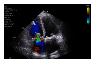
The patient was 71-year-old woman with a history of severe mitral and tricuspid regurgitation. 8 years prior, she had undergone mitral valve replacement (Perimount Magna Ease 29 mm) and tricuspid valve prosthetic annuloplasty ring (Edwards MC3-ring 32 mm). The patient complained of worsening right heart failure symptoms, with severe peripheral edema, and shortness of breath (New York Heart Association (NYHA) Functional Class III-IV). The was hospitalized several times for intravenous diuretic therapy due to right sided heart failure in the past year. Upon admission, physical examination revealed peripheral edema, jugular venous distension, and irregular heartbeat. The electrocardiogram showed atrial fibrillation. Echocardiography demonstrated normal left ventricular function. The mitral valve did not show significant abnormalities. A massive tricuspid regurgitation Grad V was documented (vena contracta width of 11 mm) (Figure 1A). The patient was evaluated by the Heart Team as to be at high risk for re-surgery of the tricuspid valve. TEER was performed by using the TriClip® device (Abbott, Vascular GmbH). During the TEER procedure, it was difficult to visualize the tricuspid leaflets in transesophageal echocardiography (TEE) due to shadowing from the prosthetic annuloplasty ring (Figure 2). This was resolved by utilizing transgastric visualisation (Figure 2) during deployment of the TriClip®. The right ventricular lead was visualized by X ray and echocardiography during the procedure (Figure 3). The device was implanted posterior to the RV lead which was in a central and anterior position into the tricuspid valve. TR was reduced to a Grad II (Figure 1B). Hemodynamic improvement was documented intraoperatively by an increase in arterial pressure up to 15 mmHg immediately after deployment of the Clip. Transvalvular gradient was 1,5 mmHg at the end of the procedure. Pacemaker interrogation did not show alteration of RV sensing or pacing.
After 1 month of follow-up, echocardiography demonstrated TR grade II without stenosis (transvalvular gradient of 1 mmHg). The patient reported that physical capacity and dyspnea had improved (NYHA functional class II).
4. Discussion
Residual tricuspid regurgitation occurs in many patients early after surgical tricuspid annuloplasty [1]. In particularly, non-ring annuloplasty as the DeVega technique, is associated with worsening of tricuspid regurgitation in up to 30% of all patients [1, 2]. In contrast and despite substantial signs of right sided heart failure and severe tricuspid regurgitation, surgical re-operation for tricuspid regurgitation are rarely performed due to the high interventional risk [4, 5]. Recently, TEER has demonstrated to be a decent alternative option for patients with a failed annuloplasty repair. TEER following surgical tricuspid valve repair has been reported in two cases describing the off-label use of the MitraClip® device [6, 7]. In another publication, the PASCALR device was used for TEER of recurrent TR after ring annuloplasty of the tricuspid valve [8]. Further, the TriClip® device was used for TEER in a patient after failed surgical suture annuloplasty of the tricuspid valve [9]. Recently, the results of the TRILUMINATE trial were published including a total of 350 patients [10]. One patient of this cohort had a history of tricuspid surgery. However, more details were not published. Furthermore, the presence of a pacemaker lead was defined as an exclusion criterion in this trial [10].
In the present case, the presence of a prosthetic annuloplasty ring and bioprosthetic mitral valve complicated TEER by massive shadowing in the conventional LVOT views. Therefore, a combined approach using the transgastric echocardiography views and fluoroscopy allowed device navigation and successful TEER in this particular setting. Nevertheless, echocardiography is of upmost importance to guide the TEER device through the right atrium into the right ventricle. In the TRILUMINATE study, 25% of TEER patients had prior mitral valve surgery [11]. Thus, similar to the present case these prior mitral valve interventions alone might also cause shadowing in the area of the tricuspid valve. Nevertheless, TEER is still possible in patients with prior mitral valve procedures. The use of intracardiac echocardiography might serve as an alternative tool to overcome some of shadowing issues of transesophageal echocardiography [12]. However, insertion of intracardiac echocardiography probe requires an additional access port and it remains unclear if the presence of an intracardiac echocardiography probe might mechanically interfere with the TEER device. The follow-up in the present case is rather short, which is a limitation. Thus, the longer-term outcome cannot be reported. However, for the observed first month after the procedure TEER remained unchanged with a sufficient results and TR reduction and clinical improvement.
5. Conclusion
TEER is feasible in severe TR following a prosthetic annuloplasty ring procedure of the tricuspid valve even in the presence of right ventricular pacemaker lead. Thus, TEER appears as a treatment option for patients with a failed prosthetic annuloplasty repair. Nevertheless, more data about TERR in larger cohorts and longer follow-up are needed in this specific subcohort.
Consent
The authors confirm that written informed consent for the submission and publication of this case, including images, has been obtained from the patient in the line with COPE guidance.
Conflict of Interest
Author AG has received speaker fees from Abbott, outside the submitted work. The remaining authors declare that the research was conducted in the absence of any commercial or financial relationships that could be construed as a potential conflict of interest.
References
- Rivera R, Duran E, Ajuria M. Carpentier’s flexible ring versus De Vega’s annuloplasty. A prospective randomized study. J Thorac Cardiovasc Surg 89 (1985): 196-203.
- McCarthy PM, Bhudia SK, Rajeswaran J, et al. Tricuspid valve repair: durability and risk factors for failure. J Thorac Cardiovasc Surg 127 (2004): 674-685.
- Lurz P, Stephan von Bardeleben R, Weber M, et al. Transcatheter edge-to-edge repair for treatment of tricuspid regurgitation. J Am Coll Cardiol 77 (2021): 229-239.
- Sugiura A, Tanaka T, Kavsur R, et al. Leaflet Configuration and Residual Tricuspid Regurgitation After Transcatheter Edge-to-Edge Tricuspid Repair. JACC Cardiovasc Interv 14 (2021): 2260-2270.
- Carpenito M, Cammalleri V, Vitez L, et al. Edge-to-Edge Repair for Tricuspid Valve Regurgitation. Preliminary Echo-Data and Clinical Implications from the Tricuspid Regurgitation IMAging (TRIMA) Study J Clin Med 11 (2022): 5609.
- Hwang HY, Kim KH, Kim KB, Ahn H. Reoperations after tricuspid valve repair: re-repair versus replacement. J Thorac Dis 8 (2016): 133-139.
- Carino D, Denti P, Buzzatti N, et al. MitraClip XTR implantation to treat torrential tricuspid regurgitation after failed annulopasty. Ann Thorac Surg 110 (2020): e165-e7.
- Doldi PM, NabauerM,Massberg S, Hausleiter J. Interventional tricuspid valve repair after failed surgical tricuspid valve reconstruction. Can J Cardiol 36 (2020):1832 e5–e6.
- Afzal S, Haschemi J, Bönner F, et al. Case report: Transcatheter edge-to-edge repair after prior surgical tricuspid annuloplasty. Front Cardiovasc Med 9 (2022): 1044410.
- Sorajja P, Whisenant B, Hamid N, et al. TRILUMINATE Pivotal Investigators. Transcatheter Repair for Patients with Tricuspid Regurgitation. N Engl J Med. 4 (2023).
- Nickenig G, Weber M, Lurz P, et al. Transcatheter edge-to-edge repair for reduction of tricuspid regurgitation: 6-month outcomes of the TRILUMINATE single-arm study. Lancet 394 (2019): 2002-2011.
- Wong I, Chui ASF, Wong CY, Chan KT, Lee MK. Complimentary role of ICE and TEE during transcatheter edge-to-edge tricuspid valve repair with TriClip G4. JACC Cardiovasc Interv 15 (2022): 562-563.


