Bilateral Occlusion of the Central Retinal Vein due to Excess of Factor VIII Level
Article Information
Bencharef H1*, Kouame H1, Oukkache B1,2
1Biological Hematology Department, Ibn Rochd University Hospital, Casablanca, Morocco
2Casablanca Faculty of Medicine and Pharmacy, Hassan II University Casablanca Morocco
*Corresponding Author: Hanaa Bencharef, Biological Hematology Department, Ibn Rochd University Hospital, Casablanca, Morocco.
Received: 27 April 2023; Accepted: 12 May 2023; Published: 16 June 2023
Citation:
Bencharef H, Kouame H, Oukkache B. Bilateral Occlusion of the Central Retinal Vein due to Excess of Factor VIII Level. Archives of Clinical and Medical Case Reports. 7 (2023): 271-275.
View / Download Pdf Share at FacebookAbstract
Introduction: Retinal vein occlusion (RVO) is an obstruction of the retinal venous system, frequently linked to cardiovascular risk factors in elderly; it remains rarer in young subjects, and requires a rigorous etiological investigation. In this article we report the first case of bilateral central retinal vein occlusion (CRVO) in a young patient related to excess factor VIII levels.
Case Presentation: A 22-year-old woman presented with a complaint of sudden and bilateral decreased visual acuity. The patient had no particular pathological history. The best-corrected visual acuity was reduced to light perception in both eyes. A complete fundus assessment including retinal angiography showed bilateral occlusion of the central retinal vein with areas of ischemia.
A complete assessment of thrombophilia was performed revealing an isolated excess of the plasma factor VIII level at 226.4%, checked on a second sample at 232%. The diagnosis of thrombophilia secondary to an increase in factor VIII responsible for OCRV was retained, and the patient was treated with panretinal photocoagulation in both eyes with specialized treatment in the hematology department. For her clinical course, at the visual level her state is stationary in light perception, and at the renal level, she continues her hemodialysis sessions at the rate of 3 sessions per week.
Conclusion: An elevated factor VIII level is an independent risk of venous thrombosis. Moreover, it is likely to influence the pathogenesis of central venous occlusion of the retina. These patients should be carefully evaluated to diagnose thrombophilia, including factor VIII, and initiate appropriate management as soon as possible.
Keywords
Central Retinal Vein Occlusion; Factor VIII; Thrombophilia; Venous Thrombosis
Central Retinal Vein Occlusion articles; Factor VIII articles; Thrombophilia articles; Venous Thrombosis articles
Article Details
1. Introduction
Retinal vein occlusion (RVO) is an obstruction of the retinal venous system that may involve the central retinal vein or one of its branches [1]. It is the second common cause of retinal vascular disease after diabetic retinopathy, which represents a major source of morbidity and can lead to permanent visual impairment [2]. The visual loss is due to macular edema, retinal ischemia, and ocular neovascularization [3].
RVOs are more common in the elderly [4] and are frequently linked to cardiovascular risk factors [5] including hypertension, dyslipidemia, diabetes, etc. In younger people, thrombophilia remains the main cause of retinal vascular disorders [6, 7]. Thrombophilias can be heritable – such as factor V Leiden (FVL), the prothrombin gene G20210A mutation (PGM) [8], deficiencies of the natural anticoagulants protein C (PC), protein S (PS), or antithrombin (AT), elevated factor VIII (FVIII) levels [9] – or acquired, particularly the antiphospholipid syndrome-lupus anticoagulant [10].
Elevated plasma levels of factor VIIIc are known to be a significant, independent and dose-dependent risk factor for venous thromboembolism (VTE) [11], and have been reported in patients with RVO in several investigations [12-14]. We report a case of bilateral occlusion of central retinal vein (CRVO) in a young patient associated with an excess of the plasma level of FVIII.
2. Observation
A 22-year-old woman with no specific pathological history presented to the emergency room with sudden bilateral blindness.
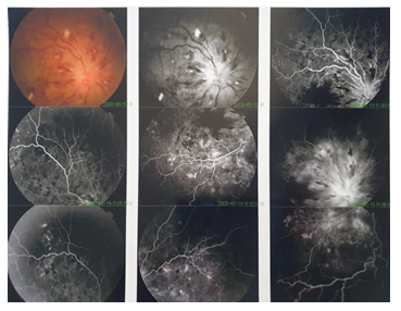
Her visual acuity was less than 1/20th, reduced to light perception in both eyes. The fundus examination showed papillary edema with dilated retinal veins and some flaming hemorrhages suggesting occlusion of the central retinal vein.
A retinal angiography was performed showing bilateral occlusion of the central retinal vein with areas of ischemia (Figure 1).
|
Antithrombine |
123 % [70-140 ] |
|
Protéine C |
150 % [70-140 ] |
|
Protéine S |
116 % [70-140 ] |
|
Factor VIII level |
232 % (First sample) [70-140 %] 226, 4 % (Second sample) |
|
Anti-phospholipid antibodies : - 'Lupus'-Type Circulating Anticoagulant - Anti-cardiolipin - Anti-beta2 glycoprotein1 |
Absence |
|
Resistance to activated protein C |
0, 77 sec [0. 58-1.10] |
|
Homocysteinemia |
9 umol/l [3, 9-10, 7] |
|
Factor V Leiden mutation |
Absence |
|
Prothrombin gene G20210A mutation |
Absence |
Table 1: Complete assessment of thrombophilia.
A standard laboratory workup was performed including a complete blood count with a normal platelet count at 230 G/L [150 – 450 G/L] and a routine hemostasis workup with a normal Prothrombin time at 105% [70-140%], Activated partial thromboplastin time at 28 seconds [23-33 sec] and a normal fibrinogen level at 2.89 g / l [2-4 g/l]. Given the bilaterality of the CRVO and the young age of the patient, a complete assessment of the thrombophilia was carried out, which was normal overall, except for the factor VIII level which was high at 232%, checked on a second sampling at 226.4% [70-40%] (see Table 1).
Immunoassay for markers of autoimmune diseases and vasculitis was free from abnormalities with absence of anti-MBG antibodies (glomerular basement membrane) and ANCA (Antineutrophil cytoplasmic antibodies) antibodies. According to this clinical presentation and laboratory results , the diagnosis of thrombophilia secondary to an increase in factor VIII; responsible for OCRV, was retained, and the patient was treated with panretinal laser photocoagulation performed in both eyes. Her clinical course at the visual level is stationary in light perception, and at the renal level, she continues her hemodialysis sessions at a rate of 3 sessions per week. For thrombophilia, the patient was referred to a hematologist to start anticoagulant therapy and for specialized follow-up.
3. Discussion
The pathogenesis of RVO is not yet fully understood. As a vein occlusion, it can be related to the Virchow triad: hemodynamic changes (venous stasis), degenerative changes in the vascular wall and blood hypercoagulability [15]. Several risk factors have been described for the development of this disease [16-18]. Strong evidence from different studies shows an increased risk of RVO in patients with arteriosclerosis [19, 20]. Over the past years, several groups have evaluated the possible role of thrombophilia in RVO pathogenesis [21, 22].
The Leiden Thrombophilia study (LETS) was the first to report an association between elevated plasma levels of FVIII and veinous thrombosis [23]. Factor VIII is a plasma glycoprotein which plays an essential role in the intrinsic coagulation pathway as a cofactor of activated factor IX (FIXa) and thus allows the activation of factor X [24]. Once activated by thrombin, activated factor VIII (FVIIIa) detaches from von Willebrand factor and forms a complex with factor IXa, leading to a marked acceleration of factor X activation [25].Then prothrombin is converted to thrombin, which in turn converts soluble fibrinogen into insoluble fibrin. Therefore, an elevated Factor VIII level stimulates thrombin formation and increases platelet activation and fibrin formation, a process which may contribute to the development of occlusive thrombi [26, 27].
The association between high levels of factor VIII: C and venous thrombosis has been confirmed by other studies which have also shown that factor VIII is an independent risk factor for thrombosis and that this relationship is a dose-dependent phenomenon [9, 23, 28-30]. It has even been included as a thrombophilia factor [31].
Our patient had a sudden visual loss due to bilateral occlusion of the retinal vein, linked to a high level of factor VIII level, as the only abnormality found in the screening of thrombophilia. Different studies have incriminated the high level of Factor VIII as a risk factor for the development of CRVO (Table 2).
|
STUDIES |
CASES |
CONTROLES |
P value |
||
|
Number (n) |
Hight FVIII: C (%) |
Number (n) |
Hight FVIII: C (%) |
||
|
Faude et al 2004 [32] |
62 CRVO |
53 |
107 |
19.7 |
0.0004 |
|
Glueck et al 2005 [3] |
44 RVO |
10 |
40 |
0 |
0.041 |
|
Glueck et al 2008 [34] |
40 CRVO |
30 |
80 |
5 |
0.0002 |
|
Glueck et al 2012 [33] |
132 CRVO |
20 (23/116) |
105 |
7 (7/98) |
0.008 |
|
Dixon 2016 [35] |
76 RVO |
42 |
62 |
11 |
< 0.0001 |
|
Bucciarelli 2017 [36] |
313 RVO |
11, 3*(28/248) |
415 |
4, 8 (20/415) |
0.032 |
* Information not available for 65 patients.
Table 2: Studies that evaluated the prevalence of elevated Factor VIII in RVO.
Faude et al [32] reported that 53% of 62 patients with CRVO had an elevated factor VIII greater than 150% (> 150 IU) compared to 19.7% of the healthy control group. This difference in factor VIII: C activity between cases and controls was very significant (p = 0.0004); suggesting that high factor VIII activity is likely to influence the pathogenesis of central retinal vein occlusion. In another study [3] , elevated levels of hereditary factor VIII were more common in RVO patients (n = 44) than in controls (n= 40) (10% vs 0%). On a larger sample including 132 CRVO and 105 controls, Glueck et al [33] concluded that CRVO cases were more likely than controls to have high Factor VIII (OR 2.47, 95% CI: 1.31-7.9), and this has also been reported by others [34, 35].
In a recent case-control study [36] involving 313 patients with confirmed RVO and 415 healthy individuals, elevated factor VIII (FVIII) levels were independently associated with an increased risk of RVO. Lately in 2019, Chang et al [37] published a case report of a patient who had a combined occlusion of the central retinal vein and artery due to elevated factor VIII levels, and suggested that these patients should be carefully assessed to diagnose underlying factors, including factor VIII, to initiate appropriate management as soon as possible. Otherwise, in 1003 acute unilateral CRVO (41 ischemic // 62 non-ischemic), elevated levels of FVIII were found as a risk factor for the ischemic form of CRVO (OR, 6.17; 95% CI, 2, 56-14, 82; p <0.001) [38] which is the case in our patient. To our knowledge, this is the first case describing « bilateral occulsion » of the central retinal vein related to elevate Factor VIII in the literature. The dosage of Factor VIII must be systematically included in the assessment of thrombophilia, its increase is a known and proven cause of venous or arterial thrombosis. It is necessary to think of this etiology especially in front of the young age and the negativity of the other parameters of the assessment of the thrombophilia.
4. Conclusion
RVO is associated with multiple contributing factors, including thrombophilia and cardiovascular risk factors. Known as a risk factor for venous thrombosis, elevated level of Factor VIII is also involved in the development of RVO. The diagnosis of an underlying thrombophilia including Factor VIII is important not only for the management of the RVO but to prevent other potentially life-threatening thrombotic events such as pulmonary embolism.
Declaration of Interest
The submitted manuscript has been approved by all the authors who declare that they have no conflict of interest in connection with this article. The manuscript has not been published elsewhere, it has not been submitted to another review, and is not being reviewed by another publication.
References
- Hayreh SS. Retinal vein occlusion. Indian J Ophthalmol 42 (1994): 109-1
- Rehak M, Wiedemann P. Retinal vein thrombosis: pathogenesis and management. J Thromb Haemost 8 (2010):1886-18
- Glueck CJ, Wang P, Bell H, et al. Associations of thrombophilia, hypofibrinolysis, and retinal vein occlusion. Clin Appl Thromb Hemost 11 (2005): 375-3
- Kuhli-Hattenbach C, Scharrer I, Lüchtenberg M, et al. Coagulation disorders and the risk of retinal vein occlusion. Thrombosis and Haemostasis 7 (2010).
- Ponto KA, Elbaz H, Peto T, et al. Prevalence and risk factors of retinal vein occlusion: the Gutenberg Health Study. J Thromb Haemost. juill 13 (2015): 1254-12
- O’Mahoney PRA, Wong DT, Ray JG. Retinal vein occlusion and traditional risk factors for atherosclerosis. Arch Ophthalmol. mai 126 (2008): 692-69
- Marcucci R, Sofi F, Grifoni E, et al. Retinal vein occlusions: a review for the internist. Intern Emerg Med 6 (2011): 307-314.
- Schockman S, Glueck CJ, Hutchins RK, et al. Diagnostic ramifications of ocular vascular occlusion as a first thrombotic event associated with factor V Leiden and prothrombin gene heterozygosity. Clin Ophthalmol 9 (2015): 591-600.
- Jenkins PV, Rawley O, Smith OP, et al. Elevated factor VIII levels and risk of venous thrombosis. Br J Haematol. juin 157 (2012): 653-663.
- Bick RL. Antiphospholipid thrombosis syndromes. Clin Appl Thromb Hemost 7 (2001): 241-258.
- Kraaijenhagen R, in’t Anker P, Koopman M, et al. High Plasma Concentration of Factor VIIIc Is a Major Risk Factor for Venous Thromboembolism. Thromb Haemost 83 (2000): 5-
- Trope GE, Lowe GD, McArdle BM, et al. Abnormal blood viscosity and haemostasis in long-standing retinal vein occlusion. Br J Ophthalmol. Mars 67 (1983): 137-1
- Lerche RC, Wilhelm C, Eifrig B, et al. [Thrombophilia factors as inducers of retinal vascular occlusion]. Ophthalmologe. juin 98 (2001): 529-5
- Faude F, Faude S, Siegemund A, et al. [Factor VIII activity in patients with central retinal vein occlusion in comparison to patients with a history of pelvic and lower limb venous thrombosis and a healthy control group]. Klin Monbl Augenheilkd 221 (2004): 862-86
- Kolar P. Risk factors for central and branch retinal vein occlusion: a meta-analysis of published clinical data. J Ophthalmol 2014 (2014): 724780.
- Hayreh SS, Zimmerman B, McCarthy MJ, et al. Systemic diseases associated with various types of retinal vein occlusion. Am J Ophthalmol. janv 131 (2001): 61-
- Marcucci R, Bertini L, Giusti B, et al. Thrombophilic risk factors in patients with central retinal vein occlusion. Thromb Haemost 86 (2001): 772-77
- Lee HBH, Pulido JS, McCannel CA, et al. Role of inflammation in retinal vein occlusion. Can J Ophthalmol. févr 42 (2007): 131-13
- Mitchell P, Smith W, Chang A. Prevalence and associations of retinal vein occlusion in Australia. The Blue Mountains Eye Study. Arch Ophthalmol 114 (1996): 1243-12
- Wong TY, Larsen EKM, Klein R, et al. Cardiovascular risk factors for retinal vein occlusion and arteriolar emboli: the Atherosclerosis Risk in Communities and Cardiovascular Health studies. Ophthalmology 112 (2005): 540-54
- Lahey JM, Tunç M, Kearney J, et al. Laboratory evaluation of hypercoagulable states in patients with central retinal vein occlusion who are less than 56 years of age. Ophthalmology 109 (2002): 126-1
- Cruciani F, Moramarco A, Curto T, et al. MTHFR C677T mutation, factor II G20210A mutation and factor V Leiden as risks factor for youth retinal vein occlusion. Clin Ter 154 (2003): 299-
- Koster T, Blann AD, Briët E, et al. Role of clotting factor VIII in effect of von Willebrand factor on occurrence of deep-vein thrombosis. Lancet 345 (1995): 152-15
- Pellegrina L, Emile C. Variation’s physiologiques et pathologiques du facteur VIII «Le facteur VIII dans tous ses états». Bio trib mag 38 (2011): 31-3
- Van Dieijen G, Tans G, Rosing J, et al. The role of phospholipid and factor VIIIa in the activation of bovine factor X. J Biol Chem 256 (1981): 3433-34
- Kawasaki T, Kaida T, Arnout J, et al. A new animal model of thrombophilia confirms that high plasma factor VIII levels are thrombogenic. Thromb Haemost 81 (1999): 306-3
- Machlus KR, Lin F-C, Wolberg AS. Procoagulant activity induced by vascular injury determines contribution of elevated factor VIII to thrombosis and thrombus stability in mice. Blood 118 (2011): 3960-396
- Kamphuisen PW, Eikenboom JCJ, Bertina RM. Elevated Factor VIII Levels and the Risk of Thrombosis. Arterioscler Thromb Vasc Biol 21 (2001): 731-73
- Kraaijenhagen RA, in’t Anker PS, Koopman MM, et al. High plasma concentration of factor VIIIc is a major risk factor for venous thromboembolism. Thromb Haemost 83 (2000): 5-9.
- Patel R, Ford E, Thumpston J, et al. Risk factors for venous thrombosis in the black population. Thromb Haemost 90 (2003): 835-83
- Roux A, Sanchez O, Meyer G. Quel bilan de thrombophilie chez un patient atteint de maladie veineuse thromboembolique ? Réanimation 17 (2008): 355-3
- Faude F, Faude S, Siegemund A, et al. Faktor-VIII-Aktivität bei Patienten mit Zentralvenenverschluss im Vergleich zu Patienten mit tiefer Becken- und Beinvenenthrombose und gesunder Kontrollgruppe. Klin Monatsbl Augenheilkd 221 (2004): 862-86
- Glueck CJ, Hutchins RK, Jurantee J, Khan Z, Wang P. Thrombophilia and retinal vascular occlusion. Clin Ophthalmol 6 (2012): 1377-13
- Glueck CJ, Ping Wang, Hutchins R, et al. Ocular Vascular Thrombotic Events: Central Retinal Vein and Central Retinal Artery Occlusions. Clin Appl Thromb Hemost 14 (2008): 286-2
- Dixon SG, Bruce CT, Glueck CJ, et al. Retinal vascular occlusion: a window to diagnosis of familial and acquired thrombophilia and hypofibrinolysis, with important ramifications for pregnancy outcomes. Clin Ophthalmol 10 (2016): 1479-14
- Bucciarelli P, Passamonti SM, Gianniello F, et al. Thrombophilic and cardiovascular risk factors for retinal vein occlusion. European Journal of Internal Medicine 44 (2017): 44-4
- Chang IB, Lee JH, Kim HW. Combined Central Retinal Vein And Artery Occlusion In A Patient With Elevated Level Of Factor VIII: A Case Report. IMCRJ 12 (2019): 309-3
- Sodi A, Giambene B, Marcucci R, et al. Atherosclerotic and thrombophilic risk factors in patients with ischemic central retinal vein occlusion. Retina 31 (2011): 724-72

