Survival Patterns of COVID-19 Pneumonia Patients with False-Negative PCR Results Transferred to the Intensive Care Unit from the Emergency Department
Article Information
Meltem Songur Kodik1*, Esin Ozturk2, Yusuf Ali Altunci1, Enver Ozcete1, Sercan Yalcinli1,
Murat Ersel1, Deniz Akyol3, Selen Bayraktaroglu4, Ozlem Goksel5, Ahmet Enes Celik6
1Department of Emergency Medicine, Faculty of Medicine, Ege University , Izmir, Turkey
2Department of Anesthesiology and Reanimation, Faculty of Medicine, Ege University, Izmir, Turkey
3Department of Infectious Diseases and Clinical Microbiology, Kagizman State Hospital, Kars, Turkey
4Department of Radiology, Faculty of Medicine, Ege University, Izmir, Turkey
5Department of Pulmonary Medicine, Faculty of Medicine, Ege University, Izmir, Turkey
6Department of Emergency Medicine, Midyat State Hospital, Mardin, Turkey
*Corresponding author: Meltem Songur Kodik, Department of Emergency Medicine, Faculty of Medicine, Ege University, Bornova-Izmir, Turkey.
Received: 27 April 2022; Accepted: 06 May 2022; Published: 19 May 2022
Citation: Meltem Songur Kodik, Esin Ozturk, Yusuf Ali Altunci, Enver Ozcete, Sercan Yalcinli, Murat Ersel, Deniz Akyol, Selen Bayraktaroglu, Ozlem Goksel, Ahmet Enes Celik. Survival Patterns of COVID-19 Pneumonia Patients with False-Negative PCR Results Transferred to the Intensive Care Unit from the Emergency Department. Archives of Clinical and Biomedical Research 6 (2022): 435-447.
View / Download Pdf Share at FacebookAbstract
Aim: This study aimed to analyze factors associated with patient mortality in COVID-19 patients hospitalized in the intensive care unit (ICU) and provide data to help prioritize the most critical patients.
Methods: A clinical retrospective cohort study was conducted in a tertiary hospital. Patients (n=289) with negative reverse transcription-polymerase chain reaction (RT-PCR) and a positive chest computed tomography (CT) scan indicating COVID-19 were included in the research. Demographics, clinical characteristics, treatment modalities, and length of hospital stay were analyzed in relation to 30-day survival outcomes.
Results: The mean age of the patients was 69.04 ± 15.10 years (range 19-100), and 41.2% (n=119) were female. The mortality rate was 48.1% (n=139). There were statistically significant differences in laboratory findings, such as hemoglobin (p=0.021), lymphocyte counts (p=0.046), d-dimer (p=0.009), and lactate (<0.001) regarding one-month survival. Additionally, those hospitalized for more than 5 days had higher one-month survival rates (hazard ratio (HR)= 0.003, CI= 10.264-155.972). Moreover, intubated patients had a higher mortality risk during this period (HR=4.15, CI=1.53-11.20).
Conclusion: The first five days of patients hospitalized in intensive care units due to COVID-19 pneumonia are critical. Besides, the need for intubation was another factor independently affecting survival. Clinicians should be alerted about the significance of these factors.
Keywords
COVID-19 Pandemic; COVID-19 RT-PCR Testing; Computed Tomography; Intensive Care Units; Mortality
COVID-19 Pandemic articles; COVID-19 RT-PCR Testing articles; Computed Tomography articles; Intensive Care Units articles; Mortality articles
Article Details
1. Introduction
The novel coronavirus disease 19 (COVID-19), which emerged in Wuhan in December 2019, has infected millions of people and evolved into a pandemic that has triggered a significant number of deaths and unprecedented global social and economic impact [1]. Although most of the patients were mild or asymp-tomatic, severe cases could challenge intensive care capacities in some periods due to the widespread prevalence of the COVID-19 infection [2]. Some patients developed moderate to severe symptoms or had other underlying medical conditions, making them more vulnerable to the coronavirus. These patients were usually treated in intensive care units (ICUs) [3]. Identifying individuals at high risk and prioritizing their care will be of great advantage in order to use limited health resources efficiently and increase survival rates [4].
Another clinical challenge is the accurate detection of COVID-19 patients. Based on a systemic review, studying negative real-time reverse transcription-polymerase chain reaction (RT-PCR) tests that were positive on a repeat test, the sensitivity of the RT-PCR tests vary between 71-98% [5]. The site and quality of sampling play a vital role in the accuracy of RT-PCR tests [6]. Also, the stage of disease and degree of viral multiplication or clearance is likely to affect the accuracy [7]. In comparison, chest computed tomo-graphy (CT) has higher proven accuracy in diagnosing COVID-19, and it may be considered a primary detection method in epidemic areas [8].Thus, when a patient is suspected of COVID 19 or showing typical symptoms but has a negative RT-PCR test, the chest CT scan is a powerful method to diagnose the disease [9].
1.1 Objective
This study aimed to analyze factors associated with patient mortality in COVID-19 patients hospitalized in the intensive care unit (ICU) and provide data to help prioritize the most critical patients.
2. Methods
2.1 Study design
This clinical retrospective cohort study was carried out by examining the files and records in the hospital automation system of patients who had a negative result on the RT-PCR test but diagnosed with COVID-19 pneumonia after observing typical positive CT findings and hospitalized in the anesthesiology and reanimation intensive care unit between March 11, 2020, and October 31, 2020. Ethical approval (Date: September 3, 2020, number: 20-9T/69) was received from the XX University Local Ethics Committee. Study reporting was done per the Strobe Guidelines [10].
2.2 Setting
This study was done at the hospital, emergency department. The research unit is a tertiary reference health center serving daily around 400 emergency admissions in …, a city at the … border with 4.1 million inhabitants. In the hospital, RT-PCR-positive patients were admitted to the intensive care unit of the chest diseases department. In contrast, RT-PCR-negative patients requiring intensive care who had typical COVID-19 CT findings were generally hospitalized to the anesthesiology and reanimation intensive care unit (A&R ICU). This is a special ICU assigned for COVID-19 patients in the study hospital. However, exceptionally, some RT-PCR-positive patients were admitted to the A&R ICU if they were transferred from another medical center or if the capacity of the other ICU was full.
2.3 Image analysis
CT images were obtained using a 160-slice-CT scanner (Aquilion Prime, Toshiba Medical Systems Tokyo, Japan). The scanning parameters were; 120 kVp, 100-200 mA, automated dose reduction, 80x0.5 mm collimation, and reconstruction with a sharp algorithm at 0.5 mm slice thickness. CT images were reviewed with respect to COVID-19 by an experienced radiologist (author SB) blinded to RT-PCR results but aware of the epidemiologic history and clinical symptoms, such as fever and/or dry cough. After the examination, the radiologist decided whether the CT findings were positive or negative. Primary CT findings associated with COVID-19 were ground-glass opacity, consolidation, reticulation, and/or thickened interlobular septa, nodules, and lesion distribution in the left lung, right lung, or bilateral lungs.
2.4 Participants
An archive search was conducted by an experienced data analyst from the hospital’s IT department using the International Statistical Classification of Diseases and Related Health Problems (ICD) code U07.3 [11], the clinical code for COVID-19. Inclusion criteria were being above 18 years old, diagnosed by COVID-19 according to the case definition given by the Ministry of Health [12], and/or having COVID-19-compatible results in the chest CT scan. As shown in Figure 1, a total of 337 COVID-19 patients were detected, from which 48 RT-PCR positives were excluded. Finally, 289 patients were included in the study, of which 139 did not survive.
2.5 Variables
The dependent variable was 30-days survival. Patients were followed up until 30 days after admission to the emergency service (even if they were discharged earlier), and the outcome was reported as survived or died. The independent numerical variables included age, results of the first clinical examination (temperature, respiratory rate, and peripheral oxygen saturation), duration of complaints (days), blood gas values, and blood test results. The categorical independent variables were sex, admission complaint, co-morbidity, symptom severity, treatments, co-infection, control PA chest radiography/CT scan, and hospital stay for more than 5 days. Evaluation of the symptom severity was performed as:
- Mild and moderate symptoms: Respiratory symptoms (cough, sputum, fever, chest pain) that do not reduce capillary oxygen saturation (SpO2) to 90% or less.
- Severe symptoms: Dyspnea and low capillary oxygen saturation (SpO2 ≤ 90%).12
2.6 Statistical analysis
Statistical analysis was performed with the Statistical Package for the Social Sciences program (SPSS for Windows, Version 25.0, Chicago, IL, USA). Results were presented as mean and standard deviations for numerical data and frequencies and percentages for categorical variables. The compatibility of variables to normal distribution was evaluated using the Kolmogorov-Smirnov test. Parametric variables were compared using the independent samples t-test. Additionally, Chi-square or Fisher's exact test was used to compare categorical variables. On the other hand, the Cox regression analysis was performed, and the effects of factors on survival were analyzed. For the statistical significance level, p<0.05 was considered sufficient.
3. Results
Data for 289 patients were analyzed. The mean age of the participants was 69.04 ± 15.10 years (19-100), and 41.2% (n=119) were female. The mortality rate was 48.1% (n=139). There were statistically significant differences in some variables, such as lymphocyte numbers, d-dimer, and lactate related to one-month survival (Table 1). The most often observed co-morbidities were cardiac failures, chronic obstructive pulmonary disease (COPD), malignancy, cardio-vascular disease (CVD), and hypertension (Table 2). Other less frequently observed co-morbidities included diabetes mellitus (DM), dementia, coronary artery disease, or chronic kidney disease. Forty-seven (62.7%) patients diagnosed with malignancy died (Table 2).
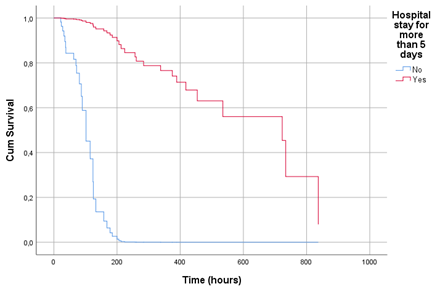
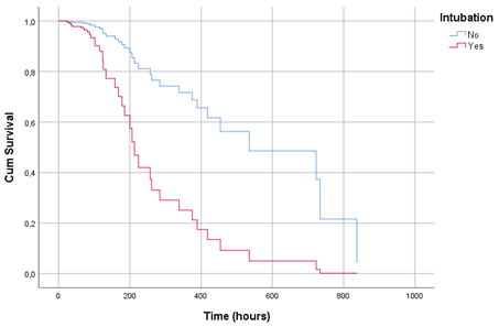
There were three main complaints: respiratory symptoms (i.e., dyspnea and cough), fever, and delirium. Less frequently observed complaints, such as nausea, diarrhea, or disordered general health, were included in the ‘other symptoms’ group. Eighteen patients had mild symptoms, and only 2 (11.1%) of those died, whereas in patients with moderate and severe symptoms, this ratio was much higher (52.7% and 50%, respectively). Intubation and inotropic treatment caused a significant difference in the one-month survival. Co-infection was present in 77 (26.6%) of the patients, and the focuses of infection were the urinary tract, sepsis, lung, or tissues (Table 2). Those hospitalized for more than 5 days were less likely to die within one month. Additionally, intubated patients had a higher mortality risk (Table 3). In the survival analysis, patients with a hospital stay of more than five days had significantly higher survival rates than those who were hospitalized five days or less (Figure 1). Also, a statistically significant difference was found between survival times in patients with and without intubation in the survival analysis (Figure 2).
3.1 Key results
There were statistically significant differences in laboratory findings, such as hemoglobin, lymphocyte counts, lactate dehydrogenase (LDH), d-dimer, and lactate related to one-month survival. Additionally, hospitalization for more than 5 days and intubation were factors that independently affected one-month survival. A hospital stay of more than five days had a positive effect on survival, while the need for intubation had a negative impact.
|
Characteristics |
Survival |
n |
Mean |
SD |
t |
p |
|
Age (year) |
Died |
139 |
70.64 |
13.88 |
1.735 |
0.084 |
|
Survived |
150 |
67.57 |
16.07 |
|||
|
Respiratory rate (/min) |
Died |
81 |
20.81 |
4.66 |
1.747 |
0.082 |
|
Survived |
86 |
22.00 |
4.10 |
|||
|
Pulse (/min) |
Died |
95 |
105.64 |
25.49 |
2.812 |
0.005 |
|
Survived |
111 |
96.42 |
21.57 |
|||
|
Temperature (°C) |
Died |
62 |
36.42 |
0.90 |
0.355 |
0.723 |
|
Survived |
57 |
36.36 |
0.77 |
|||
|
Capillary saturation (%) |
Died |
106 |
91.86 |
8.09 |
0.230 |
0.818 |
|
Survived |
125 |
91.62 |
7.90 |
|||
|
Duration of complaint (days) |
Died |
130 |
4.53 |
8.96 |
1.669 |
0.097 |
|
Survived |
130 |
3.14 |
3.19 |
|||
|
Leukocyte counts (/mm3) |
Died |
139 |
16658 |
24669 |
0.417 |
0.677 |
|
Survived |
150 |
15513 |
21970 |
|||
|
Lymphocyte counts (µL) |
Died |
139 |
2137.48 |
4644.58 |
2.003 |
0.046 |
|
Survived |
150 |
3913.07 |
9723.11 |
|||
|
Hemoglobin (g/dl) |
Died |
139 |
11.06 |
2.74 |
2.325 |
0.021 |
|
Survived |
150 |
11.78 |
2.51 |
|||
|
Hematocrit (%) |
Died |
139 |
34.78 |
8.22 |
2.702 |
0.007 |
|
Survived |
150 |
37.29 |
7.62 |
|||
|
Troponin (ug/L) |
Died |
129 |
125.82 |
363.73 |
1.137 |
0.257 |
|
Survived |
137 |
84.56 |
213.05 |
|||
|
LDH (U/L) |
Died |
78 |
579.45 |
902.45 |
2.721 |
0.008 |
|
Survived |
74 |
297.88 |
140.40 |
|||
|
D-dimer (mg/dl) |
Died |
117 |
3051.54 |
1549.44 |
2.620 |
0.009 |
|
Survived |
123 |
2535.23 |
1503.42 |
|||
|
Ferritin (ng/mL) |
Died |
7 |
445.76 |
457.13 |
1.085 |
0.306 |
|
Survived |
4 |
187.60 |
118.90 |
|||
|
Lactate (mmol/L) |
Died |
138 |
4.23 |
4.12 |
3.668 |
<0.001 |
|
Survived |
147 |
2.79 |
2.11 |
|||
|
Hospitalization (hours) |
Died |
139 |
265.07 |
287.79 |
1.219 |
0.224 |
|
Survived |
150 |
304.15 |
257.00 |
SD: standard deviation, t: Independent samples t-test value, LDH: Lactate dehydrogenase
Table 1: Comparison of numerical variables concerning one-month survival.
|
Characteristics |
One-month survival |
χ2
|
P |
||||
|
Died |
Survived |
||||||
|
n |
% |
n |
% |
||||
|
Sex |
Female |
53 |
44.5 |
66 |
55.5 |
1.026 |
0.311 |
|
Male |
86 |
50.6 |
84 |
49.4 |
|||
|
Admission Complaints |
|||||||
|
Respiratory symptoms |
No |
54 |
56.8 |
41 |
43.2 |
4.336 |
0.037 |
|
Yes |
85 |
43.8 |
109 |
56.2 |
|||
|
Fever |
No |
116 |
48.7 |
122 |
51.3 |
0.223 |
0.637 |
|
Yes |
23 |
45.1 |
28 |
54.9 |
|||
|
Delirium |
No |
123 |
46.6 |
141 |
53.4 |
2.773 |
0.096 |
|
Yes |
16 |
64 |
9 |
36 |
|||
|
Other symptoms |
No |
86 |
44.3 |
108 |
55.7 |
3.355 |
0.067 |
|
Yes |
53 |
55.8 |
42 |
44.2 |
|||
|
Co-morbidity |
No |
11 |
39.3 |
17 |
60.7 |
0.964 |
0.326 |
|
Yes |
128 |
49 |
133 |
51 |
|||
|
Cardiac failure |
No |
109 |
47.4 |
121 |
52.6 |
0.225 |
0.635 |
|
Yes |
30 |
50.8 |
29 |
49.2 |
|||
|
COPD |
No |
125 |
48.4 |
133 |
51.6 |
0.12 |
0.729 |
|
Yes |
14 |
45.2 |
17 |
54.8 |
|||
|
Malignancy |
No |
92 |
43 |
122 |
57 |
8.613 |
0.003 |
|
Yes |
47 |
62.7 |
28 |
37.3 |
|||
|
CVD |
No |
130 |
48.5 |
138 |
51.5 |
0.249 |
0.618 |
|
Yes |
9 |
42.9 |
12 |
57.1 |
|||
|
Hypertension |
No |
69 |
50.7 |
67 |
49.3 |
0.716 |
0.397 |
|
Yes |
70 |
45.8 |
83 |
54.2 |
|||
|
Other co-morbidities |
No |
52 |
48.1 |
56 |
51.9 |
0 |
0.989 |
|
Yes |
87 |
48.1 |
94 |
51.9 |
|||
|
Symptom severity |
Mild |
2 |
11.1 |
16 |
88.9 |
10.882 |
0.004 |
|
Moderate |
49 |
52.7 |
44 |
47.3 |
|||
|
Severe |
86 |
50 |
86 |
50 |
|||
|
Second PCR result |
Negative |
120 |
45.6 |
143 |
54.4 |
0.06 |
1 |
|
Positive |
4 |
50 |
4 |
50 |
|||
|
Inotrope treatment |
No |
44 |
25.7 |
127 |
74.3 |
85.59 |
<0.001 |
|
Yes |
95 |
81.2 |
22 |
18.8 |
|||
|
Intubation |
No |
37 |
23.3 |
122 |
76.7 |
88.805 |
<0.001 |
|
Yes |
102 |
79.1 |
27 |
20.9 |
|||
|
Co-infection** |
No |
91 |
42.9 |
121 |
57.1 |
8.527 |
0.003 |
|
Yes |
48 |
62.3 |
29 |
37.7 |
|||
|
Urinary track |
No |
115 |
45.8 |
136 |
54.2 |
3.976 |
0.046 |
|
Yes |
24 |
63.2 |
14 |
36.8 |
|||
|
Lung |
No |
121 |
46.2 |
141 |
53.8 |
4.114 |
0.043 |
|
Yes |
18 |
66.7 |
9 |
33.3 |
|||
|
Sepsis |
No |
120 |
45.3 |
145 |
54.7 |
10.121 |
0.001 |
|
Yes |
19 |
79.2 |
5 |
20.8 |
|||
|
Tissue |
No |
136 |
47.9 |
148 |
52.1 |
0.289 |
0.674* |
|
Yes |
3 |
60 |
2 |
40 |
|||
|
Culture positivity |
Absent |
92 |
42.8 |
123 |
57.2 |
12.792 |
0.002 |
|
Present |
47 |
65.3 |
25 |
34.7 |
|||
|
Control PA chest radiography |
Regression |
17 |
24.6 |
52 |
75.4 |
24.315 |
<0.001 |
|
Stable |
33 |
40.7 |
48 |
59.3 |
|||
|
Progression |
28 |
73.7 |
10 |
26.3 |
|||
|
Hospital stay for more than 5 days |
No |
44 |
68.8 |
20 |
31.3 |
14.046 |
<0.001 |
|
Yes |
95 |
42.2 |
130 |
57.8 |
|||
χ2: Chi-Square test value, *:Fisher’s Exact test, **: In some patients, multiple co-infections were observed
Table 2: Comparison of categorical variables regarding one-month survival.
|
Characteristics |
B |
SE |
Wald |
p |
Exp(B) |
95.0% CI for Exp(B) |
|
|
Lower |
Upper |
||||||
|
Pulse/min |
0.006 |
0.011 |
0.36 |
0.548 |
1.01 |
0.99 |
1.03 |
|
Hemoglobin (g/dl) |
0.013 |
0.103 |
0.017 |
0.896 |
1.01 |
0.83 |
1.24 |
|
Blood urea nitrogen (mg/dl) |
0.001 |
0.003 |
0.073 |
0.788 |
1 |
1 |
1.01 |
|
LDH (U/L) |
0 |
0.001 |
0.33 |
0.565 |
1 |
1 |
1 |
|
D-dimer (mg/dl) |
0 |
0 |
0.019 |
0.891 |
1 |
1 |
1 |
|
Lactate (mmol/L) |
0.092 |
0.063 |
2.131 |
0.144 |
1.1 |
0.97 |
1.24 |
|
Inotrope treatment (Yes vs. No) |
0.732 |
0.584 |
1.571 |
0.21 |
2.08 |
0.66 |
6.53 |
|
Intubation (Yes vs. No) |
1.422 |
0.507 |
7.864 |
0.005 |
4.15 |
1.53 |
11.2 |
|
Co-infection (Yes vs. No) |
0.058 |
0.474 |
0.015 |
0.902 |
1.06 |
0.42 |
2.68 |
|
Fever at admission |
3.875 |
0.144 |
|||||
|
Fever at admission (Yes vs. No) |
1.347 |
0.694 |
3.769 |
0.052 |
3.85 |
0.99 |
14.98 |
|
Fever at admission (No fever vs. No) |
0.765 |
0.678 |
1.274 |
0.259 |
2.15 |
0.57 |
8.12 |
|
Hospital stay for more than 5 days (Yes vs. No) |
-3.689 |
0.694 |
28.245 |
<0.001 |
0.03 |
0.01 |
0.1 |
|
Symptom severity |
0.337 |
0.845 |
|||||
|
Symptom severity (Moderate vs. mild) |
1.52 |
3.851 |
0.156 |
0.693 |
4.57 |
0 |
8660.53 |
|
Symptom severity (Severe vs. mild) |
1.306 |
3.846 |
0.115 |
0.734 |
3.69 |
0 |
6927.6 |
|
Malignancy (Present vs. Absent) |
0.236 |
0.443 |
0.283 |
0.595 |
1.27 |
0.53 |
3.02 |
LDH: Lactate dehydrogenase
Table 3: Cox-regression analysis computer output showing factors independently affecting one-month survival.
4. Discussion
The outbreak of novel coronavirus pneumonia is an unprecedented public health issue due to being large-scale spread that overloaded medical services and resulted in excessive usage of medical resources [1]. A widely used method to diagnose COVID-19 is the PCR tests. However, these tests can sometimes give false-negative results. Some reasons for these misdiagnoses are sampling or transport errors and mutations in probe-target regions in the SARS-CoV-2 genome [13]. If a patient is suspected of having COVID-19, but RT-PCR is negative, a chest CT scan can effectively support diagnosis [9]. In a study with 1014 patients in Wuhan that references positive RT-PCR cases, the sensitivity of chest CT for COVID-19 was estimated as 97% (580 of 601 patients).
Moreover, in a comprehensive evaluation among 308 patients with negative RT-PCR results and positive chest CT findings, 147 (48%) of these were revised as highly possible cases and 103 (33%) as probable cases [8]. This study can contribute to the literature by analyzing patients with negative PCR results and positive chest CT findings and revealing the factors affecting mortality. Thus, these findings may allow clinicians to prioritize the care for individuals, assist resource allocations, and reduce fatality rates. Mortality rates due to COVID-19 are very high in hospitalized elderly patients [14-16]. Besides, pre-existing co-morbidity and disease severity are associated with poor prognosis in these patients [17]. Moreover, in elderly patients hospitalized for COVID-19, male sex, crackles, high respiratory oxygen requirement, and bilateral and peripheral infiltrates on chest radiographs are independent risk factors for mortality [15]. In line with previous studies in this research, symptom severity, co-morbidity, having respiratory symptoms, and positive findings on chest X-ray were associated with poor prognosis. However, we found no relationship between sex and mortality.
In addition to symptoms such as dyspnea, increased LDH, D-dimer, PCT, ferritin, and leukocyte counts, and decreased lymphocyte counts have been specified as laboratory parameters that can be used to predict mortality in COVID-19 patients [18-20]. Moreover, hemoglobin (HGB) and hematocrit (HCT) values are also critical for these patients. In a study of COVID-19 cases hospitalized in an intensive care unit in Ankara, mean values of HGB and HCT were below the normal range [21]. Reportedly, the anomalies of HGB and HCT were associated with co-morbidity, and the reasons can be bone marrow being unable to produce enough red blood cells to carry oxygen or the lung damages caused by COVID-19, making gaseous exchange difficult [22]. Likewise, in our study, similar results were obtained with previous publications, and also hemoglobin and hematocrit were lower, especially in patients who did not survive for a month. Furthermore, lactate levels of the non-survivors were significantly higher than the survivors, which is reportedly a predictor of severe pneumonia [20]. In a multicenter cohort study that included only elderly emergency patients (>65, mean age 77.7) with COVID-19, 28% of the subjects had delirium at admission [23]. In our study, this rate was lower (8.7%). The main reason for this difference could be the difference in the study populations. Indeed, our study included patients older than 18 years of age, with a mean age of 69 years.
The most common co-morbidity in our study was hypertension, similar to a study in 5700 patients hospitalized with COVID-19 in New York City Area [24]. Malignancy increases the risk of death due to COVID-19 infection [25, 26]. Likewise, in our study, malignancy increased mortality, but our rate was higher than in previous studies. This difference may be due to the diversity of the departments where the studies were conducted. It is not surprising that mortality rates were high in the research performed in the ICU. Consistent with a retrospective study in which the mortality rate was estimated as 57%, another factor that increased fatality in intensive care COVID-19 patients was the presence of co-infection [27]. Therefore, antibiotic therapy should be optimi-zed in critically ill patients with COVID-19 in the ICU.
Furthermore, this study reinforced that the need for intubation should alert clinicians to the high mortality rate. In a single-center pilot study with critically ill patients, 76% of intubated patients died [28]. In another study, this rate was 81.1% [29]. Inotropic agents are frequently used in patients with concerns about severely reduced cardiac output, indicating a poor prognosis [30]. Indeed, patients requiring inotropic therapy had a lower one-month survival rate in this study. The length of stay for critically ill patients is lower for the non-survivors than for survivors [31]. The median length of ICU stay was reported between 4 and 11 days for patients who died in the ICU [4, 31]. Our findings were consistent with previous studies. Additionally, in the regression model, intubation and hospital stay were factors independently affecting survival. The higher mortality rates in those who stayed in the hospital for less than 5 days may be related to the poor condition of the patients admitted to the intensive care unit. In other words, patients may respond better to treatment after the initial critical situation has passed, and therefore mortality rates may have decreased.
This study should be interpreted in light of some limitations. Firstly, it is a retrospective investigation conducted in a single emergency service and intensive care unit. Secondly, no long-term outcomes or quality of life were tracked in the survivors. Finally, at the beginning of the coronavirus outbreak, treatments were difficult due to the facilities. Specifically, most of the frequently used COVID-19 medicines today were absent or low in supply, and plasma treatment was not possible in the first weeks or months of the pandemic.
5. Conclusion
Some of the patients hospitalized in the intensive care unit are false negative for COVID-19. The ability of clinicians to predict patients with poor prognoses can help to optimize ICU use. This study showed that the first five days of patients hospitalized in intensive care units due to COVID-19 pneumonia are critical. Additionally, the need for intubation was another factor that clinicians should be alerted about, as it is a factor independently affecting survival. Furthermore, some laboratory parameters such as high D-dimer and lactate levels, as well as clinical conditions, such as malignancy, co-infection, and co-morbidity, are factors associated with mortality. So, these should also be carefully investigated in intensive care patients with COVID-19 pneumonia. These findings can also be beneficial in providing the optimum delivery to the most in need by carefully allocating the medical resources.
Acknowledgements
I would like to express my deepest appreciation to our emergency department who provided me the possibility to complete this report.
Conflict of Interest
The authors declare no conflict of interest.
Ethical Approval
This article does not contain any studies with human participants or animals performed by any of the
authors.
References
- Serafim R B, Póvoa P, Souza-Dantas V, et al. Clinical course and outcomes of critically ill patients with COVID-19 infection: a systematic review. Clinical Microbiology and Infection 27 (2021): 47-54.
- Remuzzi A, Remuzzi G. COVID-19 and Italy: what next?. The lancet 395 (2020): 1225-1228.
- Wu C, Chen X, Cai Y, et al. Risk factors associated with acute respiratory distress syndrome and death in patients with coronavirus disease 2019 pneumonia in Wuhan, China. JAMA internal medicine 180 (2020): 934-943.
- Ñamendys-Silva S A, Alvarado-Ávila P E, Domínguez-Cherit G, et al. Outcomes of patients with COVID-19 in the intensive care unit in Mexico: A multicenter observational study. Heart & Lung 50 (2021): 28-32.
- Jessica W, Whiting Penny F, Brush John E. Interpreting a covid-19 test result. BMJ 369 (2020).
- Wang W, Xu Y, Gao R, et al. Detection of SARS-CoV-2 in different types of clinical specimens. Jama 323 (2020): 1843-1844.
- Sethuraman N, Stanleyraj S, City Y, et al. Interpreting Diagnostic Tests for SARS-CoV-2 2019 (2020): 2019-2021.
- Tao Ai, Zhenlu Yang, Hongyan Hou, et al. Correlation of Chest CT and RT-PCR Testing for Coronavirus Disease 2019 (COVID-19) in China: A Report of 1014 Cases. Radiology 296 (2020): E32-E40.
- Brogna B, Bignardi E, Brogna C, et al. Typical CT findings of COVID-19 pneumonia in patients presenting with repetitive negative RT-PCR. Radiography 27 (2021): 743-747.
- Elm E Von, Altman DG, Egger M, et al. The Strengthening the Reporting of Observational Studies in Epidemiology ( STROBE ) Statement?: Guidelines for reporting observational studies. Int J Surg 12 (2014): 1495-1499.
- ICD-10-CM coma, stroke codes require more specific documentation. JustCoding News: Inpatient (2012).
- Republic of Turkey Ministry of Health. COVID-19 (SARS-CoV-2 INFECTION) GUIDE: Study of Scientific Board 19 (2020).
- Trisnawati I, El Khair R, Puspitarani DA, et al. Prolonged nucleic acid conversion and false-negative RT-PCR results in patients with COVID-19: A case series. Ann Med Surg 59 (2020): 224-228.
- Blomaard L C, van der Linden C M, van der Bol J M, et al. Frailty is associated with in-hospital mortality in older hospitalised COVID-19 patients in the Netherlands: the COVID-OLD study. Age and ageing 50 (2021): 631-640.
- Mendes A, Serratrice C, Herrmann F R, et al. Predictors of in-hospital mortality in older patients with COVID-19: the COVIDAge study. Journal of the American Medical Directors Association 21 (2020): 1546-1554.
- Owen R K, Conroy S P, Taub N, et al. Comparing associations between frailty and mortality in hospitalised older adults with or without COVID-19 infection: a retrospective observational study using electronic health records. Age and ageing 50 (2021): 307-316.
- Chen T, Dai Z, Mo P, et al. Clinical characteristics and outcomes of older patients with coronavirus disease 2019 (COVID-19) in Wuhan, China: a single-centered, retrospective study. The Journals of Gerontology: Series A 75 (2020): 1788-1795.
- Sun H, Ning R, Tao Y, et al. Risk factors for mortality in 244 older adults with COVID-19 in Wuhan, China: a retrospective study. Journal of the American Geriatrics Society 68 (2020): E19-E23.
- Toutkaboni M P, Askari E, Khalili N, et al. Demographics, laboratory parameters and outcomes of 1061 patients with coronavirus disease 2019: a report from Tehran, Iran. New Microbes and New Infections 38 (2020): 100777.
- Ose D, Berzins A, Grigorovica K, et al. Lactate as a predictor in severe pneumonia. Acta Chirurgica Latviensis 15 (2015): 29.
- Bastug A, Bodur H, Erdogan S, et al. Clinical and laboratory features of COVID-19: Predictors of severe prognosis. International immunopharmacology 88 (2020): 106950.
- Djakpo D K, Wang Z, Zhang R, et al. Blood routine test in mild and common 2019 coronavirus (COVID-19) patients. Biosci-ence Reports 40 (2020): BSR20200817.
- Kennedy M, Helfand BKI, Gou RY, et al. Delirium in Older Patients With COVID-19 Presenting to the Emergency Department 3 (2020): 1-12.
- Richardson S, Hirsch JS, Narasimhan M, et al. Presenting Characteristics, Comorbidities, and Outcomes Among 5700 Patients Hospitalized With COVID-19 in the New York City Area. JAMA 323 (2020): 2052-2059.
- Mehta V, Goel S, Kabarriti R, et al. Case fatality rate of cancer patients with COVID-19 in a New York hospital system. Cancer discovery 10 (2020): 935-941.
- Narozniak R. No Title. Oncology Nursing News (2020).
- Soriano M C, Vaquero C, Ortiz-Fernández A, et al. Low incidence of co-infection, but high incidence of ICU-acquired infections in critically ill patients with COVID-19. The Journal of infection (2021).
- Luo M, Cao S, Wei L, et al. Intubation, mortality, and risk factors in critically ill Covid-19 patients: A pilot study. Journal of clinical anesthesia 67 (2020): 110039.
- Tu Y, Yang P, Zhou Y, et al. Risk factors for mortality of critically ill patients with COVID-19 receiving invasive ventilation. Int J Med Sci 18 (2021): 1198-1206.
- Fox S, Vashisht R, Siuba M, et al. Evaluation and management of shock in patients with COVID-19. Cleve Clin J Med (2020).
- Rees E M, Nightingale E S, Jafari Y, et al. CMMID Working Group. (2020). COVID-19 length of hospital stay: a systematic review and data synthesis. BMC medicine 18 (2020): 1-22.


