Sudden Death of an Asymptomatic Patient with Anomaly of the Circumflex Coronary Artery: An Autoptic Case
Article Information
Marzullo Andrea1*, d’Amati Antonio1, Salzillo Cecilia1, Lettini Teresa1, Scuccimarri Luciana1, Romano Daniele Egidio2, Serio Gabriella1
1Department of Emergency and Organ Transplantation-DETO, Pathology Unit, University of Bari, Piazza Giulio Cesare 11, 70121, Bari, Italy
2Department of Interdisciplinary Medicine; Internal Medicine Unit, University of Bari, University of Bari, Piazza Giulio Cesare 11, 70121, Bari, Italy
*Corresponding Author: Marzullo Andrea, Department of Emergency and Organ Transplantation-DETO, Pathology Unit, University of Bari, Piazza Giulio Cesare 11, 70121, Bari, Italy
Received: 31 March 2022; Accepted: 19 April 2022; Published: 08 June 2022
Citation: Marzullo Andrea, d’Amati Antonio, Salzillo Cecilia, Lettini Teresa, Scuccimarri Luciana, Romano Daniele Egidio, Serio Gabriella. Sudden Death of an Asymptomatic Patient with Anomaly of the Circumflex Coronary Artery: An Autoptic Case. Archives of Clinical and Medical Case Reports 6 (2022): 480-485.
View / Download Pdf Share at FacebookAbstract
Coronary artery anomalies are a rare congenital heart anomaly including a large spectrum of defects. Of all coronary anomalies the anomalous origin of the circumflex artery is relatively common and mostly clinically silent. In some patients, it is diagnosed during angiography performed for chest pain or arrhythmia or acute coronary syndrome. We report an autoptic case with an abnormal circumflex coronary originating from the right coronary artery, in a 60-years-old man submitted to hyaluronic acid injection for gonarthrosis. No other pathologies were referred. The only atherosclerosis factor risk investigated was the cigarette smoking (10-15 cigarettes/die). A subocclusive stenosis (90%) of the origin of the abnormal circumflex and a stenosis of 50% of the anterior descendent artery were detected. According to classification criteria of coronary artery anomalies it is classified as “benign” and considered not able to lead neither myocardial ischemia nor sudden death. Otherwise, our case supports the hypothesis that an anomalous right-angle origin of an artery would favour early atherosclerosis and increase the risk of sudden death also in absence of other relevant risk factors for atherosclerosis.
Keywords
Artery anomaly; Autopsy; Coronary; Cardiovascular diseases; Sudden death
Artery anomaly articles; Autopsy articles; Coronary articles; Cardiovascular diseases articles; Sudden death articles
Artery anomaly articles Artery anomaly Research articles Artery anomaly review articles Artery anomaly PubMed articles Artery anomaly PubMed Central articles Artery anomaly 2023 articles Artery anomaly 2024 articles Artery anomaly Scopus articles Artery anomaly impact factor journals Artery anomaly Scopus journals Artery anomaly PubMed journals Artery anomaly medical journals Artery anomaly free journals Artery anomaly best journals Artery anomaly top journals Artery anomaly free medical journals Artery anomaly famous journals Artery anomaly Google Scholar indexed journals COVID-19 articles COVID-19 Research articles COVID-19 review articles COVID-19 PubMed articles COVID-19 PubMed Central articles COVID-19 2023 articles COVID-19 2024 articles COVID-19 Scopus articles COVID-19 impact factor journals COVID-19 Scopus journals COVID-19 PubMed journals COVID-19 medical journals COVID-19 free journals COVID-19 best journals COVID-19 top journals COVID-19 free medical journals COVID-19 famous journals COVID-19 Google Scholar indexed journals Autopsy articles Autopsy Research articles Autopsy review articles Autopsy PubMed articles Autopsy PubMed Central articles Autopsy 2023 articles Autopsy 2024 articles Autopsy Scopus articles Autopsy impact factor journals Autopsy Scopus journals Autopsy PubMed journals Autopsy medical journals Autopsy free journals Autopsy best journals Autopsy top journals Autopsy free medical journals Autopsy famous journals Autopsy Google Scholar indexed journals Coronary articles Coronary Research articles Coronary review articles Coronary PubMed articles Coronary PubMed Central articles Coronary 2023 articles Coronary 2024 articles Coronary Scopus articles Coronary impact factor journals Coronary Scopus journals Coronary PubMed journals Coronary medical journals Coronary free journals Coronary best journals Coronary top journals Coronary free medical journals Coronary famous journals Coronary Google Scholar indexed journals Cardiovascular diseases articles Cardiovascular diseases Research articles Cardiovascular diseases review articles Cardiovascular diseases PubMed articles Cardiovascular diseases PubMed Central articles Cardiovascular diseases 2023 articles Cardiovascular diseases 2024 articles Cardiovascular diseases Scopus articles Cardiovascular diseases impact factor journals Cardiovascular diseases Scopus journals Cardiovascular diseases PubMed journals Cardiovascular diseases medical journals Cardiovascular diseases free journals Cardiovascular diseases best journals Cardiovascular diseases top journals Cardiovascular diseases free medical journals Cardiovascular diseases famous journals Cardiovascular diseases Google Scholar indexed journals treatment articles treatment Research articles treatment review articles treatment PubMed articles treatment PubMed Central articles treatment 2023 articles treatment 2024 articles treatment Scopus articles treatment impact factor journals treatment Scopus journals treatment PubMed journals treatment medical journals treatment free journals treatment best journals treatment top journals treatment free medical journals treatment famous journals treatment Google Scholar indexed journals CT articles CT Research articles CT review articles CT PubMed articles CT PubMed Central articles CT 2023 articles CT 2024 articles CT Scopus articles CT impact factor journals CT Scopus journals CT PubMed journals CT medical journals CT free journals CT best journals CT top journals CT free medical journals CT famous journals CT Google Scholar indexed journals atherosclerosis articles atherosclerosis Research articles atherosclerosis review articles atherosclerosis PubMed articles atherosclerosis PubMed Central articles atherosclerosis 2023 articles atherosclerosis 2024 articles atherosclerosis Scopus articles atherosclerosis impact factor journals atherosclerosis Scopus journals atherosclerosis PubMed journals atherosclerosis medical journals atherosclerosis free journals atherosclerosis best journals atherosclerosis top journals atherosclerosis free medical journals atherosclerosis famous journals atherosclerosis Google Scholar indexed journals Skin grafts articles Skin grafts Research articles Skin grafts review articles Skin grafts PubMed articles Skin grafts PubMed Central articles Skin grafts 2023 articles Skin grafts 2024 articles Skin grafts Scopus articles Skin grafts impact factor journals Skin grafts Scopus journals Skin grafts PubMed journals Skin grafts medical journals Skin grafts free journals Skin grafts best journals Skin grafts top journals Skin grafts free medical journals Skin grafts famous journals Skin grafts Google Scholar indexed journals SARS-CoV2 articles SARS-CoV2 Research articles SARS-CoV2 review articles SARS-CoV2 PubMed articles SARS-CoV2 PubMed Central articles SARS-CoV2 2023 articles SARS-CoV2 2024 articles SARS-CoV2 Scopus articles SARS-CoV2 impact factor journals SARS-CoV2 Scopus journals SARS-CoV2 PubMed journals SARS-CoV2 medical journals SARS-CoV2 free journals SARS-CoV2 best journals SARS-CoV2 top journals SARS-CoV2 free medical journals SARS-CoV2 famous journals SARS-CoV2 Google Scholar indexed journals
Article Details
1. Introduction
Coronary artery anomalies (CAAs) are a relatively uncommon group of congenital disorders of coronary arterial anatomy with a prevalence of 0.2–2% in the general population and with a wide variety of clinical presentations. The frequency increases in angiographic series ranging from 0.6 to 5.64% and is about 0.3% in autopsy series [1]. CAAs encompass a spectrum of anomalies including abnormalities of origin, course, number, structure, and termination of the coronary arteries [2, 3]. Most patients with CAAs are asymptomatic but, sometimes the CAAs have been linked to ischemic heart disease, syncope, and sudden cardiac death [2, 3]. Today, these anomalies have generated considerable clinical interest because they constitute the second common cause of sudden cardiac death in young competitive athletes with hypertrophic heart [1]. Nomenclature and morphological classification system proposed by Angelini et al [2] and supported by Cademartiri et al [3] have been widely accepted. They proposed the following definition of coronary artery patterns: 1) Normal: any morphological pattern seen in > 1% of an unselected general population; 2) Normal variant: an alternative, relatively uncommon morphological pattern seen in > 1% of the same population; 3) Anomaly: a morphological pattern seen in < 1% of the same population. Rigatelli et al [4] proposed the classification of CAAs on the basis of seven angiographic patterns (1: hypoplasia/atresia, 2: hyperdominance, 3: fistula, 4: originating from other arteries, 5: originating from the wrong sinus, 6: splitting, and 7: tunneling) and four clinical significance classes (benign--class A, relevant--class B, severe--class C, and critical--class D). With the same risk factors for general population, an increased risk of coronary atherosclerosis has been observed in patients with congenital coronary artery anomalies [3-5]. Click et al (6) reported that stenosis is significantly greater in an anomalous circumflex artery than in a non-anomalous circumflex artery in control subjects matched for age, gender, symptoms, and degree of atherosclerosis of non-anomalous coronary arteries.
2. Case Presentation
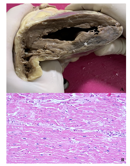
A 65-years-old-man, blacksmith, was hospitalized at the Policlinico Hospital of Bari, to undergo gonarthrosis treatment. At admission the patient did not refer any cardiovascular disease but only a mild hypercholesterolemia (210 mg/dl). He was a smoker of about 10-15 cigarettes/day. At the beginning of the procedure, the patient suddenly collapsed and died for cardiac arrest. The autopsy was performed. The heart showed a transmural dyschromic area of the posterior lateral wall of the left ventricle, which, at the histopathological evaluation revealed an acute myocardial ischemia (wavy fibers and fragmentation of cardiomyocytes) along with the morphological signs (fibrosis) of a previous posterolateral infarction of the left ventricle (Figure 1).

Figure 1: Transmural posterolateral infarction of the left ventricle indicated by a white asterisk (A). Histologically, myocardium shows diffuse wavy fibers suggestive of acute ventricular fibrillation because of ischemia (B).
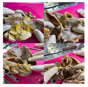
After a closer investigation, an anomaly of the Left Circumflex Coronary Artery (LCX), which originated at right angles from the initial tract of the Right Coronary Artery (RCA), in proximity (<1 cm) of Valsalva sinus was observed. The artery in question was directed posteriorly between the aorta and right atrium, and then, crossing anteriorly to the left atrium, moved towards the obtuse margin of the heart (Figure 2).

Figure 2: Anomalous origin and direction of the Left Circumflex Artery (LCX), which originates from the proximal tract of the right coronary artery (RCA), passes between the aortic root and the right atrium (A) and then between the aorta and the left atrium (B) to enter the left atrio-ventricular groove (C) and the lateral wall of the left ventricle (D). Ao Aorta; RA: Right Atrium; LA Left Atrium; A: Aortic Valve; P: Pulmonary Artery; * Circumflex Coronary Artery.
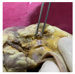
In section, the presence of a sub-occlusive stenosis (80%) of the proximal tract of the abnormal circumflex artery was detected (Figure 3). In addition, moderate coronary arteriosclerosis of the remaining epicardial coronary branches was found, with a maximum stenosis of 50% of the anterior descendant.

Figure 3: Sub-occlusive stenosis (80%) of anomalous left circumflex artery (LCX) proximal tract, due to coronary arteriosclerosis, indicated with a white asterisk.
3. Discussion
Different studies report that the anomalous origin of LCX from RCS/RCA was the most common Coronary Arteries Anomaly (CAA), but in others its prevalence was 0.4% of all CAA, being split right coronary artery the most frequent [5, 6]. In literature, this anomaly has been included in various classifications based on different criteria. The most used is based on the possibility of being able to generate heart attack or sudden cardiac death (clinical relevance). According to these data, the ectopic origin of the LCX from RCA is classified as "benign", since it is considered an anomaly not able, by itself, to lead neither to myocardial ischemia, nor to sudden death, but only to determine various degrees of atherosclerosis. In fact, the unusual angle of takeoff and more tortuous course of the proximal portion of the anomalous coronary artery predisposes it to accelerated atherosclerosis. This is because the junction point of the bound portion of the anomalous artery and the free portion as it wraps around the aorta is an area susceptible to lipid accumulation because of the mechanical stress (5, 6). Our reported case of sudden death, in an asymptomatic patient, highlights that a coronary obstruction is more frequent in abnormal coronaries even if these anomalies fall into a category considered “clinically benign”. Furthermore, in a sudden death of patients without evident cardiovascular risk factors, the accurate study of the course of coronary arteries is highly recommended. The strong discrepancy between the number of cases reported in angiographic studies and those described in in autopsy findings [7] should encourage to search such anomalies even in cases in which the death could be easily explained because of the occurrence of other risk factors.
Funding
This research received no external funding.
Informed Consent Statement
Informed consent for publication was obtained from the wife of the patient.
Acknowledgments
In this section, you can acknowledge any support given which is not covered by the author contribution or funding sections. This may include administrative and technical support, or donations in kind (e.g., materials used for experiments).
Conflicts of Interest
The authors declare no conflict of interest.
References
- Sidhu NS, Wander GS, Monga A, et al. Incidence, Characteristics and Atherosclerotic Involvement of Coronary Artery Anomalies in Adult Population Undergoing Catheter Coronary Angiography. Cardiol Res 10 (2019): 358-368.
- Angelini P, Villason S, Chan AV, et al. Normal and anomalous coronary arteries in humans in: Angelini P (ed) Coronary Arteries Anomalies: A Comprehensive Approach. Lippincott Williams & Wilkins, Philadelphia (1999): 27-150.
- Cademartiri F, La Grutta L, Malagò R, et al. Prevalence of anatomical variants and coronary anomalies in 543 consecutive patients studied with 64-slice CT coronary angiography. Eur Radiol 18 (2008): 781-791.
- Rigatelli G, Rigatelli G. Congenital coronary artery anomalies in the adult: a new practical viewpoint. Clin Cardiol 28 (2005): 61-65.
- Niwa K. Metabolic syndrome and coronary artery disease in adults with congenital heart disease. Cardiovasc Diagn Ther 11 (2021): 563-576.
- Click RL, Holmes DR, Jr, Vlietstra RE, et al. Anomalous coronary arteries: location, degree of atherosclerosis and effect on survival: a report from the coronary artery surgery study. J Am Coll Cardiol 13 (1989): 531-537.
- Alexander RW, Griffith GC, Anomalies of the coronary arteries and their clinical significance. Circulation 14 (1956): 800-805.
