Subclinical Carditis in Acute Rheumatic Fever: A Single Center Experience
Article Information
Md. Saidul Alam1*, Mohammad Abdul Hye2, Shuperna Ahmed3, Mohammad Aminul Islam4, Mustanshirah Lubna5, Biplob Bhattacharjee6, Md. Arifur Rahman7, Mohammad Jobayer8
1Associate Professor, Department of Cardiology, National Center for Control of Rheumatic Fever & Heart Diseases, Dhaka, Bangladesh.
2Assistant Professor, Department of Pediatrics, Magura Medical College, Bangladesh.
3Associate Professor, Department of Pharmacology, Magura Medical College, Magura. Bangladesh.
4Assistant Professor, Department of Cardiology, National Center for Control of Rheumatic Fever & Heart Diseases, Dhaka, Bangladesh.
5Medical Officer, National Center for Control of Rheumatic Fever & Heart Disease, Dhaka, Bangladesh.
6Assistant Professor, Department of Cardiology, Noakhali Abdul Malek Ukil Medical College, Bangladesh.
7Assistant Professor, Department of Cardiology, National Center for Control of Rheumatic Fever & Heart Diseases, Dhaka, Bangladesh.
8Assistant Professor, Department of Microbiology, National Center for Control of Rheumatic Fever & Heart Diseases, Dhaka, Bangladesh.
*Corresponding author: : Dr. Md. Saidul Alam. Associate Professor, Department of Cardiology, National Center for Control of Rheumatic Fever & Heart Diseases, Dhaka, Bangladesh.
Received: 16 August 2023; Accepted: 21 August 2023; Published: 28 August 2023
Citation: Md. Saidul Alam, Mohammad Abdul Hye, Shuperna Ahmed, Mohammad Aminul Islam, Mustanshirah Lubna, Biplob Bhattacharjee, Md. Arifur Rahman, Mohammad Jobayer. Subclinical Carditis in Acute Rheumatic Fever: A Single Center Experience. Cardiology and Cardiovascular Medicine. 7 (2023): 311-315.
View / Download Pdf Share at FacebookAbstract
Background: Acute rheumatic fever (ARF) is an important public health problem in developing countries. Subclinical carditis (SCC) that is detected only by echocardiogram without audible heart murmurs is relatively common in ARF. The aim of this study was to determine the pattern of SCC in patients of ARF in a specialized center in Bangladesh.
Methods: This cross-sectional study was conducted from April 2019 to May 2021 at the National Center for Control of Rheumatic Fever and Heart Diseases. Hundred consecutive diagnosed patients of acute rheumatic fever with SCC were included in the study. Diagnosis of ARF was done according to the revised Jones criteria in 2015. A total of 362 clinically suspected patients of ARF were screened and among them, 100 patients were detected of having SCC by Doppler echocardiography.
Results: Mean age of patients with ARF and SCC was 11.8 ±3.6 years and 10.8 ±3.3 years respectively and female was predominant (52.6% in ARF and 57.7% in SCC). Majority of patients (94%) with SCC had a mitral valve involvement and isolated mitral regurgitation was the most common (84%) valvular lesion. Detected valvular lesions mostly were not severe; all the aortic regurgitation and almost all mitral regurgitation (98.8%) were mild and trivial in nature of severity.
Conclusion: Common presence of SCC among ARF patients in our study agreed with the recommendations of revised Jones Criteria. Therefore, it is suggested that echocardiography should be done in every suspected patient with ARF for early detection of subclinical carditis and to reduce morbidity.
Keywords
Acute rheumatic fever; Subclinical carditis; Mitral regurgitation; Aortic regurgitation; Echocardiography
Article Details
1. Introduction
Acute rheumatic fever (ARF) is a multisystem disease that results from autoimmune reaction to group A β-hemolytic streptococcal pharyngitis in genetically susceptible individual [1], It is estimated that globally about 500,000 new cases of ARF occur annually and more than 230,000 people die from this disease [2]. Mean incidence of ARF is 19 per 100000 school-aged children worldwide [3]. The rate is extremely high among indigenous Australians (193 cases per 100 000 school-aged children) [4]; relatively higher in Eastern Europe, Asia and Middle East (>10/100,000) while lowest in America and Western Europe (10/100,000) [3]. Major systemic inflammatory manifestations of ARF include carditis, arthritis, Sydenhams’ chorea, erythema marginatum and subcutaneous nodules [5]. Carditis in ARF may range from mild sub-clinical involvement to severe carditis with congestive heart failure [6]. Subclinical carditis (SCC) is defined as positive findings of mitral or aortic valvitis on an echocardiogram without heart murmurs or other clinical signs. Subclinical carditis is relatively common in ARF and its prevalence in ARF is reported to be 18.1% (95% CI 11.1 to 25.2) in a metaanalysis [6]. Studies suggested that as a major criterion SCC influences diagnosis of ARF in about 16% of patients [7]. In revised Jones criteria 2015, SCC is considered as a major manifestation in all patients regardless of countries with low or high prevalence of ARF [5].
Echocardiography is sensitive and reliable in diagnosing valvular involvement in ARF [8]. Introduction of Doppler echocardiography in studies on ARF leads to detection of previously unrecognized cases of SCC [9]. Regardless of risk stratification, the latest updates recommend using Doppler echocardiography to diagnose carditis and subclinical carditis in all patients and also to repeat it in case of uncertainty [5], We believe that, 2015 update on Jones criteria might impact the clinical practice in ARF especially in developing countries like Bangladesh and the incorporation of SCC as major criterion will lead to a modest increase in the diagnosis of ARF cases. Diagnosis of rheumatic fever is a big challenge, especially in developing countries. Early diagnosis and prophylactic treatment of ARF are essential to reduce morbidity and mortality. In Bangladesh no known study was conducted recently on subclinical carditis. Therefore, the purpose of our study was to analyze subclinical carditis present in patients with acute rheumatic fever in an outdoor-based hospital.
2. Materials and Methods
This was a cross-sectional study conducted in National Center for Control of Rheumatic Fever and Heart Diseases (NCCRF&HD), Bangladesh during the period of 2 years and 3 months from January 2019 to April 2021.
2.1 Inclusion Criteria:
A total of 100 consecutive diagnosed patients of acute rheumatic fever with subclinical carditis were included in this study.
2.2 Exclusion Criteria:
Patients with pregnancy; b) Patients with uncontrolled DM; and c) Patients with any history of acute illness (e.g., ischemic heart disease, renal or pancreatic diseases).
2.3 Study procedure:
Patients were attended by specialist physicians and laboratory investigations (CBC with ESR, ASO titer, CRP), ECG was done and cardiologists performed standard color Doppler echocardiography (Philips, Affinity 30, Taiwan). Collaborating clinical history, physical examination, and investigation reports a clinical diagnosis of ARF was made according to the Jones Criteria 2015.5
Table 1: Echocardiographic criteria for diagnosis of subclinical carditis [10].
|
Pathological mitral regurgitation (meets all four criteria) |
Pathological aortic regurgitation (meets all four criteria) |
|
Seen in at least two views |
Seen in at least two views |
|
Jet length ≥2 cm in at least one view |
Jet length ≥1 cm in at least one view |
|
Peak velocity >3 m/s |
Peak velocity >3 m/s |
|
Pansystolic jet in at least one envelope |
Pandiastolic jet in at least one envelope |
Grading of cardiac valvular regurgitations was done according to color flow MR jet, continuous wave signal of jet, vena contracta width and flow convergence zone. Trivial regurgitation was classified according to jet length into jets <10mm, ≥10 mm and ≥20 mm and also jet velocity into jets <2.5mm/sec, ≥2.5mm/sec and ≥3 m/sec. Severe mitral regurgitation was considered when had a large jet with vena contracta width ≥7mm; moderate regurgitation had intermediate jet with vena contracta width 3-6 [9]. and mild regurgitation had small jet, with vena contracta width ~3mm [10-12].
2.4 Statistical Analysis:
All data were recorded systematically in preformed data collection form and quantitative data was expressed as mean and standard deviation and qualitative data was expressed as frequency distribution and percentage. Statistical analysis was performed by using SPSS 21 (Statistical Package for Social Sciences). Probability value <0.05 was considered as level of significance. The study was approved by Ethical Review Committee of National Center for Control of Rheumatic Fever and Heart Diseases, Dhaka, Bangladesh.
3. Results
A total of 100 diagnosed patients with acute rheumatic fever with subclinical carditis were enrolled in this study. The mean age of patients with acute rheumatic fever was 11.86 ±4.18 years and patients with subclinical carditis were 10.75 ±3.37 years. History of sore throat in the last 1 month was present in 59% of patients with SCC and 63% of them were residing in urban areas (Table 2).
Table 2: Comparison of demographic profile between patients of acute rheumatic fever and patients with subclinical carditis.
|
Demographic variables |
ARF |
Subclinical carditis |
P-value |
||
|
N=201 |
P(%) |
N=100 |
P(%) |
||
|
Mean age (years) |
11.86 ± 4.18 |
10.75 ± 3.37 |
0.267 |
||
|
Range |
22 (3-25) |
18 (4-22) |
|||
|
Sex |
|||||
|
Male |
85 |
42.3 |
39 |
39 |
0.421 |
|
Female |
116 |
57.7 |
61 |
61 |
|
|
Male female ratio |
0.73:1 |
0.64:1 |
|||
|
Residence |
|||||
|
Urban |
120 |
59.7 |
63 |
63 |
0.128 |
|
Rural |
81 |
40.3 |
37 |
37 |
|
|
History of sore throat in last 1 month |
121 |
60.2 |
59 |
59 |
0.387 |
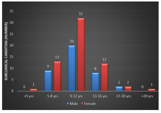
Ninety-four percent of patients with SCC were between 5 to 16 years with 52% in 9-12 years age group. Female was predominant; 57.7% and 61% of patients with ARF and patients with SCC were female respectively (Figure 2).
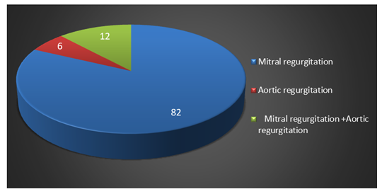
Among detected valvular lesions isolated mitral regurgitation was 82%, 6% was aortic regurgitation and a combination of mitral regurgitation with aortic regurgitation was 12% (Figure 3).
According to severity, 66(70.2%) of mitral regurgitation and 12(66.7%) of aortic regurgitation were detected as trivial (10-20 mm). One (1.1%) of the mitral regurgitation was moderate whereas none of aortic and mitral regurgitation was found severe (Table 3).
Table 3: Grades of valvular lesions in mitral regurgitation (N=94) and aortic regurgitation (N=18)
|
Grade of mitral regurgitation |
n(%) |
Grade of aortic regurgitation |
n(%) |
|
Severe MR |
0(0.0) |
Severe AR |
0(0.0) |
|
Moderate MR |
1(1.1) |
Moderate AR |
0(0.0) |
|
Mild MR ≥20 mm |
18(19.1) |
Mild AR ≥20 mm |
2(11.1) |
|
Trivial MR: 10-20 mm |
66(70.2) |
Trivial AR: 10-20 mm |
12(66.7) |
|
Trivial MR: 7-9 mm |
9(9.6) |
Trivial AR: 7-9 mm |
4(22.2) |
4. Discussion
Subclinical carditis was common among patients with acute rheumatic fever in our study population and the predominant valvular lesion was mitral regurgitation.
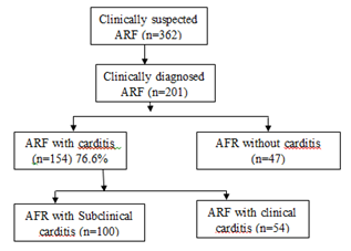
During assessing patients with ARF in this center, we observed that 154 (76.6%) patients had carditis among which 100 (49.7%) had subclinical rheumatic carditis. The detection rate of SCC is quite high in this study which is in accordance with several studies that reported such a higher detection rate of SCC in patients with ARF ranging from 31 to 51% [13-16]. These studies were done in countries of the Middle East and Asia where RF is endemic like our countries. Several studies mentioned that the use of Doppler echocardiography helps in the detection of previously unrecognized SCC during screening of ARF and therefore detection rate of ARF increases several times [17,18]. Nowadays, both conventional and portable echocardiography are used in most parts of our country. Therefore, many new cases of subclinical carditis are now diagnosed [19]. Being an outdoor-based hospital NCCRF&HD mostly deals with patients with earlier stages of carditis whereas those with obvious manifestations of carditis normally seek management in centers with indoor facilities.
ARF is a disease of children and young adults. In this study, more than half of patients of ARF with SCC were between the age of 9 to 12 years. The mean age of the patients of ARF and SCC was 11.8 ±3.6 years and 10.8 ±3.3 years respectively which is very much in accordance with the report of Carvalho and also agrees with the results of studies in Bangladesh [20-22]. Among the clinically diagnosed patients with ARF and SCC female was predominant (52.6% in ARF and 57.7% in SCC). A slight predominance of female patients in ARF was also reported in other studies in Bangladesh and other parts of the world [14,20,21].
In this study population diagnosed with subclinical carditis, 94% had mitral valve involvement among which isolated mitral regurgitation was detected in 84% of cases. Predominant mitral valve involvement was reported in several other studies done on SCC [9,14]. There were a few cases of aortic regurgitation also and a combination of mitral regurgitation with aortic regurgitation was 12%. No case of either mitral stenosis or aortic stenosis was detected in this group of patients. Mitral stenosis is not very common in younger children and rheumatic fever is an uncommon cause of aortic stenosis [23,24].
Among the detected valvular lesions majority were mild in severity in echocardiographic evaluation indicating provably the very early stage of the disease. All the aortic regurgitation and almost all mitral regurgitation were either mild or trivial in nature. This finding is very consistent with the data of two recent studies done in Egypt and Brazil where the authors found the majority of mitral regurgitation and aortic regurgitation to be mild or trivial [14,20]. Both countries have high incidences of rheumatic fever in their populations.
In the majority of patients who adhere well to antibiotic prophylaxis, valvular lesions of mild mitral regurgitation resolve indicating that secondary prophylaxis may alter the natural history of SCC [6]. Therefore it is very important to diagnose SCC in an early stage of ARF so that secondary antibiotic prophylaxis may effectively halt the progress of SCC. Subclinical carditis may be the tip of the iceberg in areas where the risk factors for developing ARF are prevailing. SCC is not diagnosed clinically in centers where echocardiography is not available. Therefore, emphasis should be given on detection of SCC by echocardiography as they can progress to carditis and cause much morbidity. We, the authors strongly agree with the recommendation of the World Health Federation, Australian guideline, and updated revised Jones Criteria that included subclinical carditis as a major criterion in the diagnosis of rheumatic fever in high-risk populations like Bangladesh.
Unfortunately, there is no single laboratory test that definitely establishes the diagnosis of acute rheumatic fever which makes the diagnosis difficult and mainly based on clinical criteria. We adhered strictly to the updated Jones criteria in the diagnosis of proven rheumatic fever patients. Several studies concluded that subclinical carditis may persist for a long time, therefore the significance of SCC should be evaluated and most importantly follow-up of these patients are very important to halt the progression to clinical carditis [13,25,26].
5. Conclusions:
Finding of our study strongly agreed with the updated recommendations of Jones Criteria 2015 revised by American Heart Association that included subclinical carditis as major criteria in diagnosis of rheumatic fever. We concluded that echocardiography should be performed in every suspected patient with rheumatic fever for early diagnosis and to reduce the morbidity
Limitations of the study
Our study was an outdoor based single center study, so the results did not represent general population with RHD of Bangladesh. This might also have under-represented the number of patients with advanced stages of disease. After evaluating once those patients we did not follow-up them and have not known other possible interference that may happen in the long term with these patients.
Ethical Consideration
Protocol of this study was approved by Ethical Review Committee of NCCRF&HD. Informed written consent was taken from each patient or authorized legal guardian. Anonymity of patients and confidentiality of information was maintained strictly.
Funding of the work
No financial support was taken for this study.
Acknowledgement
We thankfully acknowledge NCCRF&HD for providing data collection and entire laboratory facilities.
Conflict of interest
We do not have any conflicts of interest.
References
- Engel ME, Stander R, Vogel J, et al. Genetic Susceptibility to Acute Rheumatic Fever: A Systematic Review and Meta-Analysis of Twin Studies. Plos One 6 (2011): 25326.
- Webb RH, Grant C, Harnden A. Acute rheumatic fever. BMJ 351 (2015): 3443.
- Tibazarwa KB, Volmink JA, Mayosi BM. Incidence of acute rheumatic fever in the world: a systematic review of population-based studies. Heart 94 (2008): 1534-1540.
- Lawrence JG, Carapetis JR, Griffiths K, et al. Acute rheumatic fever and rheumatic heart disease: incidence and progression in the Northern Territory of Australia, 1997 to 2010. Circulation 128 (2013): 492-501.
- Gewitz MH, Baltimore RS, Tani LY, et al. American Heart Association Committee on Rheumatic Fever, Endocarditis, and Kawasaki Disease of the Council on Cardiovascular Disease in the Young. Revision of the Jones Criteria for the diagnosis of acute rheumatic fever in the era of Doppler echocardiography: a scientific statement from the American Heart Association. Circulation 131 (2015): 1806-1818.
- Tubridy-Clark M, Carapetis JR. Subclinical carditis in rheumatic fever: a systematic review. Int J Cardiol 119 (2007): 54-58.
- Vijayalakshmi IB, Vishnuprabhu RO, Chitra N, et al. The efficacy of echocardiographic criterions for the diagnosis of carditis in acute rheumatic fever. Cardiol Young 18 (2008): 586-592.
- Ramakrishnan S. Echocardiography in acute rheumatic fever. Ann Pediatr Cardiol 2 (2009): 61-64.
- Marijon E, Ou P, Celermajer DS, et al. Prevalence of rheumatic heart disease detected by echocardiographic screening. N Engl J Med 357 (2007): 470-476.
- Reményi B, Wilson N, Steer A, et al. World Heart Federation criteria for echocardiographic diagnosis of rheumatic heart disease--an evidence-based guideline. Nat Rev Cardiol 9 (2012): 297-309.
- WHO Study Group. Rheumatic Fever and Rheumatic Heart Disease: Report of a WHO Expert Consultation; 20 October-1 November 2001, Geneva, Switzerland. Technical Report Series No. 923. Geneva: World Health Organization (2004).
- RHD Australia (ARF/RHD Writing Group), National Heart Foundation of Australia, Cardiac Society of Australia and New Zealand. The Australian Guideline for Prevention, Diagnosis, and Management of Acute Rheumatic Fever and Rheumatic Heart Disease (2nd ed). Casuarina, Australia: RHD Australia (2012).
- Figueroa FE, Fernández MS, Valdés P, et al. Prospective comparison of clinical and echocardiographic diagnosis of rheumatic carditis: long term follow up of patients with subclinical disease. Heart 85 (2001): 407-410.
- Aty-Marzouk PA, Hamza H, Mosaad N, et al. New guidelines for diagnosis of rheumatic fever; do they apply to all populations? Turk J Pediatr 62 (2020): 411-423.
- Elevli M, Celebi A, Tombul T, et al. Cardiac involvement in Sydenham's chorea: clinical and Doppler echocardiographic findings. Acta Paediatr 88 (1999): 1074-1077.
- Folger GM Jr, Hajar R, Robida A, et al. Occurrence of valvar heart disease in acute rheumatic fever without evident carditis: colour-flow Doppler identification. Br Heart J 67 (1992): 434-438.
- Malla R, Thapaliya S, Gurung P, et al. Patterns of Valvular Involvement in Rheumatic Heart Disease patients taking Benzathine Penicillin at Shahid Gangalal National Heart Centre, Kathmandu, Nepal. Nepalese Heart Journal 13 (2016): 25-27.
- Lilyasari O, Prakoso R, Kurniawati Y, et al. Clinical Profile and Management of Rheumatic Heart Disease in Children and Young Adults at a Tertiary Cardiac Center in Indonesia. Front Surg 7 (2020): 47.
- Islam AKMM, Majumder AAS. Rheumatic fever and rheumatic heart disease in Bangladesh: A review. Indian Heart Journal 68 (2016): 88-98.
- Carvalho CLMA, Araújo FDR, Meira ZMA. Doppler Echocardiographic Follow-Up of Mitral and Aortic Regurgitation in Children and Adolescents with Subclinical and Mild RheumaticCarditis. Int J Cardiovasc Sci 30 (2017).
- Jobayer M, Alam MS, Rana RA, et al. Rheumatic fever and Rheumatic heart disease among clinically suspected patients with joint pain in a specialized hospital. Bangladesh Med Res Council Bull 48 (2022): 83-89.
- Zaman MM, Choudhury SR, Rahman S, et al. Prevalence of rheumatic fever and rheumatic heart disease in Bangladeshi children. Indian Heart Journal 67 (2015): 45-49.
- Chandrashekhar Y, Westaby S, Narula J. Mitral stenosis. Lancet 374 (2009): 1271-1283.
- Alkhalifa MS, Ibrahim SA, Osman SH. Pattern and severity of rheumatic valvular lesions in children in Khartoum, Sudan. East Mediterr Health J 14 (2008): 1015-1021.
- Ozkutlu S, Hallioglu O, Ayabakan C. Evaluation of subclinical valvar disease in patients with rheumatic fever. Cardiol Young 13 (2003): 495-499.
- Pekpak E, Atalay S, Karadeniz C, et al. Rheumatic silent carditis: echocardiographic diagnosis and prognosis of long-term follow up. Pediatr Int 55 (2013): 685-689.


