Small Bowel Obstruction Caused by Carciniod Tumor in Ileum
Article Information
Nor Abdi Yasin*, Ikram Abdikarim Ibrahim MD
Department of General Surgery, Mogadishu Somali Turkish Training and Research Hospital, Mogadishu, Somalia
*Corresponding Authors: Nor Abdi Yasin, Department of General Surgery, Mogadishu Somali Turkish Training and Research Hospital, Mogadishu, Somalia
Received: 03 February 2021; Accepted: 08 March 2021; Published: 22 March 2021
Citation: Nor Abdi Yasin, Department of General Surgery, Mogadishu Somali Turkish Training and Research Hospital, Mogadishu, Somalia
View / Download Pdf Share at FacebookAbstract
Carcinoid tumor is one of the rare gastrointestinal tumors. We present here; a case of 54 years old woman with complete stricture in the Small Bowel at the ileum level, caused by a carcinoid tumour who underwent a laparotomy. After resection of the tissue, histopathology was confirmed carcinoid tumor as the casual stricture. A such a typical presentation in patients not having a mass in radiological studies except signs and symptoms of bowel obstruction should raise suspicion of a carcinoid tumor.
Keywords
Carciniod Tumor; Abdominal pain; Abdomen
Carciniod Tumor articles; Abdominal pain articles; Abdomen articles
Carciniod Tumor articles Carciniod Tumor Research articles Carciniod Tumor review articles Carciniod Tumor PubMed articles Carciniod Tumor PubMed Central articles Carciniod Tumor 2023 articles Carciniod Tumor 2024 articles Carciniod Tumor Scopus articles Carciniod Tumor impact factor journals Carciniod Tumor Scopus journals Carciniod Tumor PubMed journals Carciniod Tumor medical journals Carciniod Tumor free journals Carciniod Tumor best journals Carciniod Tumor top journals Carciniod Tumor free medical journals Carciniod Tumor famous journals Carciniod Tumor Google Scholar indexed journals Abdominal pain articles Abdominal pain Research articles Abdominal pain review articles Abdominal pain PubMed articles Abdominal pain PubMed Central articles Abdominal pain 2023 articles Abdominal pain 2024 articles Abdominal pain Scopus articles Abdominal pain impact factor journals Abdominal pain Scopus journals Abdominal pain PubMed journals Abdominal pain medical journals Abdominal pain free journals Abdominal pain best journals Abdominal pain top journals Abdominal pain free medical journals Abdominal pain famous journals Abdominal pain Google Scholar indexed journals Abdomen articles Abdomen Research articles Abdomen review articles Abdomen PubMed articles Abdomen PubMed Central articles Abdomen 2023 articles Abdomen 2024 articles Abdomen Scopus articles Abdomen impact factor journals Abdomen Scopus journals Abdomen PubMed journals Abdomen medical journals Abdomen free journals Abdomen best journals Abdomen top journals Abdomen free medical journals Abdomen famous journals Abdomen Google Scholar indexed journals laparotomy articles laparotomy Research articles laparotomy review articles laparotomy PubMed articles laparotomy PubMed Central articles laparotomy 2023 articles laparotomy 2024 articles laparotomy Scopus articles laparotomy impact factor journals laparotomy Scopus journals laparotomy PubMed journals laparotomy medical journals laparotomy free journals laparotomy best journals laparotomy top journals laparotomy free medical journals laparotomy famous journals laparotomy Google Scholar indexed journals gastrointestinal tumors articles gastrointestinal tumors Research articles gastrointestinal tumors review articles gastrointestinal tumors PubMed articles gastrointestinal tumors PubMed Central articles gastrointestinal tumors 2023 articles gastrointestinal tumors 2024 articles gastrointestinal tumors Scopus articles gastrointestinal tumors impact factor journals gastrointestinal tumors Scopus journals gastrointestinal tumors PubMed journals gastrointestinal tumors medical journals gastrointestinal tumors free journals gastrointestinal tumors best journals gastrointestinal tumors top journals gastrointestinal tumors free medical journals gastrointestinal tumors famous journals gastrointestinal tumors Google Scholar indexed journals enterochromaffin cells articles enterochromaffin cells Research articles enterochromaffin cells review articles enterochromaffin cells PubMed articles enterochromaffin cells PubMed Central articles enterochromaffin cells 2023 articles enterochromaffin cells 2024 articles enterochromaffin cells Scopus articles enterochromaffin cells impact factor journals enterochromaffin cells Scopus journals enterochromaffin cells PubMed journals enterochromaffin cells medical journals enterochromaffin cells free journals enterochromaffin cells best journals enterochromaffin cells top journals enterochromaffin cells free medical journals enterochromaffin cells famous journals enterochromaffin cells Google Scholar indexed journals Lieberkühn articles Lieberkühn Research articles Lieberkühn review articles Lieberkühn PubMed articles Lieberkühn PubMed Central articles Lieberkühn 2023 articles Lieberkühn 2024 articles Lieberkühn Scopus articles Lieberkühn impact factor journals Lieberkühn Scopus journals Lieberkühn PubMed journals Lieberkühn medical journals Lieberkühn free journals Lieberkühn best journals Lieberkühn top journals Lieberkühn free medical journals Lieberkühn famous journals Lieberkühn Google Scholar indexed journals autopsy articles autopsy Research articles autopsy review articles autopsy PubMed articles autopsy PubMed Central articles autopsy 2023 articles autopsy 2024 articles autopsy Scopus articles autopsy impact factor journals autopsy Scopus journals autopsy PubMed journals autopsy medical journals autopsy free journals autopsy best journals autopsy top journals autopsy free medical journals autopsy famous journals autopsy Google Scholar indexed journals obstruction articles obstruction Research articles obstruction review articles obstruction PubMed articles obstruction PubMed Central articles obstruction 2023 articles obstruction 2024 articles obstruction Scopus articles obstruction impact factor journals obstruction Scopus journals obstruction PubMed journals obstruction medical journals obstruction free journals obstruction best journals obstruction top journals obstruction free medical journals obstruction famous journals obstruction Google Scholar indexed journals metastasis articles metastasis Research articles metastasis review articles metastasis PubMed articles metastasis PubMed Central articles metastasis 2023 articles metastasis 2024 articles metastasis Scopus articles metastasis impact factor journals metastasis Scopus journals metastasis PubMed journals metastasis medical journals metastasis free journals metastasis best journals metastasis top journals metastasis free medical journals metastasis famous journals metastasis Google Scholar indexed journals
Article Details
1. Introduction
Carcinoids tumors are slow-growing well-differentiated tumor, which arises in the enterochromaffin cells (Kulchitsky cells), found in the crypts of Lieberkühn. These tumors were first described by Lubarsch in 1888; in 1907, Oberndorfer coined the term Karzinoide to indicate the carcinoma-like appearance and the presumed lack of malignant potential. The prevalence of carcinoid tumors ranges from 1-2 cases per 100,000 persons and slightly increased in the African American population [1]. A 25-30% of carcinoid tumor seen in the small bowel and more common in the ileum in 91% of the cases. Henrietta m Wilson reported that carcinoid tumor had seen 1 in 300 cases of small bowel carcinoma at autopsy. Although common in the ileum and it can be Multicentricity in 26-30% of the cases. Carcinoid tumor can found in the colon, but it is more common in the appendix nearly 80%, and usually presented with abdominal pain, obstruction, and metastasis that need surgical intervention. Ganesan and colleagues described the metastasis of these tumors are late and mostly metastasis in the liver. We present a rare case of carcinoid tumor with complete stricture in the Small Bowel at the ileum identified early and no metastasis seen in the radiological studies [2].
2. Case Presentation
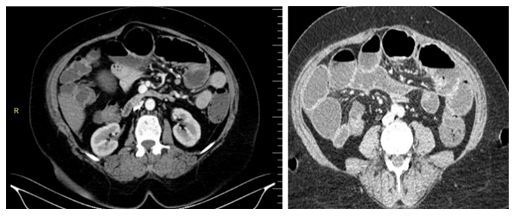
A 54 years old woman came to the emergency department with generalized dull and colicky abdominal pain accompanied by nausea, vomiting, and obstipation started four days before admission. On physical examination, the abdomen was soft with mild tenderness in the periumbilical, right lower quadrant, and left lower quadrant without guarding, rebound tenderness, and a palpable mass. Digital rectal examination showed an empty rectum. Laboratory findings revealed normal WBC count, platelet count, liver function tests, serum electrolytes, amylase, and lipase tests. The patients showed mild renal failure (BUN- 55mg/dl and Cr-2.5mg/dl). On erect Abdominal X-ray, there was an air-fluid level. Abdominal ultrasound showed significant dilatation in the bowel secondary to obstruction. Computed tomography of the abdomen confirmed the distended small bowel and fluid (Figure 1).
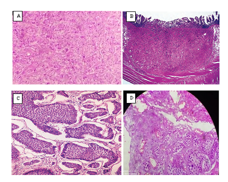
After clinical and imaging studies, laparotomy was performed, and intra-abdominal organs were exposed, 220 CM from tries ligament there was a complete obstruction by stricture and 10cm resection of small intestine and end to end anastomosis by a linear stapler, and excisional biopsy had performed. The diagnosis of carcinoid tutor was confirmed by the histopathologic report (Figure 2).

Figure 1: Computed tomography of the abdomen with intravenous contrast revealed distension of small bowel and fluid in the abdomen.

Figure 2: (A) Low power photomicrograph of a typical carcinoid tumor. Reproduced with permission from Ex-Digfer hospital. Pathology of small bowel malignancies (Accessed on December 26, 2020); (B) High power view of ileal carcinoid tumor showing full-thickness involvement with a largely preserved mucosa; (C) Microscopically intermediate power showing tumor cells embedded in dense fibrous tissue; (D) Low power photomicrograph of a typical carcinoid tumor infiltrating into serosal surface.
3. Discussion
Carcinoid tumours is one of the rare gastrointestinal tumors. These tumors are identified as metastasis at the time of diagnosis due to their late presentations. Small bowel obstruction mainly related to abdominal pain, nausea, vomiting, constipation, and abdominal distention such presented our case. Jennifer Matulich et al. described that adhesions and hernia are the most common cause of small bowel obstruction. ?n our case, the primary aetiology was stricture from the carcinoid tumor. They can be seen in the respiratory and Gastrointestinal tract, over 90% seen in the Gastrointestinal tract, and accounted for 1.5% Gastrointestinal neoplasms. They are common in the ileum and appendix. Sayed Hamdi and associates reported that Carcinoid tumor is the most primary tumor of the small bowel and mesentery accounted about 95% of all carcinoid tumors and 1.5% of all Gastrointestinal tumors, where Khaled M. Moghazy and colleagues reported that carcinoid tumor in small bowel constitutes 20% of all cases and 90% seen in the ileum. Clinical presentation varies in hormonal and non-hormonal findings where non-hormonal is mainly from the mass effect of the tumor that causes obstruction or local reaction like stricture.
Our patient presented abdominal pain and distension related to the stricture associated with the local tumor reaction [3]. The hormonal symptoms are usually related to the tumor production of serotonin and bradykinin, tachykinins, and prostaglandins. The midgut carcinoid tumors cause carcinoid syndrome due to vasoactive hormones released in the systemic circulation. Our case had not presented with symptoms of carcinoid syndrome. Although it is not common, Douglas Jun Kamei et al. reported that carcinoid syndrome affects around 5-7% of the patients. Urinary excretion of 5HIAA is useful for the diagnosis of carcinoid tumors for patients with hormonal symptoms.
A CT scan and MRI imaging are useful for a patient with bowel dysmotility as our patient has symptoms of bowel dysmotility, and we preferred a CT scan that identifies a dilation of the bowel with fluid showing obstruction [4]. In general, less than 1cm primary tumors of the small bowel can be managed with local resection in contrast to that tumors more than 1.5 cm segmental resection with an extensive clearance of mesenteric drainage are suitable due to the high risk of recurrence. In our patients, stricture was noted intraoperatively and managed with segmental resection about 10 cm at the jejunum. In cases of liver metastasis can be managed by either liver resection or HAE. Both provide palliative management for hormonal and pain symptoms. Surgical resection has a prolonged survival rate compared to HAE, but is not curative [5]. Our patient was discharged home with a follow up of a CT scan every three months to check if there is any recurrence or metastasis.
4. Conclusion
Carcinoid tumor is one of the rare tumors in the Gastrointestinal tract, although it is one of the most primary tumors in the small bowel. Instances with constitutional symptoms and there are no specific radiological findings it is important to keep in mind the possibilities of carcinoid as the causal obstruction and to examine carefully the bowel inch to inch during operation.
References
- Rodrigues G, Prabhu R, Ravi B. Small bowel carcinoid: a rare cause of bowel obstruction. BMJ Case Rep (2013).
- William J Geiger, Nancy B Davis. Medical College of Wisconsin, Milwaukee, Wisconsin. Am Fam Physician 74 (2006): 429-434.
- Wilson HM. Chronic subacute bowel obstruction caused by carcinoid tumour misdiagnosed as irritable bowel syndrome: a case report. Cases Journal 78 (2009).
- Seyed Hamid Moosavy, Yasir Andrabi, Sepehr Esmaeeli, et al. Small Bowel Obstruction by a Terminal Ileum Carcinoid Tumor: a Case Report. Medical journal of the Islamic Republic of Iran 25 (2011): 165-169.
- Chamberlain RS, Canes D, Brown KT, et al. Hepatic neuroendocrine metastases: does intervention alter outcomes?. J Am Coll Surg 190 (2000): 432-445.
