Role of Non-Rigid Minimal Intervention Surgery in the Treatment of Degenerative Spondylolisthesis
Article Information
Bañuls-Pattarelli Miguel1*, García-Ortiz María Tíscar1, Atienza-Vicente Carlos2, 3, López-Prats Fernando1, 4, Mulholland Robert C5
1Department of Orthopaedics and Trauma, Elche University General Hospital, Elche (Alicante), Spain
2Associated Professor of Mechanical and Materials Engineering, Master of Biomedical Engineering, Polytechnic University of Valencia, Valencia, Spain
3Biomechanics Institute of Valencia, Health Technology Group, Polytechnic University of Valencia, Valencia, Spain
4Department of Orthopaedics and Trauma, Miguel-Hernández University, San Juan de Alicante, Spain
5Nottingham University Hospitals NHS, Nottingham, United Kingdom
*Corresponding Author: Bañuls-Pattarelli Miguel, Department of Orthopaedics and Trauma, Elche University General Hospital, Carrer Almazara 11, 03203, Elche (Alicante), Spain
Received: 19 July 2021; Accepted: 27 July 2021; Published: 04 August 2021
Citation:
Bañuls-Pattarelli Miguel, García-Ortiz María Tíscar, Atienza-Vicente Carlos, López-Prats Fernando, Mulholland Robert C. Role of Non-Rigid Minimal Intervention Surgery in the treatment of Degenerative Spondylolisthesis. Journal of Spine Research and Surgery 3 (2021): 071-080.
View / Download Pdf Share at FacebookAbstract
Introduction: Degenerative spondylolisthesis produces abnormal intervertebral movement associated with back pain. Standard surgical treatment consists of decompression with or without fusion. There is no consensus about the method of choice.
Purpose of this study: avoiding decompression, a semi-rigid, minimally invasive device that reduces movement was used, removing the necessity for fusion and reducing fixation-loosening, or breakage.
Methods: Analytical prospective observational study. The clinical assessment included the Oswestry Disability Index (ODI) and SF-12 (Short Form-12 Health Survey), X-Rays and MRIs (Magnetic Resonance Imaging) were taken preoperatively and at follow-up. Overall, the mean postoperative follow-up was 3.8 years. It is about a posterior intrapedicular device introduced percutaneously under X-Ray control. The device consists of two semi-rigid bars, allowing 5º multiplanar movement, and 1mm compressive-spring movement. Through manipulation and distraction, reduction of listhesis is possible, increasing disc and lateral recesses height.
Results: At final follow-up, ODI and SF-12 scores significantly improved. ODI from 47.4 ± 14.9 to 22.8 ± 19.7 (p<0.001). In terms of SF-12, PHS (Physical Health Score) improved from 27.9 ± 6.3 to 37.5 ± 11.1 (p<0.001) and MHS (Mental Health Score) from 34.4 Keywords
Minimal Invasive Surgery, Degenerative Spondylolisthesis, Percutaneous Posterior Transpedicular Fixation, Non-Rigid Dynamic Stabilisation
Minimal Invasive Surgery articles Minimal Invasive Surgery Research articles Minimal Invasive Surgery review articles Minimal Invasive Surgery PubMed articles Minimal Invasive Surgery PubMed Central articles Minimal Invasive Surgery 2023 articles Minimal Invasive Surgery 2024 articles Minimal Invasive Surgery Scopus articles Minimal Invasive Surgery impact factor journals Minimal Invasive Surgery Scopus journals Minimal Invasive Surgery PubMed journals Minimal Invasive Surgery medical journals Minimal Invasive Surgery free journals Minimal Invasive Surgery best journals Minimal Invasive Surgery top journals Minimal Invasive Surgery free medical journals Minimal Invasive Surgery famous journals Minimal Invasive Surgery Google Scholar indexed journals Degenerative Spondylolisthesis articles Degenerative Spondylolisthesis Research articles Degenerative Spondylolisthesis review articles Degenerative Spondylolisthesis PubMed articles Degenerative Spondylolisthesis PubMed Central articles Degenerative Spondylolisthesis 2023 articles Degenerative Spondylolisthesis 2024 articles Degenerative Spondylolisthesis Scopus articles Degenerative Spondylolisthesis impact factor journals Degenerative Spondylolisthesis Scopus journals Degenerative Spondylolisthesis PubMed journals Degenerative Spondylolisthesis medical journals Degenerative Spondylolisthesis free journals Degenerative Spondylolisthesis best journals Degenerative Spondylolisthesis top journals Degenerative Spondylolisthesis free medical journals Degenerative Spondylolisthesis famous journals Degenerative Spondylolisthesis Google Scholar indexed journals Percutaneous Posterior Transpedicular Fixation articles Percutaneous Posterior Transpedicular Fixation Research articles Percutaneous Posterior Transpedicular Fixation review articles Percutaneous Posterior Transpedicular Fixation PubMed articles Percutaneous Posterior Transpedicular Fixation PubMed Central articles Percutaneous Posterior Transpedicular Fixation 2023 articles Percutaneous Posterior Transpedicular Fixation 2024 articles Percutaneous Posterior Transpedicular Fixation Scopus articles Percutaneous Posterior Transpedicular Fixation impact factor journals Percutaneous Posterior Transpedicular Fixation Scopus journals Percutaneous Posterior Transpedicular Fixation PubMed journals Percutaneous Posterior Transpedicular Fixation medical journals Percutaneous Posterior Transpedicular Fixation free journals Percutaneous Posterior Transpedicular Fixation best journals Percutaneous Posterior Transpedicular Fixation top journals Percutaneous Posterior Transpedicular Fixation free medical journals Percutaneous Posterior Transpedicular Fixation famous journals Percutaneous Posterior Transpedicular Fixation Google Scholar indexed journals Non-Rigid Dynamic Stabilisation articles Non-Rigid Dynamic Stabilisation Research articles Non-Rigid Dynamic Stabilisation review articles Non-Rigid Dynamic Stabilisation PubMed articles Non-Rigid Dynamic Stabilisation PubMed Central articles Non-Rigid Dynamic Stabilisation 2023 articles Non-Rigid Dynamic Stabilisation 2024 articles Non-Rigid Dynamic Stabilisation Scopus articles Non-Rigid Dynamic Stabilisation impact factor journals Non-Rigid Dynamic Stabilisation Scopus journals Non-Rigid Dynamic Stabilisation PubMed journals Non-Rigid Dynamic Stabilisation medical journals Non-Rigid Dynamic Stabilisation free journals Non-Rigid Dynamic Stabilisation best journals Non-Rigid Dynamic Stabilisation top journals Non-Rigid Dynamic Stabilisation free medical journals Non-Rigid Dynamic Stabilisation famous journals Non-Rigid Dynamic Stabilisation Google Scholar indexed journals spine surgeons articles spine surgeons Research articles spine surgeons review articles spine surgeons PubMed articles spine surgeons PubMed Central articles spine surgeons 2023 articles spine surgeons 2024 articles spine surgeons Scopus articles spine surgeons impact factor journals spine surgeons Scopus journals spine surgeons PubMed journals spine surgeons medical journals spine surgeons free journals spine surgeons best journals spine surgeons top journals spine surgeons free medical journals spine surgeons famous journals spine surgeons Google Scholar indexed journals BMI articles BMI Research articles BMI review articles BMI PubMed articles BMI PubMed Central articles BMI 2023 articles BMI 2024 articles BMI Scopus articles BMI impact factor journals BMI Scopus journals BMI PubMed journals BMI medical journals BMI free journals BMI best journals BMI top journals BMI free medical journals BMI famous journals BMI Google Scholar indexed journals Kolmogorov-Smirnov articles Kolmogorov-Smirnov Research articles Kolmogorov-Smirnov review articles Kolmogorov-Smirnov PubMed articles Kolmogorov-Smirnov PubMed Central articles Kolmogorov-Smirnov 2023 articles Kolmogorov-Smirnov 2024 articles Kolmogorov-Smirnov Scopus articles Kolmogorov-Smirnov impact factor journals Kolmogorov-Smirnov Scopus journals Kolmogorov-Smirnov PubMed journals Kolmogorov-Smirnov medical journals Kolmogorov-Smirnov free journals Kolmogorov-Smirnov best journals Kolmogorov-Smirnov top journals Kolmogorov-Smirnov free medical journals Kolmogorov-Smirnov famous journals Kolmogorov-Smirnov Google Scholar indexed journals anteroposterior articles anteroposterior Research articles anteroposterior review articles anteroposterior PubMed articles anteroposterior PubMed Central articles anteroposterior 2023 articles anteroposterior 2024 articles anteroposterior Scopus articles anteroposterior impact factor journals anteroposterior Scopus journals anteroposterior PubMed journals anteroposterior medical journals anteroposterior free journals anteroposterior best journals anteroposterior top journals anteroposterior free medical journals anteroposterior famous journals anteroposterior Google Scholar indexed journals Oswestry Disability Index articles Oswestry Disability Index Research articles Oswestry Disability Index review articles Oswestry Disability Index PubMed articles Oswestry Disability Index PubMed Central articles Oswestry Disability Index 2023 articles Oswestry Disability Index 2024 articles Oswestry Disability Index Scopus articles Oswestry Disability Index impact factor journals Oswestry Disability Index Scopus journals Oswestry Disability Index PubMed journals Oswestry Disability Index medical journals Oswestry Disability Index free journals Oswestry Disability Index best journals Oswestry Disability Index top journals Oswestry Disability Index free medical journals Oswestry Disability Index famous journals Oswestry Disability Index Google Scholar indexed journals facet joints articles facet joints Research articles facet joints review articles facet joints PubMed articles facet joints PubMed Central articles facet joints 2023 articles facet joints 2024 articles facet joints Scopus articles facet joints impact factor journals facet joints Scopus journals facet joints PubMed journals facet joints medical journals facet joints free journals facet joints best journals facet joints top journals facet joints free medical journals facet joints famous journals facet joints Google Scholar indexed journals
Article Details
1. Introduction
Degenerative spondylolisthesis is commoner in middle age women, mostly at L4-L5. This is due to failure of the disc that increases loads to the facets that generates cartilage changes and facet subluxation [1]. The cause is probably multifactorial, but weakness of the abdominal muscles, related to pregnancy and obesity may explain the female preponderance [2]. The anatomical distortion may produce pain due to spinal stenosis, and abnormal movement of the disordered facet joints. In severely affected patients surgical treatment is appropriate. Posterior decompression with laminectomy and undercutting of the facet joints is an established technique [3]. Whether spinal fusion should be done at the same time as decompression is still debated [4]. In the past it was established the role of fusion when decompressing a patient with spinal stenosis due to spondylolisthesis improved the results significantly [5]. At present whether instrumentation is of value to improve the chances of a solid fusion is uncertain [6]. Modern tendencies suggest fusion is recommended [7].
Anterior fusion with reduction is now an established technique, now including many minimally invasive procedures of various interbody fusions [8]. However, the morbidity associated with the interbody fusions should be considered [9]. A recent paper [10] shows that extreme lateral interbody fusion (XLIF) is effective in decompressing the neural elements, independently of the degree of decompression. It is likely that stopping movement, by reducing the inflammatory change around the disorganized joints is of equal importance. It was clear from the literature that stopping movement was an important factor in the relief of symptoms, and with various percutaneous techniques this could be achieved, with indirect reduction. The unknown factor was that a rigid fixation would in time loosen if unaccompanied by a fusion. Loosening of fixation is related to the load that the fixation has to take. If the fixation was so designed that it took no load, but at the same time stopped painful movement, then it might be secure in the long term. The authors identified a device, Dynabolt® part of a Silverbolt® System (Vertiflex, Exactech, ChoiceSpine), which could be inserted percutaneously, it allowed a very restricted range of movement. However, biomechanical testing confirmed that most of the load was transferred to the disc. With the advent of Minimal Intervention Surgery (MIS), clearly it may be possible to stop movement and decompress [11]. Using these technique, the present study develops this concept. It involves doing a dynamic pedicle fixation introduced percutaneously [12].
2. Materials and Methods
This analytical observational study has a prospective design and it has been approved by the institutional review board. Informed consent was required from every patient prior to operation. The data protection law it is properly followed. The sampling is consecutive, not probabilistic, which consists of selecting the individuals who meet the selection criteria, as they come to the consultation in the given period. From August 2015 until May 2019, 93 patients were operated of lumbar degenerative spondylolisthesis at the single affected level (Table 1) using a minimally invasive non-rigid percutaneous posterior intrapedicular system. Level L3-L4 was addressed in 7 cases (7.5%), L4-L5 in 80 patients (86%) and finally L5-S1 in 6 cases (6,5%). The system is comprised of cannulated standard polyaxial pedicular screws and a semi-rigid bar. It consists of 4 self-tapping polyaxial cannulated screws united by 5mm diameter rods. Each rod allows 5º angular movement in all planes and 1mm of compression (Figure 1).
Preoperatively and postoperatively ODI and SF-12 tests were done together with dynamic flexion-extension X-rays and MRI annually. Post-operative length of stay and operative time was recorded. Body mass index (BMI) was recorded preoperatively (Figure 2).
- Inclusion criteria: Adults with degenerative lumbar spondylolisthesis whose symptoms did not improve with conservative treatment.
- Exclusion criteria: Patients with symptomatic major diseases like hip or/and knee pathology, patients where decompression was performed together with the fixation, patients where the fixation was performed in more than one level, symptomatic Fibromyalgia or if questionnaires were not answered.
2.1 Evaluations
Follow-up was performed annually after the operation (range 2-5 years). On each revision the ODI and SF-12, and flexion-extension dynamic X-rays and MRI were performed. Radiographs at the final follow-up were evaluated by 2 independent observers who were blinded to clinical outcomes. We made the follow-up of 93 patients. Sixty-five were women (69,9%) and twenty-eight (30,1%) men. Mean age was 63.8 years (range, 26-79). Using the Meyerding criteria [13], 75 (80.6%) were grade I and 18 (19.3%) grade II spondylolisthesis.
2.2 Biomechanics
The biomechanical characteristics of the non-rigid system was tested against its rigid version. The implants were mechanically tested by means of a validated method using artificial discs and polyethylene blocks resembling vertebral bodies. Both simulate the lumbar spine (ISO 12189:2008). Axial and anteroposterior tests were performed applying forces in the axial and antero-posterior direction resembling the human body transmission of forces (Figure 3). The analysis of the resistance curve shows that the non-rigid system fails at 320N against 420N for the rigid. Both, rigid and non-rigid systems need antero-posterior forces above the 300N mark to produce displacements of more than 1mm. Those forces are within the normal applied anteroposterior load of the human spine [14-17]. The performed biomechanical tests suggest that the non-rigid system does not control loading but acts like a garden stake, controlling antero-posterior gliding movement, this is avoiding lysthesic movement in the sagittal plane.
2.3 Surgical technique
The surgeries were performed by experienced spine surgeons; the procedures were standardized in our department performed under general anaesthesia in prone position. Prophylaxis with 2g of cefazolin was given preoperatively to all patients (except allergies). Through four 1.5mm incisions and under X-ray control a Jamshidi type cannula is introduced into the pedicles. Guide wires are placed through the cannulas. The screws placed in assembly towers are then introduced following the guide wires. Once the screws are in place the non-rigid bars are deployed by means of an introductory tool which allows the bar to be guided vertically first, and then lowered to reach the next screw (Figure 4). Through manipulation of the assembly towers, it is possible to reduce the lysthesis and then fix it in place with distraction of the elements.
|
Affected level |
N |
|
L3-L4 |
7 (7.5%) |
|
L4-L5 |
80 (86%) |
|
L5-S1 |
6 (6,5%) |
Table 1: Lumbar segments affected.
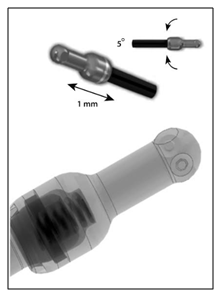
Figure 1: Each rod allows 5º angular movement in all planes and 1mm of compression.
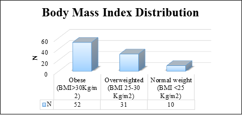
Figure 2: Graphic diagram showing BMI distribution.
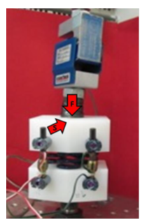
Figure 3: Flexo-compression test configuration and load applied.
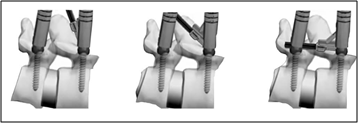
Figure 4: Introductory tool which allows the bar to be guided vertically first, and then lowered to reach the next screw.
2.4 Statistical analysis
The descriptive analysis of the data is presented as mean ± standard deviation (sd) and the proportions as percentages. With the Kolmogorov-Smirnov test, it is verified whether the variables have a normal distribution. With the Student's T test, the differences between the continuous variables for related samples were analysed. All statistical evaluations were bilateral and values ??with p <0.05 were considered statistically significant To perform the statistical analysis, Statistical Package for the Social Science (SPSS v22.0) was used.
3. Results
After applying inclusion and exclusion criteria, 93 patients were included. Minimum follow up was two years (range 2-5). The mean duration of the surgical intervention is 65.1 ± 18.3 minutes, (range 30-120 minutes). Finally, the mean stay was 1.7 ± 0.9 days, with the median being 2 days and the mode being 1 day of hospitalization. No patient was hospitalized for more than a week.
3.1 Clinical evaluation
Clinical scores significantly improved. ODI diminished from 47.4 ± 14.9 to 22.8 ± 19.7 at the final follow-up (p<0.001). In terms of SF-12, PHS (Physical Health Score) improved from 27.9 ± 6.3 to 37.5 ± 11.1 (p<0.001) and MHS (Mental Health Score) from 34,4 ± 11,3 to 42,7 ± 13,2 (p<0.002) (Table 2). Revision surgery was not required in any case. Post-surgical complications rate was really low, we only had one postoperatively hematoma solved conservatively, and one mal position of a screw inside the lumbar canal, causing no symptoms in that lady.
3.2 Radiological assessment
In flexion-extension x-rays no instability was observed, meaning that in the normal anteroposterior movements the system acts like a rigid fixation one. We observed either complete reduction of the lysthesis 62 %, or improvement of the degree of lysthesic displacement 31%, together with increase height of the discal space because of the locking of the device in distraction when introduced. In addition, the influence of age divided in two groups (under 65, working people, and over 65 years, retired people), sex, level of intervention (L3-L4, L4-L5, L5-S1) and diagnosis (Lysthesis I or II) was assessed with the results of ODI, and SF-12 obtained without finding statistically significant differences between them (p> 0.05).
|
Preop ODI |
Postop ODI |
p-value |
|
47.4 ± 14.9 |
22.8 ± 19.7 |
<0.001 |
|
Preop SF-12 (PHS) |
Postop SF-12 (PHS) |
|
|
27.9 ± 6.3 |
37.5 ± 11.1 |
<0.001 |
|
Preop SF-12 (MHS) |
1 year SF-12 (MHS) |
|
|
34.4 ± 11.3 |
42.7 ± 13.2 |
<0.002 |
Table 2: Oswestry Disability Index (ODI) and SF-12 results (mean ± standard deviation).
4. Discussion
Degenerative spondylolisthesis is due to changes in the facet joints so that the upper vertebrae progressively moves ventrally on the pedicle towards the vertebral body. This explains two important features, firstly the slip can never be more than about 30%, as the joint would reach the back of the body and secondly the root emerging below the level of the spondylolisthesis is compressed by the superior facet of the lower vertebrae, not by the facet joint of the upper vertebrae. Hence an L4-L5 degenerative spondylolisthesis compresses the 4th root however, the 5th root (traversing) can also be compressed by flavum and exuberant capsule of L4-L5 [18]. The altered anatomy produces spinal stenosis, presenting clinically as back and root pain. Described classically by Herkowitz et al [19, 20], they established that decompression alone was successful in 60% of patients, but an added uninstrumented fusion improved the results to some 90%. Instrumentation did not affect the initial results, but a subsequent paper from that department demonstrated that the addition of instrumentation had benefit in the long term. However, a recent paper by Abdu et al [21, 22] demonstrated that the method of fusion, even anterior fusion showed no differences. It is the case therefore that direct decompression alone, involving partial or total laminectomy and undercutting facetectomy [3] although successful is not the only method of achieving a satisfactory result. The current tendency is that decompression alone is not recommended, and instrumented fusion is the standard [7].
The authors accept that a contributing cause of pain in spondylolisthesis is due to multiple factors (osteoporosis, multilevel involvement and sagittal balance specially at the L5-S1 level in lytic and istmic spondylolisthesis). The literature [19, 20] established that stopping movement that is fusion and neural decompression were key procedures in this disorder. The problem is that those procedures do also denervate the joints, remove most of the ligamentous tissues, and synovium, all potential pain sources [23]. An anterior fusion removes the disc, again a potential source of pain. On those bases we cannot with absolute certainty conclude that stopping movement and decompression are the main reasons for alleviation of pain. This minimal intervention procedure could be regarded as an experiment to see if just stopping movement and indirectly decompressing without major tissue damage are the reasons for the relief of symptoms. Those were the reasons why dynamic x-rays and MRI were taken and demonstrated that abnormal movement present before was not present after fixation. In summary we believe the literature establishes that in this disorder, stopping movement and indirect decompression is an acceptable form of treatment. Our case is that this is achieved by this procedure.
In our series we have compared in each patient pre- and post-operative MRI scans and established that on visual examination reduction does increase space for the neural tissue (Figure 5). We believe that is well established that reduction increases spinal diameters and decompresses, the neural elements. Indeed, it is the whole basis of anterior fusion and indirect decompression. Clearly indirect decompression, achieved by an anterior fusion will reduce the spondylolisthesis, but will not decompress the root lying below the superior facet joint of the lower vertebrae. Yet it does cure L4 root pain. The likely reason for this is revealed if we study the pre- and post-operative MRI scans. In the pre-operatively scans it is clear that around the facet joint is exuberant inflammatory tissue related to the moving degenerate joints. The exuberant tissue, of capsule and synovium is an important factor in compressing the nerve. Post-operatively on the MRI scan this tissue is gone (Figure 5). The reduction contributes to increase diameters as the redundant capsule is pulled back, but the diminished space for the nerve beneath the superior facet of the lower vertebrae remain. This suggests that it was the movement of the degenerate joint that produced the exuberant tissue and stopping movement caused it to regress. These considerations are of relevance to the success of this minimal intervention technique. The lamina of the upper vertebrae is reduced solving the central canal stenosis and any L5 root compression but, by the stopping of movement reduces the irritation on the root, by inflammatory synovium and capsule although it still remains entrapped below the superior facet of the lower vertebrae. Our results are comparable to anterior fusion [24, 25] as the one described by Takahashi [9], 76% of satisfaction. Similar to the ones described by Oliveira et al [10] 86.5% by XLIF. In summary the results in those 93 patients demonstrated that in 79 of the patients stopping movement, and indirectly reducing compression, both confirmed by dynamic x-rays and MRI scans at review stopped both their back pain and claudication. Because of the nature of the intervention, we can be confident that no other access related factors could explain this.
In the present study, the pain was related either to the stenosis, or the facet degeneration not to an abnormal loading pattern. Adequate reduction of the spondylolisthesis is important to ensure decompression of the neural elements. This is a device that is only appropriate when the pain is related to stenosis or clearly facet degeneration as is seen in degenerative spondylolisthesis. Although implant failures have been reported in recent papers [26, 27] we did not record implant failure in our series, not in the dynamic rod and only one screw breakage with no clinical significance in the patient. No adjacent segment degeneration was noticed in the present study. The authors believe that abnormal movement inflammation and stenosis in degenerative spondylolisthesis can be treated by a minimally invasive device without the need of decompression and fusion. We looked at the results of this series and it was clear that stopping movement, and reduction of the lysthesis even to some degree was of clinical benefit.
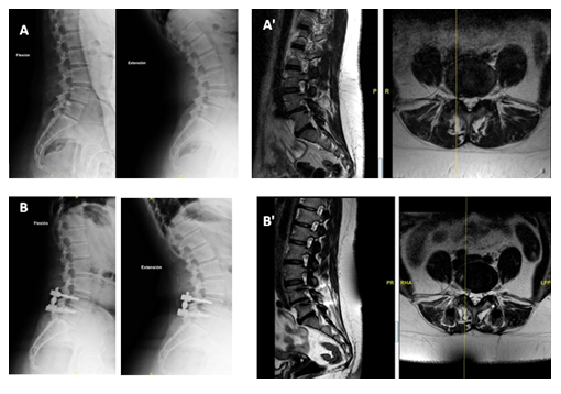
Figure 5: A) Preoperative flexion-extension X-ray; A') MRI; B) postoperative flexion-extension X-ray; B') postoperative MRI. Shows reduction of lysthesis and increase of diameters of central canal and recesses.
5. Conclusion
We conclude that in this group of patients, that is elderly, mainly women who may have generalized lumbar degeneration, but who have spinal stenosis due to a degenerative spondylolisthesis, can be treated very effectively, with little likelihood of operative related complications. This appliance deals effectively with pain due to movement or stenosis, but will not deal with pain due to abnormal loading, as it does not unload the disc. Hence this series does not validate its use more generally for back pain considered to be due to abnormal loading. The authors conclude that the non-rigid system is a good option for the treatment of demonstrated mobile single segment degenerative spondylolisthesis. There is minimal access related injury, a short operating time, short stay in hospital and good recorded 3–4 years results.
Declaration of Conflicting Interests
The author(s) declared no potential conflicts of interest with respect to the research, authorship, and/or publication of this article.
Funding
The author(s) received no financial support for the research, authorship, and/or publication of this article.
References
- Benoist M. Natural history of the aging spine. In The aging spine (2005): 4-7.
- Fraser RD, Brooks F, Dalzell K. Degenerative spondylolisthesis: a prospective cross-sectional cohort study on the role of weakened anterior abdominal musculature on causation. Eur Spine J 28 (2019): 1406-1412.
- Getty CJ, Johnson JR, Kirwan EO, et al. Partial undercutting facetectomy for bony entrapment of the lumbar nerve root. The Journal of bone and joint surgery 63 (1981): 330-335.
- Dijkerman ML, Overdevest GM, Moojen WA, et al. Decompression with or without concomitant fusion in lumbar stenosis due to degenerative spondylolisthesis: a systematic review. Eur Spine J 27 (2018): 1629-1643.
- Fischgrund J S. Degenerative lumbar spondylolisthesis with spinal stenosis: a prospective, randomized study comparing decompressive laminectomy and arthrodesis with and without spinal instrumentation. Spine 22 (1997): 2807-2812.
- Kornblum MB, Fischgrund JS, Herkowitz HN, et al. Degenerative lumbar spondylolisthesis with spinal stenosis: a prospective long-term study comparing fusion and pseudarthrosis. Spine 29 (2004): 726-733.
- Ferrero E, Guigui P. Current trends in the management of degenerative lumbar spondylolisthesis. EFORT open reviews 3 (2018): 192-199.
- Spiker WR, Goz V, Brodke DS. Lumbar interbody fusions for degenerative spondylolisthesis: review of techniques, indications, and outcomes. Global spine journal 9 (2019): 77-84.
- Takahashi T, Hanakita J, Ohtake Y, et al. Current status of lumbar interbody fusion for degenerative spondylolisthesis. Neurologia medico-chirurgica (2016).
- Oliveira L, Marchi L, Coutinho E, et al. A radiographic assessment of the ability of the extreme lateral interbody fusion procedure to indirectly decompress the neural elements. Spine 35 (2010): S331-S337.
- Arts MP, Wolfs JF, Kuijlen JM, et al. Minimally invasive surgery versus open surgery in the trea-tment of lumbar spondylolisthesis: study proto-col of a multicentre, randomised controlled trial (MISOS trial). BMJ open 7 (2017): e017882.
- Qian W, Yin H, Yang HL, et al. Pedicle screw?based dynamic stabilisation systems versus pedicle screw?based rigid fusion system for lumbar degenerative diseases. Cochrane Database of Systematic Reviews (2015).
- Meyerding HW. Spondylolisthesis. Surg Gynecol Obstet 54 (1932): 371-377.
- Nachemson ALF, Morris JM. In vivo measurements of intradiscal pressure: discometry, a method for the determination of pressure in the lower lumbar discs. JBJS 46 (1964): 1077-1092.
- Panjabi M M. The stabilizing system of the spine. Part II. Neutral zone and instability hypothesis. Journal of spinal disorders 5 (1992): 390-390.
- Biomechanics of the spine and fixation systems IBV. Mario Comín Clavijo (1995): 110
- Peul WC, Van Houwelingen HC, van den Hout WB, et al. Surgery versus prolonged conservative treatment for sciatica. N Engl J Med 356 (2007): 2245-2256.
- Rosenberg NJ. Degenerative spondylolisthesis: surgical treatment. Clin Orthop Relat Res 117 (1976): 112-120.
- Herkowitz HN, Kurz LT. Degenerative lumbar spondylolisthesis with spinal stenosis. J Bone Joint Surg Am 73 (1991): 802-808.
- Fischgrund JS, Mackay M, Herkowitz HN, et al. Volvo Award winner in clinical studies: Degenerative lumbar spondylolisthesis with spinal stenosis: a prospective, randomized study comparing decompressive laminectomy and arthrodesis with and without spinal instrumenta-tion. Spine 22 (1997): 2807-2812.
- Abdu WA, Lurie JD, Spratt KF, et al. Degenerative spondylolisthesis: does fusion method influence outcome? Four-year results of the spine patient outcomes research trial (SPORT). Spine 34 (2009): 2351.
- Abdu WA, Sacks OA, Tosteson ANA, et al. Long-Term Results of Surgery Compared with Nonoperative Treatment for Lumbar Degenerative Spondylolisthesis in the Spine Patients Outcome Research Trial (SPORT). Spine 43 (2018): 1619-1630.
- Edgar MA, Ghadially JA. Innervation of the lumbar spine. CORR 115 (1976): 35-41.
- Eismont FJ, Norton RP, Hirsch BP. Surgical management of lumbar degenerative spondylolisthesis. J Am Acad Orthop Surg 22 (2014): 203-213.
- Blumenthal C, Curran J, Benzel EC, et al. Radiographic predictors of delayed instability following decompression without fusion for degenerative grade I lumbar spondylolisthesis. J Neurosurg Spine 18 (2013): 340-346.
- Oikonomidis S, Sobottke R, Wilke HJ, et al. Material failure in dynamic spine implants: are the standardized implant tests before market launch sufficient?. Eur Spine J 28 (2019): 872-882.
- Oikonomidis S, Ashqar G, Kaulhausen T, et al. Clinical experiences with a PEEK-based dynamic instrumentation device in lumbar spinal surgery: 2 years and no more. J Orthop Surg Res 13 (2018): 1-10.
