Refractory Thrombocytopenia in Antiphospholipid Syndrome: Case Report and Mini Literature Review
Article Information
Maria Gloria Aversano1*, Alice Botta2, Matteo Sbattella1, Luca Giuseppe Balossi2, Fabrizio Colombo1
1Department of Internal Medicine, ASST Grande Ospedale Metropolitano Niguarda, Piazza Ospedale Maggiore 3, 20162, Milan, Italy
2Department of Allergology and Immunology, ASST Grande Ospedale Metropolitano Niguarda, Piazza Ospedale Maggiore 3, 20162, Milan, Italy
*Corresponding Author: Maria Gloria Aversano, Department of Internal Medicine, ASST Grande Ospedale Metropolitano Niguarda, Piazza Ospedale Maggiore 3, 20162, Milan, Italy.
Received: 25 April 2022; Accepted: 05 May 2022; Published: 20 May 2022
Citation: Maria Gloria Aversano, Alice Botta, Matteo Sbattella, Luca Giuseppe Balossi, Fabrizio Colombo. Refractory Thrombocytopenia in Antiphospholipid Syndrome: Case Report and Mini Literature Review. Archives of Clinical and Medical Case Reports 6 (2022): 404-410.
View / Download Pdf Share at FacebookAbstract
Antiphospholipid Syndrome (APS) is a condition characterized by clinical features of thrombosis and repeated miscarriages (criteria manifestations) accompanied by presence of antiphospholipid antibodies on laboratory tests. APS is a disease that can also present different clinical manifestations (non-criteria manifestations), including livedo reticularis, cutaneous ulcerations, thrombocytopenia, hemolytic anemia, valvular heart disease, and nephropathy, for which the gold standard treatment is often not known. Most of the APS management guidelines focus on the prevention and treatment of thrombotic and obstetrical manifestations, by anticoagulant therapy. However there is less consensus on the gold standard treatment for other clinical manifestations, which can have a serious impact on the health of the affected patient. Here we present a case report that emphasizes the importance of identifying correct therapy for non-criteria clinical manifestations of APS triggered by post-traumatic left fronto-temporal hematoma, subsequent neurosurgical intervention complicated by subdural empyema.
Keywords
Antiphospholipid syndrome; Thrombocytopenia; Purpura; Intravenous immunoglobulins
Antiphospholipid syndrome articles; Thrombocytopenia articles; Purpura articles; Intravenous immunoglobulins articles
Antiphospholipid syndrome articles Antiphospholipid syndrome Research articles Antiphospholipid syndrome review articles Antiphospholipid syndrome PubMed articles Antiphospholipid syndrome PubMed Central articles Antiphospholipid syndrome 2023 articles Antiphospholipid syndrome 2024 articles Antiphospholipid syndrome Scopus articles Antiphospholipid syndrome impact factor journals Antiphospholipid syndrome Scopus journals Antiphospholipid syndrome PubMed journals Antiphospholipid syndrome medical journals Antiphospholipid syndrome free journals Antiphospholipid syndrome best journals Antiphospholipid syndrome top journals Antiphospholipid syndrome free medical journals Antiphospholipid syndrome famous journals Antiphospholipid syndrome Google Scholar indexed journals syndrome articles syndrome Research articles syndrome review articles syndrome PubMed articles syndrome PubMed Central articles syndrome 2023 articles syndrome 2024 articles syndrome Scopus articles syndrome impact factor journals syndrome Scopus journals syndrome PubMed journals syndrome medical journals syndrome free journals syndrome best journals syndrome top journals syndrome free medical journals syndrome famous journals syndrome Google Scholar indexed journals Case Report articles Case Report Research articles Case Report review articles Case Report PubMed articles Case Report PubMed Central articles Case Report 2023 articles Case Report 2024 articles Case Report Scopus articles Case Report impact factor journals Case Report Scopus journals Case Report PubMed journals Case Report medical journals Case Report free journals Case Report best journals Case Report top journals Case Report free medical journals Case Report famous journals Case Report Google Scholar indexed journals Thrombocytopenia articles Thrombocytopenia Research articles Thrombocytopenia review articles Thrombocytopenia PubMed articles Thrombocytopenia PubMed Central articles Thrombocytopenia 2023 articles Thrombocytopenia 2024 articles Thrombocytopenia Scopus articles Thrombocytopenia impact factor journals Thrombocytopenia Scopus journals Thrombocytopenia PubMed journals Thrombocytopenia medical journals Thrombocytopenia free journals Thrombocytopenia best journals Thrombocytopenia top journals Thrombocytopenia free medical journals Thrombocytopenia famous journals Thrombocytopenia Google Scholar indexed journals Purpura articles Purpura Research articles Purpura review articles Purpura PubMed articles Purpura PubMed Central articles Purpura 2023 articles Purpura 2024 articles Purpura Scopus articles Purpura impact factor journals Purpura Scopus journals Purpura PubMed journals Purpura medical journals Purpura free journals Purpura best journals Purpura top journals Purpura free medical journals Purpura famous journals Purpura Google Scholar indexed journals treatment articles treatment Research articles treatment review articles treatment PubMed articles treatment PubMed Central articles treatment 2023 articles treatment 2024 articles treatment Scopus articles treatment impact factor journals treatment Scopus journals treatment PubMed journals treatment medical journals treatment free journals treatment best journals treatment top journals treatment free medical journals treatment famous journals treatment Google Scholar indexed journals CT articles CT Research articles CT review articles CT PubMed articles CT PubMed Central articles CT 2023 articles CT 2024 articles CT Scopus articles CT impact factor journals CT Scopus journals CT PubMed journals CT medical journals CT free journals CT best journals CT top journals CT free medical journals CT famous journals CT Google Scholar indexed journals surgery articles surgery Research articles surgery review articles surgery PubMed articles surgery PubMed Central articles surgery 2023 articles surgery 2024 articles surgery Scopus articles surgery impact factor journals surgery Scopus journals surgery PubMed journals surgery medical journals surgery free journals surgery best journals surgery top journals surgery free medical journals surgery famous journals surgery Google Scholar indexed journals Intravenous immunoglobulins articles Intravenous immunoglobulins Research articles Intravenous immunoglobulins review articles Intravenous immunoglobulins PubMed articles Intravenous immunoglobulins PubMed Central articles Intravenous immunoglobulins 2023 articles Intravenous immunoglobulins 2024 articles Intravenous immunoglobulins Scopus articles Intravenous immunoglobulins impact factor journals Intravenous immunoglobulins Scopus journals Intravenous immunoglobulins PubMed journals Intravenous immunoglobulins medical journals Intravenous immunoglobulins free journals Intravenous immunoglobulins best journals Intravenous immunoglobulins top journals Intravenous immunoglobulins free medical journals Intravenous immunoglobulins famous journals Intravenous immunoglobulins Google Scholar indexed journals Pulmonary Artery articles Pulmonary Artery Research articles Pulmonary Artery review articles Pulmonary Artery PubMed articles Pulmonary Artery PubMed Central articles Pulmonary Artery 2023 articles Pulmonary Artery 2024 articles Pulmonary Artery Scopus articles Pulmonary Artery impact factor journals Pulmonary Artery Scopus journals Pulmonary Artery PubMed journals Pulmonary Artery medical journals Pulmonary Artery free journals Pulmonary Artery best journals Pulmonary Artery top journals Pulmonary Artery free medical journals Pulmonary Artery famous journals Pulmonary Artery Google Scholar indexed journals
Article Details
Abbreviations:
APS: Antiphospholipid syndrome; CAPS: Catastrophic APS; aPL: Antiphospholipid antibodies; aCL: Anticardiolipin antibodies; LAC: Lupus anticoagulant; anti-β2GPI: Anti-β2 glycoprotein-I antibody; INR: International normalised ratio; DVT: Deep venous thrombosis; MSSA: Methicillin-susceptible Staphylococcus aureus; SLE: Systemic lupus erythematosus; IVIg: Intravenous immunoglobulin
1. Introduction
Antiphospholipid syndrome, also known as “Hughes Syndrome”, is a systemic autoimmune disease characterized by recurrent thrombosis and pregnancy morbidity along with persistent Anti-Phospholipid Antibodies (aPL) including lupus anticoagulant (LA), anti-β2-glycoprotein I (anti-β2GPI) and/or Anti-Cardiolipin (aCL) antibodies. APS is one of the most common acquired thrombophilic conditions, and unlike most cases of the genetic thrombophilia, is associated with both venous and arterial thrombosis. Indeed clinical manifestations are in most cases direct or indirect sequelae of a state of hypercoagulability which involves both venous and arterial vessels [1, 2]. APL can be either an isolated primary disease or associated with underlying autoimmune diseases, mainly systemic lupus erythematosus [3]. Although the first description of APS was made in 1983 under the name of anticardiolipin syndrome [4] several cases of patients with recurrent miscarriages, thromboembolic events and positive serology had been previously described [5-7]. In the 1980s, thanks to the use of molecular biology techniques, it was possible to identify the anticardiolipin antibodies and better understand the role of the mechanisms involved in the clinical manifestations of APS [8]. Clinical presentation of APS is highly variable (Table 1). The two hallmarks are thrombosis and obstetric diseases; these manifestations belong to the clinical classification criteria, along with the laboratory classification criteria (presence of LA, aCL, and/or anti-β2GPI) [9].
|
Criteria Manifestations |
|
|
1. Vascular thrombosis |
a. One or more clinical episodes of arterial, venous, or small vessel thrombosis, in any tissue or organ. |
|
2. Pregnancy morbidity |
a. One or more unexplained deaths of a morphologically normal fetus at or beyond the 10th week of gestation, or |
|
b. One or more premature births of a morphologically normal neonate before the 34th week of gestation because of eclampsia, severe preeclampsia or recognized features of placental insufficiency, or |
|
|
c. Three or more unexplained consecutive spontaneous abortions before the 10th week of gestation (excluded maternal anatomic or hormonal abnormalities and chromosomal causes) |
|
|
Non Criteria Manifestations |
|
|
1. Haematological |
thrombocytopenia, haemolytic anaemia with or without schistocytes, Evans syndrome |
|
2. Cutaneous |
livedo reticularis and livedo vasculitis, with purpuric macules, skin nodules, painful ulcers and necrosis, splinter haemorrhages |
|
3. Neurological |
migraine, epilepsy and seizures, cognitive dysfunction, delirium, chorea, transverse myelitis, multiple sclerosis, acute encephalopathy |
|
4. Cardiovascular |
heart valve lesions, coronary artery disease, cardiomyopathy, accelerated atherosclerosis, arterial stenosis |
|
5. Renal |
glomerulonephritis, renal thrombotic microangiopathy |
|
6. Orthopaedic |
avascular necrosis of bones, non-traumatic fractures, arthritis, arthralgia |
Table 1: Criteria manifestations and non-criteria manifestations of APS.
The most frequent thrombotic manifestations are represented by deep vein thrombosis of the lower limbs and pulmonary embolism. Regarding the involvement of arterial vessels, the ones of the central nervous system are the most affected, resulting in cerebral ischemia stroke and transient ischemic attack. Obstetrical manifestations include recurrent miscarriages, eclampsia, and preeclampsia that can result in premature delivery and fetal death [3,9]. APS should be suspected in patients with thrombotic events or obstetrical morbidity, without other risk factors. APS is then confirmed by the presence of antiphospholipid antibodies. For the diagnosis the positivity of at least one of the tests for aPL on two separate occasions at 12 weeks apart is needed [3]. In addition to the criteria manifestations, there are a series of other signs defined as "non-criteria", which may be present even in the absence of criteria manifestations. Non criteria manifestations include haematological (thrombocytopenia, haemolytic anaemia with or without schistocytes), cutaneous (livedo reticularis and livedo vasculitis, with purpuric macules, skin nodules, painful ulcers and necrosis), neurological (migraine, seizures, cognitive dysfunction, delirium, chorea, transverse myelitis, multiple sclerosis), cardiovascular (heart valve lesions, coronary artery disease), renal (glomerulonephritis, renal thrombotic microangiopathy) and orthopaedic (avascular necrosis of bones, non-traumatic fractures) manifestations [5,8,9]. There is also the so-called catastrophic APS (CAPS), which is characterized by diffuse thrombosis of small vessels resulting in multiorgan failure and high mortality. CAPS is the most severe form of APS, fortunately infrequent. In our patient we observed the concomitant appearance of several non-criteria manifestations: thrombocytopenia, hemolytic anemia, purpura and delirium. The presence of non-criteria manifestations is usually complicated by the fact that in most cases patients are already undergoing chronic corticosteroid therapy. Moreover, there isn’t a systematic, well-described approach in the guidelines for the treatment of these manifestations [2]. Therapy is based on primary and secondary antithrombotic prophylaxis. Low-dose aspirin proved to be effective as primary prophylaxis in asymptomatic aPL-positive individuals, as has hydroxychloroquine, particularly in patients with secondary APS and systemic lupus erythematosus. Regarding the treatment of thrombotic events, patients with venous thromboembolism are treated with long-term oral anticoagulants to maintain a target International Normalised Ratio (INR) of 2.0-3.0. In case of arterial thrombosis, the target INR increases to INR of 3.0-4.0, or a therapy with low dose aspirin plus anticoagulation can be set [5,10]. The anticoagulant medications used are lifetime warfarin or an alternative vitamin K antagonist. The literature regarding direct oral anticoagulants is not extensive. Alternatively, other anticoagulant options include low molecular weight heparin, unfractionated heparin and fondaparinux. Regarding non-criteria manifestations there are no standard treatment recommendations. Patients with thrombocytopenia and/or autoimmune hemolytic anemia may benefit from glucocorticoids with or without intravenous immunoglobulins as first-line treatment, whereas azathioprine, cyclophosphamide, and mycophenolate mofetil may be used as second-line therapies [5, 11]. Our patient only initially responded to isolated steroid therapy, and later intravenous immunoglobulin therapy was effective to correct severe thrombocytopenia. APS continues to be a cause of significant morbidity and mortality, so future treatment approaches should be tailored to meet individual patient characteristics.
2. Case Presentation
A 56-year-old female with a history of Budd-Chiari syndrome, epilepsy, Antiphospholipid Syndrome (APS) associated with recurrent Deep Venous Thrombosis (DVT) in treatment with warfarin, was brought to the emergency department because of an accidental fall at home followed by altered mental status. Subsequent instrumental investigations revealed the presence of a post-traumatic left fronto-temporal hematoma successfully treated with surgical evacuation on January 23rd. On February 19th the patient returned to emergency room for fever and purulent secretions from the wound.
2.1. Clinical evaluation
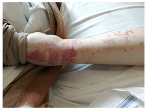
A brain CT with contrast medium was performed with evidence of a subdural empyema. This was treated with surgical evacuation and antibiotic therapy based on meropenem and linezolid. After isolation of Methicillin-Susceptible Staphylococcus Aureus (MSSA) on surgical piece, antibiotic therapy was modified by introducing oxacillin 3 g every 6 hours. During hospitalization postoperative delirium appeared, resolved after treatment with haloperidol and promazine and progressive decalage of steroid therapy with dexamethasone. On February 26th a sudden drop in platelet count was observed (from 140000/mm3 to 13000/mm3), dexamethasone was again increased from 1 mg daily to 4 mg daily and prophylactic dose of fondaparinux was discontinued. Subsequent finding of haemolytic anemia and purpura on the lower extremities (Figure 1, 2, 3) required a further increase in dexamethasone to 8 mg daily. In the suspicion of Systemic Lupus Erythematosus (SLE), anti-double stranded DNA antibodies were performed and resulted negative, confirming the picture of primitive APS, determining severe thrombocytopenia, hemolysis and purpura. At the next attempt of steroid transfer, further worsening of thrombocytopenia appeared (Platelet count < 20000/ mm3) and required therapy with Intravenous Immunoglobulin (IVIg) 800 mg/kg for two days. Subsequent progressive increase in platelet values was observed, stabilizing on a value of 80000/mm3 in the last days of hospitalization. Anticoagulant dose of fondaparinux was gradually reintroduced without further problems.

Figure 1: Purpura on the lower extremities.

Figure 2: Purpura on the lower extremities.

Figure 3: Purpura on the lower extremities.
3. Discussion
Antiphospholipid syndrome is an autoimmune systemic disorder that represents one of the most common causes of acquired hypercoagulable state. The main clinical presentations are vascular thrombosis and pregnancy morbidity, associated with the persistent presence of antiphospholipid antibodies (criteria manifestations). There are also several non-criteria manifestations that may be present in patients with APS and whose treatment can be challenging. These are hematological, neurological, dermatological, cardiovascular, and renal manifestations that compromise the health of the affected individual. Treatment’s guidelines give indications about management of criteria manifestations. Long-term warfarin or other vitamin K antagonist therapy are used as antithrombotic prophylaxis. Low dose aspirin and prophylactic heparin are used to prevent recurrent obstetric complications. What is currently lacking is a consensus regarding the management of non-criteria manifestations. In patients with severe thrombocytopenia and/or hemolytic anemia, a beneficial effect of steroid therapy has been observed, which can be implemented with intravenous immunoglobulins, up to the use of immunosuppressant therapy as a second line of treatment. In our patient known for APS in general good control, we observed the development of an important hematological (thrombocytopenia, hemolytic anemia), cutaneous (purpura) and neurological (delirium) failure after a neurosurgical intervention complicated by subdural empyema. These non-criteria manifestations threatened the clinical stability of the patient, who was moreover frail. In fact, the patient's clinical conditions were worsened by the onset of thrombocytopenia and delirium, which also negatively affected the duration of hospital stay. In this case, the patient responded only partially to increase in steroid therapy and modification of anticoagulant. Only after the introduction of intravenous immunoglobulins we observed a gradual increase in platelet count and an improvement in patient's general clinical conditions.
4. Conclusion
This case exemplifies the need for a tailored approach to patients with APS, with treatment strategies that should consider different clinical manifestations and individual risk factors.
References
- Chaturvedi S, McCrae KR. Diagnosis and management of the antiphospholipid syndrome. Blood Rev 31 (2017): 406-417.
- Cervera R, Piette JC, Font J, et al. Antiphospholipid syndrome: clinical and immunologic manifestations and patterns of disease expression in a cohort of 1,000 patients. Arthritis Rheum 46 (2002): 1019-1027.
- Vaidya B, Nakarmi S, Joshi R, et al. A Simplified Understanding of the Black Swan: Anti-phospholipid Antibody Syndrome. J Nepal Med Assoc 57 (2019): 133-143.
- GR Hughes. Thrombosis, abortion, cerebral disease, and the lupus anticoagulant. Br Med J 287 (1983): 1088-1089.
- Uthman I, Noureldine MHA, Ruiz-Irastorza G, et al. Management of antiphospholipid syndrome. Ann Rheum Dis 78 (2019): 155-161.
- Nilsson IM, Astedt B, Hedner U, et al. Intrauterine death and circulating anticoagulant ("antithromboplastin"). Acta Med Scand 197 (1975):153-159.
- Firkin BG, Howard MA, Radford N. Possible relationship between lupus inhibitor and recurrent abortion in young women. Lancet 2 (1980): 366.
- Sammaritano LR. Antiphospholipid syndrome. Best Practice & Research Clinical Rheumatology (2020).
- Miyakis S, Lockshin MD, Atsumi T, et al. International consensus statement on an update of the classification criteria for definite antiphospholipid syndrome (APS). J Thromb Haemost 4 (2006): 295-306.
- Ruiz-Irastorza G, Cuadrado MJ, Ruiz-Arruza I, et al. Evidence-based recommendations for the prevention and long-term management of thrombosis in antiphospholipid antibody-positive patients: report of a task force at the 13th International Congress on antibodies. Lupus 20 (2011): 206-218.
- Garcia D, Erkan D. Diagnosis and management of the antiphospholipid syndrome. N Engl J Med 378 (2018): 2010-2021.
