Pulmonary Thromboembolism in COVID-19 Patients on CT Pulmonary Angiography - A Single-Centre Retrospective Cohort Study in the United Arab Emirates
Article Information
Ghufran Aref Saeed1*, Waqar H Gaba2, Abd Al Kareem Adi2, Reima Al Marshoodi2, Safaa Al Mazrouei1, Asad R Shah1
1Radiology Department, Shiekh Khalifa Medical City, Abu Dhabi, U.A.E
2Internal Medicine Department, Shiekh Khalifa Medical City, Abu Dhabi, U.A.E.
*Corresponding Author: Ghufran Aref Saeed, Radiology Department, Shiekh Khalifa Medical City, Abu Dhabi, U.A.E
Received: 13 August 2022; Accepted: 20 August 2022; Published: 26 August 2022
Citation:
Ghufran Aref Saeed. Pulmonary thromboembolism in COVID-19 Patients on CT Pulmonary Angiography - A Single-Centre Retrospective Cohort Study in the United Arab Emirates. Journal of Surgery and Research 5 (2022): 472-476.
View / Download Pdf Share at FacebookAbstract
Purpose
Our aim is to identify the prevalence and distribution of pulmonary thromboembolism in COVID-19 infected patients in our hospital. Materials and
Methods
Data of all patients with COVID-19 infection either on RT-PCR testing or non-contrast high resolution CT(HRCT) who had CT pulmonary angiography (CTPA) from April to June 2020 were included. 133 patients were initially included in the study, 7 were excluded leaving a total number of 126 patients.
Results
Twenty (15.8%) patients had evidence of pulmonary embolism (PE) on CTPA with mean age of 50 years (ranging 31-85) with 95% males. The mean D-dimer was 5.61mcg/mL among the PE-negative and 14.49 mcg/mL in the PE-positive groups respectively. Among the patients with evidence of pulmonary embolism on CTPA almost half required admission to intensive care unit in comparison to only one-fifth with negative CTPA. One-fourth died among the PE positive group with only 5% died among the PE negative group. There was a 33% reduction in the development of PE in the COVID-19 patients who had received low molecular weight heparin (LMWH) prior to their CTPA study versus those who had not.
Conclusion
D dimers correlate well with the incidence of pulmonary embolism among COVID-19 patients. Our data suggest that majority of our patients, developed pulmonary embolisms within 5 days into their hospital stay, accounting to almost two thirds of all positive cases diagnosed by CTPA. Those with PE among COVID-19 patients have high chances of ICU admission and mortality. Use of thromboprophylaxis early on might reduce the incidence of PE.
Keywords
Pulmonary Thromboembolism, COVID-19, COVID-19
COVID-19 articles
Pulmonary Thromboembolism articles Pulmonary Thromboembolism Research articles Pulmonary Thromboembolism review articles Pulmonary Thromboembolism PubMed articles Pulmonary Thromboembolism PubMed Central articles Pulmonary Thromboembolism 2023 articles Pulmonary Thromboembolism 2024 articles Pulmonary Thromboembolism Scopus articles Pulmonary Thromboembolism impact factor journals Pulmonary Thromboembolism Scopus journals Pulmonary Thromboembolism PubMed journals Pulmonary Thromboembolism medical journals Pulmonary Thromboembolism free journals Pulmonary Thromboembolism best journals Pulmonary Thromboembolism top journals Pulmonary Thromboembolism free medical journals Pulmonary Thromboembolism famous journals Pulmonary Thromboembolism Google Scholar indexed journals COVID-19 articles COVID-19 Research articles COVID-19 review articles COVID-19 PubMed articles COVID-19 PubMed Central articles COVID-19 2023 articles COVID-19 2024 articles COVID-19 Scopus articles COVID-19 impact factor journals COVID-19 Scopus journals COVID-19 PubMed journals COVID-19 medical journals COVID-19 free journals COVID-19 best journals COVID-19 top journals COVID-19 free medical journals COVID-19 famous journals COVID-19 Google Scholar indexed journals CT pulmonary angiography articles CT pulmonary angiography Research articles CT pulmonary angiography review articles CT pulmonary angiography PubMed articles CT pulmonary angiography PubMed Central articles CT pulmonary angiography 2023 articles CT pulmonary angiography 2024 articles CT pulmonary angiography Scopus articles CT pulmonary angiography impact factor journals CT pulmonary angiography Scopus journals CT pulmonary angiography PubMed journals CT pulmonary angiography medical journals CT pulmonary angiography free journals CT pulmonary angiography best journals CT pulmonary angiography top journals CT pulmonary angiography free medical journals CT pulmonary angiography famous journals CT pulmonary angiography Google Scholar indexed journals RT-PCR testing articles RT-PCR testing Research articles RT-PCR testing review articles RT-PCR testing PubMed articles RT-PCR testing PubMed Central articles RT-PCR testing 2023 articles RT-PCR testing 2024 articles RT-PCR testing Scopus articles RT-PCR testing impact factor journals RT-PCR testing Scopus journals RT-PCR testing PubMed journals RT-PCR testing medical journals RT-PCR testing free journals RT-PCR testing best journals RT-PCR testing top journals RT-PCR testing free medical journals RT-PCR testing famous journals RT-PCR testing Google Scholar indexed journals non-contrast high resolution CT articles non-contrast high resolution CT Research articles non-contrast high resolution CT review articles non-contrast high resolution CT PubMed articles non-contrast high resolution CT PubMed Central articles non-contrast high resolution CT 2023 articles non-contrast high resolution CT 2024 articles non-contrast high resolution CT Scopus articles non-contrast high resolution CT impact factor journals non-contrast high resolution CT Scopus journals non-contrast high resolution CT PubMed journals non-contrast high resolution CT medical journals non-contrast high resolution CT free journals non-contrast high resolution CT best journals non-contrast high resolution CT top journals non-contrast high resolution CT free medical journals non-contrast high resolution CT famous journals non-contrast high resolution CT Google Scholar indexed journals CT pulmonary angiography articles CT pulmonary angiography Research articles CT pulmonary angiography review articles CT pulmonary angiography PubMed articles CT pulmonary angiography PubMed Central articles CT pulmonary angiography 2023 articles CT pulmonary angiography 2024 articles CT pulmonary angiography Scopus articles CT pulmonary angiography impact factor journals CT pulmonary angiography Scopus journals CT pulmonary angiography PubMed journals CT pulmonary angiography medical journals CT pulmonary angiography free journals CT pulmonary angiography best journals CT pulmonary angiography top journals CT pulmonary angiography free medical journals CT pulmonary angiography famous journals CT pulmonary angiography Google Scholar indexed journals intensive care unit articles intensive care unit Research articles intensive care unit review articles intensive care unit PubMed articles intensive care unit PubMed Central articles intensive care unit 2023 articles intensive care unit 2024 articles intensive care unit Scopus articles intensive care unit impact factor journals intensive care unit Scopus journals intensive care unit PubMed journals intensive care unit medical journals intensive care unit free journals intensive care unit best journals intensive care unit top journals intensive care unit free medical journals intensive care unit famous journals intensive care unit Google Scholar indexed journals therapeutic anticoagulation articles therapeutic anticoagulation Research articles therapeutic anticoagulation review articles therapeutic anticoagulation PubMed articles therapeutic anticoagulation PubMed Central articles therapeutic anticoagulation 2023 articles therapeutic anticoagulation 2024 articles therapeutic anticoagulation Scopus articles therapeutic anticoagulation impact factor journals therapeutic anticoagulation Scopus journals therapeutic anticoagulation PubMed journals therapeutic anticoagulation medical journals therapeutic anticoagulation free journals therapeutic anticoagulation best journals therapeutic anticoagulation top journals therapeutic anticoagulation free medical journals therapeutic anticoagulation famous journals therapeutic anticoagulation Google Scholar indexed journals
Article Details
1. Introduction
In December 2019, cases of a new virus causing pneumonia have been identified in Wuhan, China [1]. Ever since then, researchers and healthcare workers all around the world have started studying, analyzing, and publishing data on this novel coronavirus. One of the more heavily studied aspects was the virus’s induced prothrombotic state [2] that was strongly associated with increased mortality [3]. Among the notable pathologies caused by the virus’s induced hypercoagulable state was the development of pulmonary emboli. There was an increased prevalence of pulmonary thromboembolism in patients with moderate to severe COVID-19 pneumonia [4]. One meta-analysis reported the incidence of pulmonary embolism in COVID-19 pneumonia patients to be up to 15%, and urged the need to evaluate the roles of both prophylactic and therapeutic anticoagulation [5], as developing pulmonary embolism was shown to increase the morbidity, mortality, prolong the duration of invasive ventilation, and ICU stay [6]. The main objective of our study is to identify the prevalence and distribution of pulmonary thromboembolism in COVID-19 infected patients in Abu Dhabi, UAE; and analyze whether there is a notable difference between the PE positive and PE negative groups regarding their age, gender, CT severity score, D-dimer levels, thromboprophylaxis, and mortality rates.
2. Methods
2.1. Data Collection. We obtained ethical approval from the Institutional Review Board (IRB) and Department of Health (DOH), Abu Dhabi, United Arab Emirates (UAE). Waiver of informed consent was allowed by the ethics committee. We collected clinical and laboratory data for analysis derived from an electronic medical record system, from April to June 2020 of patients who were suspected to have COVID-19 infection and underwent a CT pulmonary angiography (CTPA) scan. The results for the CTPA images were collected and evaluated using the Picture Archiving and Communication Systems (PACS).
2.2. CTPA Inspection. All CT Pulmonary Angiograms were performed on the SIEMENS 128 Somatom scanner. Patients were placed in a supine position with a single breath hold. Omnipaque 350 was used as IV contrast with an average volume of 40-50 mL, using a power injector with a flow rate of 4-5 cc/s using a peripheral venous cannula of 18-20 G. Scanning parameters were as follows: topogram length of 512cm, scan direction was craniocaudal, tube voltage of 100 kV and a tube current of 75 mA - smart mA dose modulation. The region-of-interest was the main pulmonary artery with a trigger threshold of 100HU. The monitoring delay was 3 sec with a diagnostic delay of 4 sec, slice thickness was 0.6 mm, pitch was 1.9, rotation time was 0.25 s, and scan time was 0.25 s. Sagittal, and Coronal reformats were also obtained with additional maximum intensity projection images.
2.3. CTPA Image Analysis. The images were evaluated and reviewed by two radiologists with more than 8 years of experience to judge the presence of typical COVID-19 pneumonia findings (bilateral, multilobed, posterior peripheral ground-glass opacities) and judge upon the severity of the infection radiologically.
2.4. Statistical Analysis. Data was analyzed using SPSS 21.0. Demographic, clinical and laboratory data was reported as numbers where each value pertained to a particular subgroup based on their findings (1 for male, 2 for female, etc.). We used the Pearson correlation coefficient for correlations and regarded a p-value less than 0.05 as statistically significant.
2.5. Inclusion criteria consisted of patients having either a positive COVID-19 RT PCR nasal swab or showing typical clinical and radiological pictures highly suggestive of COVID-19 pneumonia. Excluded were patients with an underlying malignancy, and those with a non-diagnostic opacification of the main pulmonary artery on CTPA.
2.6. Thromboprophylaxis. We identified the patients who received low molecular weight heparin (LMWH) prior to their CTPA amongst everyone included in the study, then compared the incidence of PE in them against those who had not. The decision on whether to give thromboprophylaxis, along with the dose of LMWH was decided by the Consultant Physician, guided by a hospital locally approved algorithm (see Appendix A).
3. Results
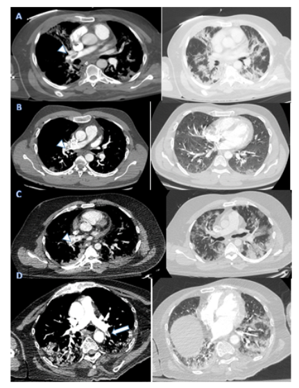
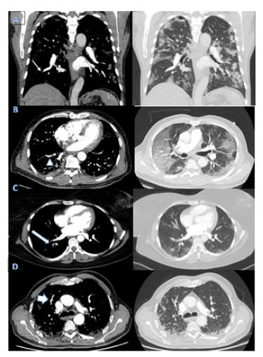
A total of 133 patients underwent CT Pulmonary Angiography during the 3 months period of the COVID-19 pandemic from April to June 2020 at our institution. 126 of those patients were included for the final analysis. 7 were excluded as their COVID-19 PCR was negative and they had no radiological evidence of COVID-19 pneumonia, or they had inconclusive CT scans suggesting the likely presence of a malignancy. The mean age was 51 years (ranging 29-85). 103 (82%) of patients were males, while the remaining 23 (18%) were females. Twenty (15.8%) patients had evidence of pulmonary embolism (PE) on CTPA. Most pulmonary emboli [16/20 (80%)] were in the lobar and distal branches and only 4/20 (20%) were in the main pulmonary arteries (Fig1 & 2, Appendix B). Among those patients with PE, mean age was 50 years (ranging 31-85). As for their genders, 19 (95%) were male, and only 1 (5%) was female. 34 (26.9 %) CTPA were done upon presentation to the emergency department with a positive incidence of 6/34 (17.6%). 39 (30.9%) CTPA were done 1-5 days into admission with a positive incidence of 7/39 (17.9%), and 53 (42%) CTPA were done 6+ days into admission with a positive incidence of 7/53 (13.2%). (See Appendix C). It was also evident that almost half (45%) of patients with evidence of pulmonary embolism on CTPA required admission to the intensive care unit during their hospital stay, while only one-fifth (20%) of the 106 patients with no evidence of pulmonary embolism on CTPA did. The mean D-dimer was 5.61mcg/mL among the PE-negative and 14.49 mcg/mL in the PE-positive groups respectively. As for mortality rates, there were a total of ten deaths out of all patients studied, with 5% mortality rate in the PE-negative group compared to the 25% mortality rate among the PE-positive group. There was no significant difference in the severity of parenchymal lung involvement radiologically amongst the groups, with most cases being of either the moderate or severe categories almost equally. Regarding thromboprophylaxis; 83 patients had LMWH prior to CTPA and incidence of PE among those was 12 (14%). On the other hand, 43 patients had not received LMWH or any other type of anticoagulation, and 9 (21%) of those patients had evidence of PE on their scans. As for the dosing, 29(35%) patients received prophylactic dose, 22(26%) received intermediate dose, and 32(38%) received therapeutic dose with incidence of PE being 14%, 13% and 6% respectively (See Appendix D). All doses were adjusted according to patients’ kidney function (creatinine clearance).

Figure 1: Four Cases of Central Pulmonary Embolism presenting to the ED. Axial soft tissue window (left pane) showing right main pulmonary artery emboli (Arrow-heads in A-C) and small proximal left lower lobar pulmonary artery embolus (Arrow in D). Corresponding lung windows demonstrating the typical sub-pleural Covid pneumonia changes.

Figure 2: Coronal and axial soft tissue window images (left pane) showing segmental emboli in the right lower lobe (Thin arrow in A, Arrow head in B and Long arrow in C) and right upper lobe (Broad arrow in D). Corresponding lung window images showing typical changes of Covid pneumonia.
4. Discussion
Numerous studies of the COVID-19 pandemic have advised early caution and testing against patients developing coagulopathies and venous thromboembolisms including PE [7,8]. It has proven to be a significant burden over the morbidity and mortality rates, reaching mortality rate increases of up to 45% when compared to other general causes [9]. In our study, that risk is evident, and what we would like to highlight in our results is that the majority of our patients who developed pulmonary embolisms did so early on, 0 to 5 days into their hospital stay, accounting to almost two thirds of all positive cases diagnosed by CTPA. That emphasizes the need to be on guard and look for it as a cause early on once patients show any signs of clinical deterioration or have any symptoms suggesting a pulmonary embolism during their hospital stay. We believe early diagnosis and treatment initiation against pulmonary emboli would potentially lower both morbidity and mortality rates, since the subjects studied showed that those who developed pulmonary emboli had substantially higher ICU admission rates and are at a higher risk of death. We also suggest that, like other studies have shown regarding the issue, D-dimer acts as a decent indicator of hypercoagulability [10,11], given that all our patients who developed pulmonary embolisms on CTPA had positive D-dimers, and their mean D-dimer level was much higher than that of the PE-negative group. Even though it’s been shown through evidence that increasing CT severity score did to an extent predict patient’s clinical status, particularly their oxygen requirement [12], we could not demonstrate that it had any predictive value on patients developing pulmonary embolism with a moderate to severe chest HRCT score. [See Appendix E for more information on how HRCT severity scoring was calculated]. Overall, acute pulmonary embolism developed in 20/126 (15.8%) of the cases studied over the 3-month period. which is more or less consistent with the currently available worldwide statistic [5]. Moreover, regarding thromboprophylaxis; when the effect of receiving LMWH prior to the patients’ CTPA was analyzed, even though both groups are not identical in number and lack information regarding certain control parameters (such as severity and duration of illness) there was a 33% reduction in the development of PE in the COVID-19 patients who had received LMWH prior to their CTPA study versus those who had not.
5. Conclusion
In patients diagnosed with COVID-19 infection either by RT-PCR or high clinical and radiological suspicion, we should be aware of this evident risk of hypercoagulability and embolism to the lung’s vasculature, whose symptoms could be masked as a mere deterioration of the patient’s respiratory status. Being vigilant in diagnosing and treating such incidents could potentially reduce morbidity and mortality rates of the COVID-19 pandemic. Our study also shows benefit in giving thromboprophylaxis (LMWH at prophylactic doses or higher), leading to a potential decrease in the incidence of PE among such patients.
References
- Tan BK, Kang GC, Tay EH. Subunit principle of vulvar reconstruction: algorithm and outcomes. Arch. Plast. Surg 41 (2014): 379-386.
- Di Donato V, Bracchi C, Cigna E. Vulvo-vaginal reconstruction after radical excision for treatment of vulvar cancer: Evaluation of feasibility and morbidity of different surgical techniques. Surg. Oncol 26 (2017): 511-521.
- Cordeiro PG, Pusic AL, Disa JJ. A classification system and reconstructive algorithm for acquired vaginal defects. Plastic and reconstructive surgery 110 (2002): 1058-1065.
- Morley GW, Lindenauer SM, Youngs D. Vaginal reconstruction following pelvic exenteration: surgical and psychological considerations. American journal of obstetrics and gynecology 116 (1973): 996-1002.
- Li JS, Crane CN, Santucci RA. Vaginoplasty tips and tricks. International braz j urol : official journal of the Brazilian Society of Urology 47 (2021): 263-273.
- Boccara D, Serro RK, Lefevre J, et al. Reconstruction of abdo-perineal resection by Taylor flap: about 68 patients. Ann. Chir. Plast. Esthet 63 (2018): 222-228.
- Cordeiro PG, Pusic AL, Disa JJ. A classification system and reconstructive algorithm for acquired vaginal defects. Plast Reconstr Surg 110 (2002): 1058-1065.
- Block LM, Hartmann EC, King J, et al. Outcomes Analysis of Gynecologic Oncologic Reconstruction. Plastic and reconstructive surgery. Global open 7 (2019): e2015.
- Casey WJ, Tran NV, Petty PM, et al. A comparison of 99 consecutive vaginal reconstructions: an outcome study 52 (2004).
- Barbara G, Facchin F, Buggio L, et al. Vaginal rejuvenation: current perspectives. International journal of women's health 9 (2017): 513-519.
- Erdogan G. Experience of Vaginoplasty for Enhancement of Sexual Functioning in a Center in Turkey: A Before and After Study. Cureus 13 (2021): e14767.
- Desai SA, Kroumpouzos G, Sadick N. Vaginal rejuvenation: From scalpel to wands. International journal of women's dermatology 5 (2019): 79-84.
- Gupta V, Lennox GK, Covens A. The rectus abdominus myoperitoneal flap for vaginal reconstruction. Gynecologic oncology reports 32 (2020): 100567.
- Wang TN, Whetzel T, Mathes SJ, et al. A fasciocutaneous flap for vaginal and perineal reconstruction. Plast Reconstr Surg 80 (1987): 95-103.
- Dobbeleir JM, Landuyt KV, Monstrey SJ. Aesthetic surgery of the female genitalia. Seminars in plastic surgery 25 (2011): 130-141.
- Arkoulis N, Kearns C, Deeny M. Abstract: Vaginal Reconstruction with Interdigitating Y-flaps in Women with Transverse Vaginal Septa. Plastic and Reconstructive Surgery Global Open 4 (2016): 108-109.
- Zak PW, Chow I, Zhu X, et al. The Use of a Hartmann's Pouch for Bowel Vaginoplasty: A Case Report. Plastic and reconstructive surgery. Global open 9 (2021): e3546.
- Wee JT, Joseph VT. A new technique of vaginal reconstruction using neurovascular pudendal-thigh flaps: a preliminary report. Plast Reconstr Surg 83 (1989): 701-709.
- Gleeson NC, Baile W, Roberts WS, et al. Pudendal thigh fasciocutaneous flaps for vaginal reconstruction in gynecologic oncology. Gynecologic oncology 54 (1994): 269-274.
- Lee RC, Rotmensch J. Rectovaginal radiation fistula repair using an obturator fasciocutaneous thigh flap. Gynecol Oncol 94 (2004): 277-282.
- Woods JE, Alter G, Meland B, et al. Experience with vaginal reconstruction utilizing the modified Singapore flap. Plast Reconstr Surg 90 (1992): 270-274.
- Berger JL, Westin SN, Fellman B, et al. Modified vertical rectus abdominis myocutaneous flap vaginal reconstruction: an analysis of surgical outcomes. Gynecologic oncology 125 (2012): 252-255.
- Behan FC. The keystone design perforator island flap in reconstructive surgery. ANZ J. Surg 73 (2003): 112–120.
- Sinna R, Qassemyar Q, Benhaim T. Perforator flaps: a new option in perineal reconstruction. J. Plast. Reconstr. Aesthet. Surg 63 (2010): e766-e774.
- Friedman J, Dinh T, Potochny J. Reconstruction of the perineum. Semin Surg Oncol 19 (2000): 282-293.
- Shepherd JH, Van Dam PA, Jobling TW, et al. The use of rectus abdominis myocutaneous flaps following excision of vulvar cancer. Br J Obstet Gynaecol 97 (1990): 1020-1025.
- Pusic AL, Mehrara BJ. Vaginal reconstruction: an algorithm approach to defect classification and flap reconstruction. J Surg Oncol 94 (2006): 515-521.
- McCraw JB, Massey FM, Skanklin KD, et al. Vaginal reconstruction with gracilis myocutaneous flaps. Plast. Reconstr. Surg 58 (1976): 176-183.
- Soper JT, Rodriguez G, Berchuck A, et al. Long and short gracilis myocutaneous flaps for vulvovaginal reconstruction after radical pelvic surgery: comparison of flap-specific complications. Gynecol Oncol 56 (1995): 271-275.
- Chen SH, Hentz VR, Wei FC, et al. Short gracilis myocutaneous flaps for vulvoperineal and inguinal reconstruction. Plast Reconstr Surg 95 (1995): 372-377.
- Soper JT, Larson D, Hunter VJ, et al. Short gracilis myocutaneous flaps for vulvovaginal reconstruction after radical pelvic surgery. Obstetrics and gynecology 74 (1989): 823-827.
- Achauer BM, Braly P, Berman ML, et al. Immediate vaginal reconstruction following resection for malignancy using the gluteal thigh flap. Gynecol Oncol 19 (1984): 79-89.
- Kwun Kim S, Hoon Park J, Cheol Lee K, et al. Long-term results in patients after rectosigmoid vaginoplasty. Plast Reconstr Surg 112 (2003): 143-151.
- Ozkan O, Akar ME, Ozkan O, et al. The use of vascularized jejunum flap for vaginal reconstruction: clinical experience and results in 22 patients. Microsurgery 30 (2010): 125-131.
- Pratt JH, Smith GR. Vaginal reconstruction with a sigmoid loop. Am J Obstet Gynecol 96 (1966): 31-40.
- O'Connor JL, DeMarco RT, Pope JC, et al. Bowel vaginoplasty in children: a retrospective review. J Pediatr Surg 39 (2004): 1205-1208.
- Kwun Kim S, Hoon Park J, Cheol Lee K, et al. Long-term results in patients after rectosigmoid vaginoplasty. Plastic and reconstructive surgery 112 (2003): 143-151.
- Kim SK, Park JW, Lim KRet al. Is Rectosigmoid Vaginoplasty Still Useful?. Archives of plastic surgery 44 (2017): 48-52.
- Kim JK, Na W, Cho JH, et al. Refinement of recto-sigmoid colon vaginoplasty using a three-dimensional laparoscopic technique. Medicine, 100 (2021): e27042.
