Protective Effects of Exercise Training Against Aged-Induced the Reduction of Cardiac Angiogenic Capacity in Middle-Aged Rats
Article Information
Titiporn Mekrungruangwong1, Pimpetch Kasetsuwan2, Sheepsumon Viboolvorakul3, Suthiluk Patumraj4*
1PhD Program in Medical Science, Faculty of Medicine, Chulalongkorn University, Bangkok, 10330, Thailand
2Medical Student, Faculty of Medicine, Chulalongkorn University, Bangkok, 10330, Thailand
3Department of Medical Science, Faculty of Science, Rangsit University, Pathum-Thani 12000, Thailand
4Center of Excellence for Microcirculation, Department of Physiology, Faculty of Medicine, Chulalongkorn University, Bangkok,10330, Thailand
*Corresponding author: Suthiluk Patumraj, Center of Excellence for Microcirculation, Department of Physiology, Faculty of Medicine, Chulalongkorn University, Bangkok, 10330, Thailand
Received: 29 October 2019; Accepted: 15 November 2019; Published: 23 January 2020
Citation: Titiporn Mekrungruangwong, Pimpetch Kasetsuwan, Sheepsumon Viboolvorakul, Suthiluk Patumraj. Protective Effects of Exercise Training Against Aged-Induced the Reduction of Cardiac Angiogenic Capacity in Middle-Aged Rats. Cardiology and Cardiovascular Medicine 4 (2020): 058-065.
View / Download Pdf Share at FacebookAbstract
Objective: To investigate the protective effects of exercise training against aged-induced the reduction of cardiac angiogenic capacity associated with VEGF, phospho-Akt1, and eNOS in middle-aged rat hearts.
Methods: Rats were divided into three groups: Sedentary - young group (aged 4 months), Sedentary - middle-aged sham group (aged 14 months), and Exercise-trained middle-aged group. In the younger group, rats were subjected to the same swim environment as the trained-aged animals, except they were remained freely in their cages. In sham group, rats were immersed in cylindrical tanks filled with water to a depth of 5 cm that controlled temperature at 33-36 oC for 30 minutes/day, 5 days/week for 8 weeks. In Exercise trained – middle-aged group, rats swam in cylindrical tanks filled with water to depth of 50-55 cmcontrolled temperature at 33-36 oC for 60 minutes/day, 5 days/week for 8 weeks. To evaluate the malondialdehyde level (MDA), the thiobarbituric acid reactive substances assay (TBARS) were used in this study. The level of phospho-Akt1, eNOS, and VEGF in heart tissue homogenate were determined by sandwich immunoassays.
Results: There were significant difference in systolic blood pressure, diastolic blood pressure and mean arterial blood pressure among the sedentary- young group and sedentary-middle aged. However, the exercise trained middle aged group showed the lower value than in the sedentary middle-aged group but not significant. The levels of phospho-Akt1, eNOS, and VEGF were significantly increased in the exercise trained middle group when compare with sedentary - middle aged groups.
Conclusion: The present study demonstrated that the protective effect of exercise training on age-induced cardiac microvascular changes are involved the upregulation of VEGF eNOS and phospho-Akt1 expression in association with changes in the ROS balance.<
Keywords
Exercise; VEGF eNOS; Phospho-Akt1; Middle-aged rat heart
Exercise articles, VEGF eNOS articles, Phospho-Akt1 articles, Middle-aged rat heart articles
Article Details
List of abbreviations
VEGF = vascular endothelial growth factor
eNOS = endothelial nitric oxide synthase
phospho-Akt1 = AKT1 phosphorylation on Serine 473
SE = Sedentary group
ET = Exercise-training group
MDA = malondialdehyde level
ROS = reactive oxygen species
Introduction
Cardiovascular disease is a leading cause of death in people aged 65 and older both in men and women [1]. In aging process, the reduction of angiogenic capacity leading to myocardial infarction has been described as one of major underlying causes of cardiac diseases [2]. Recent study done by Iemitsu M, et al., 2006, demonstrated that the exercise training could improve the aging- induced decrease in capillary density in the heart [3]. Moreover, they also found that mRNA and protein of vascular endothelial growth factor (VEGF), vascular endothelial growth factor receptor (VEGFR), phosphorylation Akt, eNOS protein, and capillary density in cardiomyocyte were reduced with advance aged. The recent data done by our research team indicated that the exercise training could attenuate these abnormalities of cerebral angiogenic capacity by which the increased VEGF expression in coordinated with increased capillary density were demonstrated in the aging rat model. As the result, it turned into the importance of exercise training on beneficial adaptation described by the increased capillary network that could increase oxygen supply and energy substances for aging [4]. However, little is known about the relation of exercise training in cardiac angiogenic capacity in middle aged heart. Therefore, the objective of this study is to investigate the protective effects of exercise training against aged-induced the reduction of cardiac angiogenic capacity associated with the multiple factors, VEGF, phospho-Akt1, eNOS, in middle-aged rats.
Material and methods
Animals
Animal were purchased from the National Laboratory Animal Center (Salaya, Mahidol University, Thailand). They were housed 3-4 rats per cage, in the animal room with controlled temperature in 20±2oC and 12:12 h light: dark cycle (on 7.00 a.m. off 19.00 p.m.) with free access to food and water ad libitum. The experimental protocol was conducted in accordance with the guidelines for experimental animals by National Research Council of Thailand, and authorized by the Committee of animal care and use protocol (CU-ACUP), Faculty of Medicine, Chulalongkorn University (NO.023/2561). Rats were divided into three groups: Sedentary - young group (aged 4 months; n=5): SE-Young rats were subjected to the same swim environment as the trained-middle-aged rats, except they were remained freely in their cages. Sedentary - middle-aged sham group (aged 14 months; n=5) SE - Middle-aged were immerse in cylindrical tanks filled with water to depth of 5 cm that controlled temperature at 33-36 oC for 30 minutes/day, 5 days/week for 8 weeks. Exercise trained – middle-aged group (aged 14 months; n=5) ET – Middle-aged were subjected to swim in cylindrical tanks filled with water to depth of 50-55cm with the controlled temperature at 33-36 oC for 60 minutes/day, 5 days/week for 8 weeks.
Exercise training protocol
The swimming exercise protocol modified from the method of Iemitsu et al. [3] and Viboolvorakul et al. [4] was a non-impact endurance exercise with moderate intensity. Each day, animals were transported to an exercise training room, and individually swam in cylindrical tanks with a diameter and height of 44 and 63 cm, respectively, in water at a depth of 50-55 cm. Rats were exercise once per day, between 9.00-12.00 a.m., 5 days/week. The rats swam for 15 min/day for the first 2 days, and then the swimming time was gradually increased by 1-week periods from 15 to 60 min/day. After that, the trained middle -aged group continued the swim training for 7 weeks. Therefore, the exercise training groups received 8 weeks of swim training. To minimize stress associated with cold or hot water exposure, water temperature was monitored at 33-36 °C. At the end of each training session, rats were dried with towel and hairdryer and returned to their cages. After 8-weeks, all animals were rested for at least 24 hours before they were conducted to the experiment.
Lipid peroxidation
To evaluate the malondialdehyde level (MDA), the thiobarbituric acid reactive substances assay (TBARS) were used in this study. The TBARS assay were performed according to the procedure described by the kit (Cayman, USA). The MDA concentration were measured at a wavelength of 540 nm. The difference in absorbance were calculated and MDA concentration were determined from a standard curve which expressed as mmol/mg protein.
Sandwich immunoassay
The level of phospho-Akt1, eNOS, and VEGF in heart tissue homogenate were determined by commercial ELISA kits (DYC2289C-2, DY950-05, MMV00, DY1679-05, R&D system, USA, respectively). All techniques were performed under the manufacture’ protocol. All values were determined from a standard curve which expressed as pg/mg protein.
Statistics
Data were represented as mean ± standard error of mean (SE). Comparisions between groups were statistically assessed by one-way analysis of variance (ANOVA), and the difference between means were evaluated by Least significant difference (LSD) test. P-value ≤ 0.05 were considered to be statistically significant. Data were analyzed by SPSS 22 for Windows (SPSS Inc.,USA)
Results
Exercise training effects on hemodynamic parameters
Table 1 shows the significant difference in systolic blood pressure, diastolic blood pressure and mean arterial blood pressure among the sedentary- young group and sedentary-middle aged. However, the exercise trained- middle- aged group showed the sign of lowering values than in the sedentary middle-aged group with no significant.
|
SE-Young (n=5) |
SE - Middle-aged (n=5) |
ET – Middle-aged (n=5) |
|
|
Systolic blood pressure, mmHg |
112.65±2.51 |
124.07±1.97** |
120.07±2.19* |
|
Diastolic blood pressure, mmHg |
87.33±2.39 |
102.40±3.64* |
102.41±5.28* |
|
Mean arterial blood pressure, mmHg |
95.72±2.35 |
109.56±2.35** |
108.20±3.51** |
|
Heart rate, beats/min |
362±5.83 |
350±9.49 |
358±11.58 |
Values are means±SE.
SE-Young = sedentary - young group, SE-Middle aged = sedentary - middle-aged sham group. ET-middle aged groups= exercise trained –middle aged rats.
* = P<0.05 compared to the SE-Young, ** = P<0.01 compared to the SE-Young,
Table 1: Hemodynamic parameters in sedentary-young, sedentary middle aged, and exercise trained –middle aged rats.
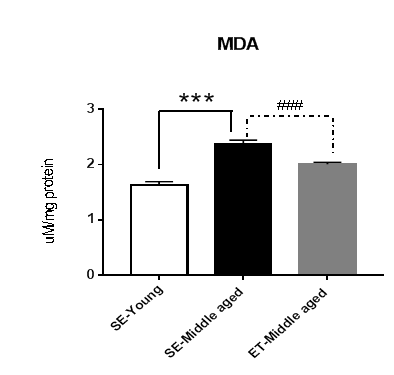
The level of MDA in heart was significantly increased with increasing aged, and the level of MDA was significantly decreased after exercise training (Figure 1)
*** = P<0.001 compared to the SE-Young
### = P<0.001 compared to the SE- Middle-aged group
Exercise training effects on cardiac angiogenic capacity
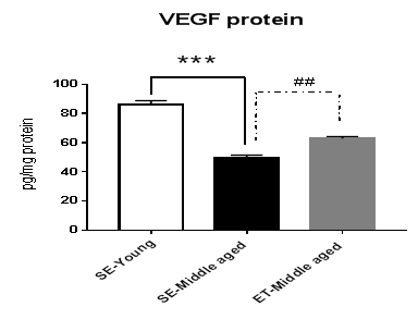
The protein level of VEGF in heart was significantly increase in the sedentary –young rats compare with sedentary-middle and sedentary-aged rats. The level of VEGF was significantly increased in the exercise trained middle group when compared with sedentary group control (Figure 2).
*** = P<0.001 compared to the SE-Young; ## = P<0.01 compared to the SE- Middle-aged group
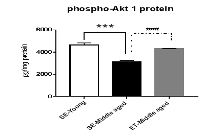
The protein levels of phospho-Akt1 in heart was significantly increase in the sedentary –young rats compared with sedentary-middle aged rat. The level of phospho-Akt1 protein was significantly increased in the exercise trained middle group compared with sedentary group control (Figure 3).
*** = P<0.001 compared to the SE-Young; ### = P<0.001 compared to the SE- Middle-aged group
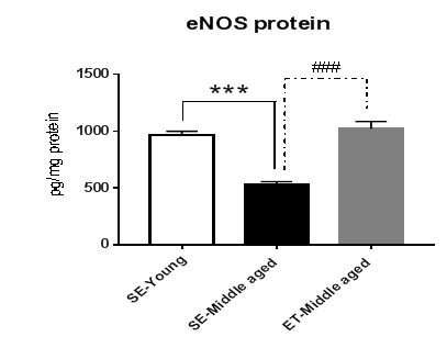
The protein levels of eNOS in heart was significantly increase in the sedentary –young rats compare with sedentary-middle-aged rats. The level of eNOS significantly increased in the exercise trained middle-aged group when compared with sedentary group control (Figure 4).
Figure 4: Expression of eNOS protein in heart of sedentary young (n=5), sedentary –middle aged (n=5), exercise trained – middle-aged (n=5). Data are expressed as mean±SE.
*** = P<0.001 compared to the SE-Young; ### = P<0.001 compared to the SE- Middle-aged group
Discussion and Conclusion
The present study demonstrated that middle-aged rats had tendency in developing hypertension as shown in Table 1. Our results showed that there was a tendency of the exercise training to prevent the developing hypertension in middle-aged group. As shown by the previous studies, they indicated that the incidence of hypertension related cardiovascular disease is higher in the aged than in the young [5-6]. The prevalence of hypertension and cardiovascular disease was confirmed as age-associated changes in cardiovascular physiology. The pathophysiology of age-associated changes in cardiovascular system appear at the cellular, extracellular, and whole-heart levels [5-7].
Moreover, our findings also demonstrated that lipid peroxidation, MDA, was significantly increased with increasing age (Figure 1). The previous study demonstrated that the elevation of ROS and impairment of antioxidant system were found in the aging heart [8]. It was also suggested that oxidative stress could be consequently caused by the decreased peripheral vascular compliance and augmented afterload leads to increased oxygen consumption, and energy deficits [9-11].
In addition, the effect of age-induce downregulation of cardiac angiogenic capacity as described by VEGF, eNOS and phospho-Akt1 in the middle-aged heart were indicated in our results (Figures 2-4). The present study demonstrated that exercise training improved the reduction of phosphorylation of Akt protein and eNOS protein in the heart, and these corresponded to the changes in VEGF protein levels. These results suggest that exercise training improves aging-induced downregulation of VEGF angiogenic signaling cascade in the middle-aged heart. Thus, the exercise training in middle aged should be recommend. The sedentary lifestyle appeared to accelerate heart aging and regular exercise especially, endurance exercise improved cardiac function both of young and senior population. The beneficial effect of exercise training in angiogenesis was observed in several studies [12-13]. Moreover, Iemitsu et al. [3] showed exercise training improved the age-induce down regulation of VEGF, eNOS and phospho-Akt1 in the heart. The result of the current study shows that lipid peroxidation, MDA, was significantly increased with increasing aged, and the level of MDA was significantly decreased after exercise training, indicating the decrease in oxidative damage. Our previous study demonstrated exercise training effects on positive correlation between MDA level and tissue capillary vascularity in aging animals [14]. This may suggest that alterations of cardiac angiogenic capacity in middle-age animals by exercise training partly relates to the reduction of oxidative stress in the heart.
In conclusion, the present study demonstrated that the protective effect of exercise training on age-induced cardiac microvascular changes are involved the up-regulation of VEGF eNOS and phospho-Akt1 expression in association with changes in the ROS balance.
Acknowledgements
This study has been supported by The 90th Anniversary of Chulalongkorn University Fund (Ratchadaphiseksomphot Endowment Fund, Grant NO. GCUGR1125622037D), and Government Budget Grant (CU-GBG-2561).
Conflicts of interest
The authors have no conflicts of interest to report.
References
- Brock DW, Guralnik JM, BrodyJ A. Demography and epide- miology of aging in the United States. In: Schneider EL, Rowe JW, ed. Handbook of the Biology of Aging. San Diego, CA: Academic Press 1990: 3-23.
- Edelberg JM, Lee SH, Kaur M, Tang L, Feirt NM, McCabe S, et al. Platelet-derived growth factor-AB limits the extent of myocardial infarction in a rat model: feasibility of restoring impaired angiogenic capacity in the aging heart. Circulation 105 (2002): 608-613.
- Iemitsu M, Maeda S, Jesmin S, Otsuki T, Miyauchi T. Exercise training improves aging-induced downregulation of VEGF angiogenic signaling cascade in hearts. Am J Physiol Heart Circ Physiol 291 (2006): H1290-H1298.
- Viboolvorakul S, Patumraj S. Exercise Training Could Improve Age-Related Changes in Cerebral Blood Flow and Capillary Vascularity through the Upregulation of VEGF and eNOS. BioMed Research International 2014: 1-12.
- Sun Z. Aging, arterial stiffness, and hypertension. Hypertension 65 (2015): 252-256.
- AlGhatrif M, Strait JB, Morrell CH, Canepa M, Wright J, Elango P, et al. Longitudinal trajectories of arterial stiffness and the role of blood pressure: the Baltimore Longitudinal Study of Aging. Hypertension 62 (2013): 934-941.
- Jiang L, Zhang J, Monticone RE, Telljohann R, Wu J, Wang M, et al. Calpain-1 regulation of matrix metalloproteinase 2 activity in vascular smooth muscle cells facilitates age-associated aortic wall calcification and fibrosis. Hypertension 60 (2012): 1192-1199.
- Gounder SS, Kannan S, Devadoss D, Miller CJ, Whitehead KJ, Odelberg SJ, et al. Impaired transcriptional activity of Nrf2 in age-related myocardial oxidative stress is reversible by moderate exercise training. PLoS One 7 (2012): e45697.
- Liao PH, Hsieh DJ, Kuo CH, Day CH, Shen CY, Lai CH, et al. Moderate exercise training attenuates aging-induced cardiac inflammation, hypertrophy and fibrosis injuries of rat hearts. Oncotarget 6 (2015): 35383-35394.
- Viboolvorakul S, Patumraj S. Exercise Training Could Improve Age-Related Changes in Cerebral Blood Flow and Capillary Vascularity through the Upregulation of VEGF and eNOS. BioMed Research International 2014: 1-12.
- Sun Z. Aging, arterial stiffness, and hypertension. Hypertension 65 (2015): 252-256.
- Hassan AF, Kamal MM. Effect of exercise training and anabolic androgenic steroids on hemodynamics, glycogen content, angiogenesis and apoptosis of cardiac muscle in adult male rats. International Journal of Health Sciences 7 (2013): 47-60.
- Bloor CM. Angiogenesis during exercise and training. Angiogenesis 8 (2005): 263-271.
- Viboolvorakul, S. Eksakulkla, N. Wongeak-in, H. Niimi and S. Patumraj. Exercise Training Could Reduce Age-Induced Microvascular Impairment Related to Its Anti-Oxidant Potential. British Journal of Medicine and Medical Research 2011: 385-396.


