Piezosurgery Removal of Mandibular Tori: A Case Series
Article Information
Pozzetti E1,2*, Garibaldi J2, Cassinotto E2, Figliomeno E2, Merlini A2
1Department of Dentistry, IRCCS San Raffaele Hospital and Dental School, Vita Salute University, Milan, Italy 2Department of Odontostomatology, Galliera Hospital, Genoa, Italy
*Corresponding Author: Enrico Pozzetti, Department of Dentistry, IRCCS San Raffaele Hospital and Dental School, Vita Salute University, Milan, Italy.
Received: 26 February 2023; Accepted: 10 March 2023; Published: 23 June 2023
Citation: Pozzetti E, Garibaldi J, Cassinotto E, Figliomeno E, Merlini A. Piezosurgery Removal of Mandibular Tori: A Case Series. Archives of Clinical and Medical Case Reports. 7 (2023): 290-294.
View / Download Pdf Share at FacebookAbstract
Introduction: Tori mandibularis are exostosis of the lingual side of the mandible, categorized as development defects. The etiology is multifactorial and the excision is not always required. Piezosurgery results as a useful device for exostosis removal because it causes less bleeding and discomfort in the postoperative healing, it is characterized by a micrometric and selective cutting for hard tissue.
Materials and methods: At Galliera Hospital (Genoa, Italy) 4 patients demanding a partial or complete denture or reporting pain, were referred for tori mandibularis removal. The surgical procedure was standardized and performed by the same surgeon, using piezoelectric technology, and accomplished with chisel and mallet for en-block removal.
Results and Conclusion: tori mandibularis removal mean measured 6.9 mm (range 5.3-7.6) in antero-posterior dimension and 18.3 (range 12.6-23.1) in bucco-lingual dimension. Piezosurgery appeared effective for exostosis removal and provides a good healing and a better comfort for the patient.
Keywords
Piezosurgery; Tori Mandibularis; Exostosis; Pre-prosthetic Surgery; Osteotomy
Piezosurgery articles; Tori Mandibularis articles; Exostosis articles; Pre-prosthetic Surgery articles; Osteotomy articles
COVID-19 articles COVID-19 Research articles COVID-19 review articles COVID-19 PubMed articles COVID-19 PubMed Central articles COVID-19 2023 articles COVID-19 2024 articles COVID-19 Scopus articles COVID-19 impact factor journals COVID-19 Scopus journals COVID-19 PubMed journals COVID-19 medical journals COVID-19 free journals COVID-19 best journals COVID-19 top journals COVID-19 free medical journals COVID-19 famous journals COVID-19 Google Scholar indexed journals Piezosurgery articles Piezosurgery Research articles Piezosurgery review articles Piezosurgery PubMed articles Piezosurgery PubMed Central articles Piezosurgery 2023 articles Piezosurgery 2024 articles Piezosurgery Scopus articles Piezosurgery impact factor journals Piezosurgery Scopus journals Piezosurgery PubMed journals Piezosurgery medical journals Piezosurgery free journals Piezosurgery best journals Piezosurgery top journals Piezosurgery free medical journals Piezosurgery famous journals Piezosurgery Google Scholar indexed journals lymphoma articles lymphoma Research articles lymphoma review articles lymphoma PubMed articles lymphoma PubMed Central articles lymphoma 2023 articles lymphoma 2024 articles lymphoma Scopus articles lymphoma impact factor journals lymphoma Scopus journals lymphoma PubMed journals lymphoma medical journals lymphoma free journals lymphoma best journals lymphoma top journals lymphoma free medical journals lymphoma famous journals lymphoma Google Scholar indexed journals Tori Mandibularis articles Tori Mandibularis Research articles Tori Mandibularis review articles Tori Mandibularis PubMed articles Tori Mandibularis PubMed Central articles Tori Mandibularis 2023 articles Tori Mandibularis 2024 articles Tori Mandibularis Scopus articles Tori Mandibularis impact factor journals Tori Mandibularis Scopus journals Tori Mandibularis PubMed journals Tori Mandibularis medical journals Tori Mandibularis free journals Tori Mandibularis best journals Tori Mandibularis top journals Tori Mandibularis free medical journals Tori Mandibularis famous journals Tori Mandibularis Google Scholar indexed journals Ultrasound articles Ultrasound Research articles Ultrasound review articles Ultrasound PubMed articles Ultrasound PubMed Central articles Ultrasound 2023 articles Ultrasound 2024 articles Ultrasound Scopus articles Ultrasound impact factor journals Ultrasound Scopus journals Ultrasound PubMed journals Ultrasound medical journals Ultrasound free journals Ultrasound best journals Ultrasound top journals Ultrasound free medical journals Ultrasound famous journals Ultrasound Google Scholar indexed journals treatment articles treatment Research articles treatment review articles treatment PubMed articles treatment PubMed Central articles treatment 2023 articles treatment 2024 articles treatment Scopus articles treatment impact factor journals treatment Scopus journals treatment PubMed journals treatment medical journals treatment free journals treatment best journals treatment top journals treatment free medical journals treatment famous journals treatment Google Scholar indexed journals CT articles CT Research articles CT review articles CT PubMed articles CT PubMed Central articles CT 2023 articles CT 2024 articles CT Scopus articles CT impact factor journals CT Scopus journals CT PubMed journals CT medical journals CT free journals CT best journals CT top journals CT free medical journals CT famous journals CT Google Scholar indexed journals Exostosis articles Exostosis Research articles Exostosis review articles Exostosis PubMed articles Exostosis PubMed Central articles Exostosis 2023 articles Exostosis 2024 articles Exostosis Scopus articles Exostosis impact factor journals Exostosis Scopus journals Exostosis PubMed journals Exostosis medical journals Exostosis free journals Exostosis best journals Exostosis top journals Exostosis free medical journals Exostosis famous journals Exostosis Google Scholar indexed journals Pre-prosthetic Surgery articles Pre-prosthetic Surgery Research articles Pre-prosthetic Surgery review articles Pre-prosthetic Surgery PubMed articles Pre-prosthetic Surgery PubMed Central articles Pre-prosthetic Surgery 2023 articles Pre-prosthetic Surgery 2024 articles Pre-prosthetic Surgery Scopus articles Pre-prosthetic Surgery impact factor journals Pre-prosthetic Surgery Scopus journals Pre-prosthetic Surgery PubMed journals Pre-prosthetic Surgery medical journals Pre-prosthetic Surgery free journals Pre-prosthetic Surgery best journals Pre-prosthetic Surgery top journals Pre-prosthetic Surgery free medical journals Pre-prosthetic Surgery famous journals Pre-prosthetic Surgery Google Scholar indexed journals Osteotomy articles Osteotomy Research articles Osteotomy review articles Osteotomy PubMed articles Osteotomy PubMed Central articles Osteotomy 2023 articles Osteotomy 2024 articles Osteotomy Scopus articles Osteotomy impact factor journals Osteotomy Scopus journals Osteotomy PubMed journals Osteotomy medical journals Osteotomy free journals Osteotomy best journals Osteotomy top journals Osteotomy free medical journals Osteotomy famous journals Osteotomy Google Scholar indexed journals
Article Details
1. Introduction
Torus mandibularis is a common exostosis that develops along the lingual aspect of the mandible above the mylohyoid line, in the region of canines and premolars. These exostoses are developmental defects that appears as localized bony protuberances arising from the cortical plate and they are composed by compact bone with small amount of trabecular bone and fibrofatty marrow [1, 2].
The prevalence of tori mandibularis is in a wide range, from 0, 54% to 64,4%, and correlates with ethnicity, in fact there is an higher prevalence in Asians and Inuit [3, 4]. The etiology of this lesion is multifactorial including both genetic with autosomal dominant inheritance and environmental influences. In a study of Eggen the genetic determination was estimated to be 30%, while the remnant was interpreted by occlusal overload as bruxism or heavy food consumption and other clinical variables as malocclusion, Angle class or curve of Spee. Another study suggests an association between these lesions and a rich-in-calcium diet, unsaturated fatty acids and vitamin D [5].
An important theory on the tori mandibularis is the functional matrix hypothesis in which compressive stresses may lead to buckling of the mandible in the mental foramen region, which has a reduced bone volume. Osteogenic periosteum in these regions is stretched and this tension leads to new bone formation and consequently of tori [6. 7].
Generally, surgical resection is not required for mandibular torus, as long as the condition remains asymptomatic. However, treatment is indicated when subjective symptoms such as discomfort, pain, articulation disorder or if this condition determinates an instability of dentures. A further indication for surgical removal is earning an adequate bone graft for bone regeneration; it could be used for en-block harvest or autogenous particles for guided bone regeneration [8-10].
Invented in 1988 by Tommaso Vercellotti, Piezosurgery is a precise and soft tissue-sparing technique for bone cutting, based on ultrasonic microvibrations and on the concept of “pressure electrification”. When electric tension is applied to certain materials like quartz and Rochelle salts, it generates an expansion and contraction of the material, producing ultrasonic vibrations. The piezoelectric device is characterized by having ultrasonic vibrations in the frequency range of 25-29 kHz, an oscillation (amplitude) of 60-210 µm, and power up to 50 W; this allows selective cutting only on hard structures without damaging soft tissue [11].
2. Materials and Methods
A sample of 4 patients (2 men, 2 women) with a mean age of 63.66 years (range: 60-68 years) at the time of recruitment referred to Galliera Hospital (Department of Odontostomatology) in Genoa (Italy) requiring a removable partial or complete denture between October 2022 and February 2023. All these patients presented two mandibular exostosis (one left sided and one right sided), known as tori mandibularis, in premolars region. This condition represents a contraindication for the removable prosthetic rehabilitation, consequently the patient’s required pre-prosthetic surgical treatment.
All the patients reported:
- Good systemic health
- Need of removable prosthetic rehabilitation
- No heavy smokers (<20 cigarettes/day)
The sample was surgically treated between December 2022 and January 2023.
2.1 Surgical procedure
The patients were informed about all the surgical method and the derivational risks of this procedure; after that, they signed an informed consent. Under local anesthesia (2% of Mepivacaine Hydrochloride with adrenaline 1:100000) it was elevated a lingual mucoperiosteal flap to exposing tori mandibularis. When the exostosis area was accessible, the surgeon (J.G.), with the auxilium of piezoelectric instrument (Mectron Piezorurgery 3, Mectron® Medical Technology, Genoa, Italy), started the cut to delimitate tori mandibularis. For the osteotomy of the tori, the authors chose an aggressive tip (Mectron OT7-a), with cortical bone modality. When the lesion was sufficiently separated from the mandibular cortical plate, the surgeon proceeded to remove the block with scalpel and mallet. Following, using the Mectron OT-9 tip the operator recontoured the lingual cortical plate in order to smooth the bone to avoid possible decubitus. Eventually the operator sutured with a resorbable suture (Vycril, Polyglactin 910, Ethicon 4/0 with a 19 mm needle), choosing sling sutures between two teeth and single sutures in edentulous area. After the surgical procedure the patient was medicated with an intramuscular injection of betametasone 4 mg and ketorolac 30 mg in order to reduce post-operative pain and swelling. Finally, the patient was educated in the post-surgical behaviours. Postoperative medication included antibiotics (Amoxicillin and Clavulanic acid 1000 mg), analgesics and anti-inflammatory medicine (paracetamol). The prosthetic phase began after the healing of the soft tissue, at least one month after the surgical procedure.
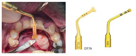
Figure 1: Osteotomy performed with Piezosurgery 3 (Mectron® Medical Technology, Genoa), in detail the tip OT7-a and OT-9.
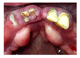
Figure 2: At the initial stage the patient presented tori mandibularis, hopless teeth and the necessity of mandibularis rehabilitation with a removable complete denture.
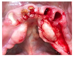
Figure 3: Extraction of teeth, elevation of lingual mucoperiosteal flap and exposition of tori.
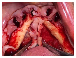
Figure 4: Excision of tori mandubularis and osteoplasty to recontour the lingual cortical plate.
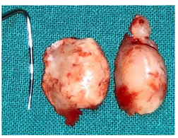
Figure 5: Measurement with periodontal probe.
3. Results
The tori mandibularis were removed en-block and they mean measured 6.9 mm (range 5.3-7.6 mm) in anteroposterior direction and 18.3 mm (range 12.6-23.1 mm) in the transverse plane. During the surgical procedure the piezoelectric technology caused less bleeding and less vibrations compared to rotating tools; although a greater amount of time was required for the surgical procedure. At the 7-day follow-up sutures were removed with the soft tissue appeared healthy and the patients reported a low grade of pain, manageable with FANS. Diagnosis of exostosis was confirmed by anatomopathological examination, which revealed compact bone tissue compatible with tori mandibularis. At the 1-month follow-up the tissue appeared stable, and it was possible to proceed to prosthetic rehabilitation taking the first impression.
4. Discussion
Tori mandibularis are a cortical bone ipergrowth on the lingual side of the mandible and they represent a development anomaly of the oral cavity. The surgical excision is not always required, but it is necessary when the patient needs a removable prosthesis or if these exostosis cause long lasting oral pain exacerbated by swallowing and mastication, presence of ulceration of the overlying mucosa or there is the necessity of autologous bone graft [12-15].
In this procedure the surgeon must be aware of the rich vascularization and innervation of this region, the presence of the submandibular and sublingual glands and their ducts. Accordingly, bone exposure of tori mandibularis require a mucoperiosteal flap without lingual release incisions. Furthermore, for osteotomy and osteoplasty the piezoelectric system, due to the selective cutting action on hard tissue, is a safer solution than rotating instruments. During the removal procedure of exostosis, the osteotomy can be performed with conventional tools such as saws and burs, that generate an important amount of heat in the cutting area. Overheating of adjacent tissue may delay the healing process causing osteonecrosis and diminished regeneration [16]. These tools are highly effective in cutting bone tissue but are not discerning for bone, and thus can produce significant damage the surrounding soft tissues, in particular nerves and vessels [17]. Bone scrapers, gouge-shaped bone chisels, trephines and ronguers can also find application [18].
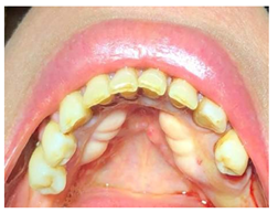
Figure 6: Patient presented tori mandibularis, posterior edentulous areas and the request of a removable partial denture.
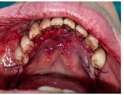
Figure 7: Tori mandibularis removed and sling sutures applied between two teeth and single sutures in posterior areas.
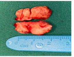
Figure 8: Completed osteotomy and measurement of tori mandibularis.
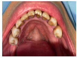
Figure 9: One month follow-up, before the beginning of prosthetic phase.
Piezosurgery technique is an alternative that overcomes the limits of traditional instrumentation in oral bone surgery. The main advantages include soft tissue protection, optimal visibility in the surgical field, decreased blood loss, less vibration and noise, increased comfort for the patient and protection of tooth structure [19].
Not only is this technique clinically effective, but histological and histomorphometric evidence of wound healing and bone formation in experimental animal models has shown that tissue response is more favorable in Piezosurgery than it is in conventional bone- cutting techniques such as diamond or carbide rotary instruments [20].
On the other hand, this methodic requires a greater amount of time in the osteotomy phase compared to classical tools. Despite Piezosurgery has become a very common technique in different fields of dentistry like maxillary sinus lift, surgery of impacted teeth, apicectomy, orthognatic surgery and many more, there’s a lack of literature about its application in exostosis removal.
5. Conclusions
Although exostosis removal is not always mandatory, it is required for prosthetic rehabilitation, pain or autogenous bone graft harvest.
With the limitations of this case series, the authors reported the utility of piezoelectric surgery in tori mandibularis removal and its clinical use.
Further investigations with a larger sample size and case-control RCT between piezoelectric and rotating instruments are needed to clarify the histological and clinical differences between these two methodics.
Funding
This research received no external funding.
Conflict of Interest
The authors have read and approved the submitted version of the paper and have contributed significantly to the work.
References
- Brad W Neville et al. Oral and Maxillofacial Pathology, Fourth edition (St. Louis, Missouri: Elsevier (2016).
- Andrés S García-García et al. Current Status of the Torus Palatinus and Torus Mandibularis, Medicina Oral, Patologia Oral Y Cirugia Bucal 15 (2010): e353-e360.
- Youna Choi A, et al. Prevalence and Anatomic Topography of Mandibular Tori: Computed Tomographic Analysis. Journal of Oral and Maxillofacial Surgery: Official Journal of the American Association of Oral and Maxillofacial Surgeons 70 (2012): 1286-1291.
- Ruprecht A, et al. The Prevalence of Radiographically Evident Mandibular Tori in the University of Iowa Dental Patients. Dento Maxillo Facial Radiology 29 (2000): 291-296.
- Adomas Auškalnis et al. Multifactorial Etiology of Torus Mandibularis: Study of Twins. Stomatologija 17 (2015): 35-40.
- Chan-Woo Jeong, et al. The Relationship between Oral Tori and Bite Force. Cranio: The Journal of Craniomandibular Practice 37 (2019): 246-253.
- Eduardo Bertazzo-Silveira, et al. Association between Signs and Symptoms of Bruxism and Presence of Tori: A Systematic Review. Clinical Oral Investigations 21 (2017): 2789-2799.
- Ganz SD. Mandibular Tori as a Source for Onlay Bone Graft Augmentation: A Surgical Procedure. Practical Periodontics and Aesthetic Dentistry: PPAD 9 (1997): 973-982.
- Inci Rana Karaca, Dilara Nur Ozturk, e Huseyin Ozan Akinci. Mandibular Torus Harvesting for Sinus Augmentation: Two-Year Follow-Up. Journal of Maxillofacial and Oral Surgery 18 (2019): 61-64.
- Khushboo Rastogi, Santosh Kumar Verma, e Rajarshi Bhushan. Surgical Removal of Mandibular Tori and Its Use as an Autogenous Graft. BMJ Case Reports 2013 (2013): bcr2012008297.
- Cassiano Costa Silva Pereira, et al. Piezosurgery Applied to Implant Dentistry: Clinical and Biological Aspects. The Journal of Oral Implantology 40 (2014): 401-408.
- Rastogi, Verma, e Bhushan. Surgical Removal of Mandibular Tori and Its Use as an Autogenous Graft.
- Karaca, Ozturk, e Akinci, Mandibular Torus Harvesting for Sinus Augmentation.
- Rastogi, Verma, e Bhushan. Surgical Removal of Mandibular Tori and Its Use as an Autogenous Graft.
- García-García, et al. Current Status of the Torus Palatinus and Torus Mandibularis.
- Horton JE, Tarpley TM, Wood eLD. The Healing of Surgical Defects in Alveolar Bone Produced with Ultrasonic Instrumentation, Chisel, and Rotary Bur. Oral Surgery, Oral Medicine, and Oral Pathology 39 (1975): 536-546.
- Sabrina Pappalardo e Renzo Guarnieri. Randomized Clinical Study Comparing Piezosurgery and Conventional Rotatory Surgery in Mandibular Cyst Enucleation. Journal of Cranio-Maxillofacial Surgery 42 (2014): e80-e85.
- Arndt Happe. Use of a Piezoelectric Surgical Device to Harvest Bone Grafts from the Mandibular Ramus: Report of 40 Cases. The International Journal of Periodontics and Restorative Dentistry 27 (2007): 241-249.
- Pavlíková G, et al. Piezosurgery in Oral and Maxillofacial Surgery. International Journal of Oral and Maxillofacial Surgery 40 (2011): 451-457.
- Tomaso Vercellotti, et al. Osseous Response Following Respective Therapy with Piezosurgery. The International Journal of Periodontics and Restorative Dentistry 25 (2005): 543-549.
