Ovarian Hemangioma Presented as an Incidental Ovarian Mass: A Rare Case Report along with Literature Review
Article Information
Daniela Nakuci1*, Erisa Kola2, Edlira Horjeti3, Ina Kola4, Blerona Shaipi5, Juna Musa6, Ali Guy7, Mehdi Alimehmeti8
1Pathologist, Department of Pathology, Vlora Regional Hospital, Albania
2Pathologist, Department of Pathology and Forensic Medicine, Albania
3Family Doctor, Department of Family Medicine, Tirane, Albania
4Department of Plastic Surgery and Burns, Mother Teresa University Hospital Center, Albania
5Medical Doctor, University of Medicine, Tirana
6Postdoctoral Research Fellow, Department of Robotic Surgery, Mayo Clinic, Minnesota
7Department of Physical Medicine and, New York University, School of Medicine-NYU, New York, USA
8Pathologyst, Professor of Anatomic-Pathology, University of Medicine, Tirana, Albania
*Corresponding Author: Dr. Erisa Kola, Pathologist, Department of Pathology and Forensic Medicine, Albania
Received: 14 July 2020; Accepted: 22 July 2020; Published: 03 September 2020
Citation: Daniela Nakuci, Erisa Kola, Edlira Horjeti, Ina Kola, Blerona Shaipi, Juna Musa, Ali Guy, Mehdi Alimehmeti. Ovarian Hemangioma Presented as an Incidental Ovarian Mass: A Rare Case Report along with Literature Review. Archives of Clinical and Medical Case Reports 4 (2020): 760-765.
View / Download Pdf Share at FacebookAbstract
Vascular malformation tumors in the female genital tract, especially those arising in the ovary are uncommon with low morbidity. Ovarian hemangioma (OH) featured as a benign rare tumor occurs among adults and children with the age ranging from infancy till octogenarian. Moreover, it presents as an obscured landscape in the backdrop of other gynecologic issues, making hard accuracy perceiving an isolated diagnosis. Usually asymptomatic cases amid patients are being investigated, remaining in dormant phase for a long time and until simultaneously cohabiting with another primary pathology. Metrorrhagia, a dysfunction uterine bleeding, might be a unique sign or association with suprapubic abdominal pain may suggest a vascular ovarian hemangioma.
Here we analyze a case of a 44-year-old female that underwent a surgical intervention of uterine leiomyoma leading to the diagnosis of infrequent latent cavernous and capillary hemangioma in the right ovary with normal tumor markers. Radiologic evaluation settled the symptomatic leiomyoma diagnosis and orientated us to presume an ambiguous investigation as a slightly right adnexa enlargement was recognized. Furthermore, a detailed microscopic examination and immunohistochemistry staining process was significant to confirm the blended type of ovarian hemangioma.
Keywords
Ovarian Hemangioma; Cavernous Hemangioma; Ovarian mass; Incidental ovarian mass
Ovarian Hemangioma articles, Cavernous Hemangioma articles, Ovarian mass articles, Incidental ovarian mass articles
Ovarian Hemangioma articles Ovarian Hemangioma Research articles Ovarian Hemangioma review articles Ovarian Hemangioma PubMed articles Ovarian Hemangioma PubMed Central articles Ovarian Hemangioma 2023 articles Ovarian Hemangioma 2024 articles Ovarian Hemangioma Scopus articles Ovarian Hemangioma impact factor journals Ovarian Hemangioma Scopus journals Ovarian Hemangioma PubMed journals Ovarian Hemangioma medical journals Ovarian Hemangioma free journals Ovarian Hemangioma best journals Ovarian Hemangioma top journals Ovarian Hemangioma free medical journals Ovarian Hemangioma famous journals Ovarian Hemangioma Google Scholar indexed journals Hemangioma articles Hemangioma Research articles Hemangioma review articles Hemangioma PubMed articles Hemangioma PubMed Central articles Hemangioma 2023 articles Hemangioma 2024 articles Hemangioma Scopus articles Hemangioma impact factor journals Hemangioma Scopus journals Hemangioma PubMed journals Hemangioma medical journals Hemangioma free journals Hemangioma best journals Hemangioma top journals Hemangioma free medical journals Hemangioma famous journals Hemangioma Google Scholar indexed journals Cavernous Hemangioma articles Cavernous Hemangioma Research articles Cavernous Hemangioma review articles Cavernous Hemangioma PubMed articles Cavernous Hemangioma PubMed Central articles Cavernous Hemangioma 2023 articles Cavernous Hemangioma 2024 articles Cavernous Hemangioma Scopus articles Cavernous Hemangioma impact factor journals Cavernous Hemangioma Scopus journals Cavernous Hemangioma PubMed journals Cavernous Hemangioma medical journals Cavernous Hemangioma free journals Cavernous Hemangioma best journals Cavernous Hemangioma top journals Cavernous Hemangioma free medical journals Cavernous Hemangioma famous journals Cavernous Hemangioma Google Scholar indexed journals Ovarian mass articles Ovarian mass Research articles Ovarian mass review articles Ovarian mass PubMed articles Ovarian mass PubMed Central articles Ovarian mass 2023 articles Ovarian mass 2024 articles Ovarian mass Scopus articles Ovarian mass impact factor journals Ovarian mass Scopus journals Ovarian mass PubMed journals Ovarian mass medical journals Ovarian mass free journals Ovarian mass best journals Ovarian mass top journals Ovarian mass free medical journals Ovarian mass famous journals Ovarian mass Google Scholar indexed journals Incidental ovarian mass articles Incidental ovarian mass Research articles Incidental ovarian mass review articles Incidental ovarian mass PubMed articles Incidental ovarian mass PubMed Central articles Incidental ovarian mass 2023 articles Incidental ovarian mass 2024 articles Incidental ovarian mass Scopus articles Incidental ovarian mass impact factor journals Incidental ovarian mass Scopus journals Incidental ovarian mass PubMed journals Incidental ovarian mass medical journals Incidental ovarian mass free journals Incidental ovarian mass best journals Incidental ovarian mass top journals Incidental ovarian mass free medical journals Incidental ovarian mass famous journals Incidental ovarian mass Google Scholar indexed journals treatment articles treatment Research articles treatment review articles treatment PubMed articles treatment PubMed Central articles treatment 2023 articles treatment 2024 articles treatment Scopus articles treatment impact factor journals treatment Scopus journals treatment PubMed journals treatment medical journals treatment free journals treatment best journals treatment top journals treatment free medical journals treatment famous journals treatment Google Scholar indexed journals CT articles CT Research articles CT review articles CT PubMed articles CT PubMed Central articles CT 2023 articles CT 2024 articles CT Scopus articles CT impact factor journals CT Scopus journals CT PubMed journals CT medical journals CT free journals CT best journals CT top journals CT free medical journals CT famous journals CT Google Scholar indexed journals surgery articles surgery Research articles surgery review articles surgery PubMed articles surgery PubMed Central articles surgery 2023 articles surgery 2024 articles surgery Scopus articles surgery impact factor journals surgery Scopus journals surgery PubMed journals surgery medical journals surgery free journals surgery best journals surgery top journals surgery free medical journals surgery famous journals surgery Google Scholar indexed journals pathology articles pathology Research articles pathology review articles pathology PubMed articles pathology PubMed Central articles pathology 2023 articles pathology 2024 articles pathology Scopus articles pathology impact factor journals pathology Scopus journals pathology PubMed journals pathology medical journals pathology free journals pathology best journals pathology top journals pathology free medical journals pathology famous journals pathology Google Scholar indexed journals Cystic Fibrosis articles Cystic Fibrosis Research articles Cystic Fibrosis review articles Cystic Fibrosis PubMed articles Cystic Fibrosis PubMed Central articles Cystic Fibrosis 2023 articles Cystic Fibrosis 2024 articles Cystic Fibrosis Scopus articles Cystic Fibrosis impact factor journals Cystic Fibrosis Scopus journals Cystic Fibrosis PubMed journals Cystic Fibrosis medical journals Cystic Fibrosis free journals Cystic Fibrosis best journals Cystic Fibrosis top journals Cystic Fibrosis free medical journals Cystic Fibrosis famous journals Cystic Fibrosis Google Scholar indexed journals
Article Details
1. Introduction
Hemangiomas are benign overgrowth vascular cells occur in the body more often located in the skin and inner structure of the organs such as liver, muscle, bone. Ovarian hemangioma, a vascular malformation originating in the female genital tract is a very rare condition often misled to a cancerous condition or diagnosed during autopsy. Recently, it has been reported very few cases in articles all over the world featuring all ages from childhood till elderly. The overall number of the well-documented cases of (OH) does not exceed 60 [1]. Frequently this vascular proliferation histologically is classified as a cavernous form compared to the capillary one or blended type. Microscopically cavernous hemangioma is formed by proliferated vessels that are fragile due to uncial endothelial sheathed layer causing drip or leakage of the blood into the cavity leading sometimes to peritonitis or massive ascites. This type of hemangioma may happen to be spread all over the abdominal cavity or involving the adnexa either [2, 3]. Other complications will be ovarian torsion or elevated (CA)-125 and CA 19-9 tumor markers resembling a malignant tumor [1]. Whereas, capillary Hemangioma are characterized as tiny vessels close to each other occupying the ovary, usually asymptomatic, not causing hemorrhage in the closer structures. Moreover, it is seen that one or two OH are non functional neoplasms [4]. They are thought to develop due to cyclic changes that ovaries undergo during the reproductive years. Hypothetically Stromal luteinization may play a key role in vascular proliferation involving here the hyperestrogenism or overproduction of androgens inciting the endothelial lesion. However, its pathogenesis is not known clear yet [2, 3, 5-7].
2. Case Presentation
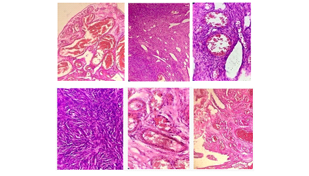
We report a case of a 44 years old woman admitted to our hospital with abdominal pain and dysfunctional uterine bleeding. After the clinical and radiological assessments, a diagnosis of uterine leiomyoma was made. The next step was the surgical procedure. She underwent a total hysterectomy along with the removal of the adnexa. Primary ovarian hemangioma was an incidental finding during the histopathological examination of the specimen. Macroscopically the right ovary presented fairly enlarged with a clean glistening outer ground displaying a purple or purplish coloration. On lessen surfaces spongy textured and honeycomb appearance due to multiloculated cystic regions crammed with frank blood or serous fluid and multiloculated cystic spaces filled with blood.

Figure 1: Microscopic examination of the ovary revealed blended type cavernous and capillary hemangioma with the cavernous one predominating. High power view of the H&E stained slides revealed multiple various sized thin-walled blood vessels, filled with red blood cells. These vessels were lined by a single, flattened endothelial cells without atypical features. The vessels may show a roughly lobular association in a variable quantity of connective tissue stroma in which inflammation, hemorrhage and hemosiderin deposits can be seen. Histologically, they show each a cavernous capillary or blended type with the cavernous one predominating.
3. Discussion
Hemangiomas are benign vascular tumors arising from failure in vascular formation, particularly in the canalizing process, resulting in abnormal vascular channels. These are three types based on the evaluation of the size of the blood vessels formed: cavernous, capillary and a mix type. Despite the rich vascularity of the ovary, the incidence of ovarian hemangioma is rare. They are usually asymptomatic and present as incidental finding especially small lesion as in our case during operation or autopsy although they may occasionally be large and symptomatic [8]. Macroscopically, ovarian hemangiomas are commonly small and the dimension of the lesion has been stated from 5 mm to 24 cm inside the first-class diameter. The etiology remains unknown, these lesions have been considered either hamartomatous or true neoplasms in which pregnancy, other hormonal effects, or infection have been implicated as risk factors in defective vascular formation [9]. Differential diagnosis of OH include tubo-ovarian mass, chocolate cyst, but the main pathological differential diagnosis are those of vascular proliferations, lymphangioma and mono-dermal teratoma composed of a prominent vascular component [10]. According to the first hypothesis hyperestrogenism became taken into consideration as a first event derived from stromal hyperthecosis or stromal hyperplasia, that could lead to spur the evolution of an ovarian hemangioma. Estrogen has proven growth stimulatory impacts on endothelial cells and most hemangiomas do explicit estrogen receptors on endothelial cells [5, 8]. Alternatively, the second hypothesis is a mechanical concept proposing that ovarian hemangioma changed into the primary occasion that led to stromal luteinization due to growth of the mass.
These luteinized stromal cells produce steroid hormones, mainly androgens, which are subsequently converted to estrogens in adipose tissue, that cause unopposed estrogen stimulation to the endometrium [5, 11]. In bilateral stromal luteinization, the changes are outstanding in the ovary containing the tumor [8, 11-13]. Acquired hemangioma by extended utilization of tamoxifen, has been pronounced in patients with preceding presence of little hemangioma that could not have been detected via ultrasound exam [12].
Cowden syndrome is a genetic syndrome usually caused by mutations in a gene known as PTEN. It has been seen an incidence of 10-40% of hemangiomas in people with Cowden syndrome [14]. Differential diagnosis of OH includes tubo-ovarian mass, chocolate cyst, but the main pathological differential diagnosis is those of vascular proliferation as angiosarcoma, lymphangioma and mono-dermal teratoma composed of a prominent vascular component [10].
Misdiagnosis with teratoma is made due to the outstanding vascular component. In cases like these, cautious sampling is important to pass over the presence of different teratomatous factors earlier than diagnosing the tumor as a pure hemangioma [6, 15].
The presence of RBC in hemangioma, excludes the possibility of lymphangioma [16]. The unilateral position, cystic, soft, friable and spongy appearance with hemorrhage and necrosis leads toward the diagnosis of angiosarcoma. Histologically stumble on marked cytologic atypia, increased mitotic activity, pleomorphism, papillary endothelial tufting, necrosis, hemorrhage [17]. Hemangioma in the ovary needs to be differentiated from proliferations of dilated blood vessels of the ovarian hilar region. Presence of circumscribed nodules or a mass strongly supports the potentiality of hemangiomas [18].
MR imaging and color Doppler ultrasound are valuable for detecting preoperative ovarian hemangioma [19, 20] showing a richly vascularized tumor with distinguished blood. Infrequently, OH is represented as nonfunctional vascular neoplasms of the ovary. Sometimes we might see them associated with ascites and highly increased serum of CA-125, clinically imitating an ovarian carcinoma. Some of ovarian hemangioma cases are interconnected with thrombocytopenia, ovarian stromal luteinization, postmenopausal bleeding, endometrial hyperplasia, or even endometrial carcinoma [5-7, 11, 12, 21]. The diminished platelet count number is considered as one of the manifestations of Kasabach and Merritt Syndrome, especially in bilateral instances related with diffuse abdominopelvic hemangiomatosis [17]. Ovarian hemangioma coexistence with non-ovarian neoplasm such as cervical carcinoma, endometrial carcinoma, rectosigmoid carcinoma, and tubal carcinoma has additionally been studied [5,10].
4. Conclusions
Hemangiomas of the ovary are very rare neoplasms with a wide age range and incidental discovery during operation or autopsy. The aim of this article is to emphasize that even though these neoplasms are very rare in the ovary, they should be considered in the differential diagnosis of a hemorrhagic ovarian lesion.
References
- Ziari K, Alizadeh K. Ovarian Hemangioma: a Rare Case Report and Review of the Literature. Iran J Pathol 11 (2016): 61-65.
- Cavernous Hemangioma Presenting as a Large Growing Mass in a Postmenopausal Woman: A Case Report and Review of the Literature. J Menopausal Med. 21 (2015): 155-159.
- Shirazi B, Anbardar MH, AzarpiraN, Robati M.A case of ovarian hemangioma with proeminent stromal luteinization.Med JDY pathol 2015 8: 227-230.
- Bolat F, Erkanli S, Kocer NE. OvarianHemangioma: Report of two Cases and Review of the Literaure.Tur J Pathol 26 (2010): 264-266.
- Miliaras D, Papaemmanouil S, Blatzas G. Ovarian capillary hemangioma and stromal luteinization: a case study with hormonal receptor evaluation. Eur J Gynaecol Oncol 22 (2001): 369-371.
- Itoh H, Wada T, Michikata K, et al. Ovarian teratoma showing a predominant hemangiomatous element with stromal luteinization: report of a case and review of the literature. Pathol Int 54 (2004): 279-283.
- Grant JW, Millward-Sadler GH. Haemangioma of the ovary with associated endometrial hyperplasia. Case report. Br J ObstetGynaecol 93 (1986): 1166-1168.
- Kim MY, Rha SE, Oh SN, et al. Case report: Ovarian cavernous haemangioma presenting as a heavily calcified adnexal mass. Br J Radiol 81 (2008): e269-e271.
- DiOrio J Jr, Lowe LC. Hemangioma of the ovary in pregnancy: a case report. J Reprod Med 24 (1980): 232-234.
- Gupta R, Singh S, Nigam S, et al. Benign vascular tumors of female genital tract. Int J Gynecol Cancer 16 (2006): 1195-1200.
- Carder PJ, Gouldesbrough DR. Ovarian haemangiomas and stromal luteinization. Histopathology. 1995 26 (6): 585-586.
- Gücer F, Ozyilmaz F, Balkanli-Kaplan P, et al. Ovarian hemangioma presenting with hyperandrogenism and endometrial cancer: A case report. Gynecol Oncol 94 (2004): 821.
- Murthy S Anand, Shakila Shetty, Vijaya V Mysorekar, et al. Ovarian hemangioma with stromal luteinization and HCG-producing mononucleate and multinucleate cells of uncertain histogenesis: a rare co-existence with therapeutic dilemma. Indian J Pathol Microbiol (2012).
- Univeristy of IOWA Hospitals and Clinics. https: //uihc.org/health-topics/cowden-syndrome-guide-patients-and-their-families.
- Prus D, Rosenberg AE, Blumenfeld A, et al. Infantile hemangioendothelioma of the ovary: a monodermal teratoma or a neoplasm of ovarian somatic cells? Am J Surg Pathol 21 (1997): 1231-1235.
- RBC - Bavikar RR, Tampi C. Lymphangioma of Ovary. Bombay Hosp J 53 (2011): 89-91.
- Uppal S, Heller DS, Majmudar B. Ovarian hemangioma-report of three cases and review of the literature. Arch Gynecol Obstet 270 (2004): 1-5.
- Talerman A. Nonspecific tumors of the ovary, including mesenchymal tumors and malignant lymphoma. In: Kurman RJ, editor. Blaustein's Pathology of the Female Genital Tract. 5th ed. New York: Springer Verlag (2002).
- Kaneta Y, Nishino R, Asaoka K, et al. Ovarian hemangioma presenting as pseudo-Meigs’ syndrome with elevated CA125. J Obstet Gynaecol Res 29 (2003): 132-135.
- Cormio G, Loverro G, Iacobellis M, et al. Hemangioma of the ovary. A case report. J Reprod Med 43 (1998): 459-461.
- Rivasi F, Philippe E, Walter P, et al. Ovarian angioma. Report of 3 asymptomatic cases. Ann Pathol 16 (1996): 439-441.
