Neglected Baby with Various Insect and Leech Bites at Cemetery: A Case Report
Article Information
Johnny Rompis*, Rocky Willar, Shekina Rondonuwu, Felicia Halim
Division of Perinatology, Department of Pediatrics, Faculty of Medicine, Sam Ratulangi University, and Prof. R.D. Kandou General Hospital, North Sulawesi, Indonesia
*Corresponding Author: Johnny Rompis, Staff in division of Perinatology, Department of Pediatrics, Faculty of Medicine, Sam Ratulangi University, and Prof. R.D. Kandou General Hospital, Jl. Raya Tanawangko No.56, Manado, North Sulawesi, Indonesia
Received: 21 February 2022; Accepted: 04 March 2022; Published: 19 March 2022
Citation:
Johnny Rompis, Rocky Willar, Shekina Rondonuwu, Felicia Halim. Neglected Baby with Various Insect and Leech Bites at Cemetery: A Case Report. Journal of Pediatrics, Perinatology and Child Health 6 (2022): 137-144.
View / Download Pdf Share at FacebookKeywords
Insect Bites, Leeches, Neglected Baby
Article Details
1. Introduction
Baby neglect is still quite high in Indonesia. One form of baby neglect is to leave the newborn in unworthy places. This is a case of newborns being abandoned by being placed in cemetery that resulted in babies suffering injuries from insect and leech bites. Erythematous and edematous eruptions along with other dermatological findings such as papules and urticaria represent the most common clinical manifestations of arthropod bites and stings. In some cases, the delivery of toxic venom can result in significant systemic reactions including autonomic instability, neurotoxicity and organ failure. The acute development of anaphylactic reactions can be rapidly fatal, most commonly due to angioedema or circulatory collapse [1]. Leech bites of the skin are common event in tropical region. However, serious consequence of leech bite injury to the internal viscera is uncommon. But when it occurs, it can cause significant morbidity [2].
3. Case Report
An unidentified baby boy, unknown date and time of birth, was found at sea cemetery on July 13, 2020, at 08.30 AM. Around the baby's body was found the presence of ants and leeches. The baby was then taken for examination at Pineleng Health Center and then referred to the central general hospital Prof. Dr. R. D Kandou. On physical examination obtained weight 2200 grams, body length 48 centimeters. New Ballard test scores were performed on the patient and obtained conformity with gestation age 40-41 weeks, and when we plotted using Lubchenco curves, we obtained weight that does not correspond to the gestational age so that the baby is a term baby with a small gestation age.
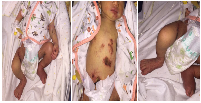
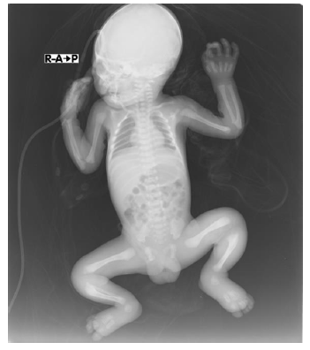
At the general examination the baby appeared to cry actively. There was a fever, 38oC on the body temperature examination when discovered, but no manifestation of fever was obtained during the next examination. At the examination of vital signs, oxygen saturation decreases to 90%. Oxygen delivered through nasal canule and saturation increases to 98%. From physical examination, erythe-matous macules, erosion, excoriation, blisters and some areas are accompanied by pus on the head, chest, stomach, legs and fingers (Figure 1) was found. The left leg appeared erythematous, swollen, appears unable to move actively, even difficult to straighten. At the examination of the genitalia there were wounds on the scrotum area. From the results of laboratory tests obtained an increase of C-Reactive Protein that is 48 mg/dL, while the results of other laboratory tests obtained within normal limits. Infants were given ampicillin antibiotics, intravenously every 12 hours, and gentamicin antibiotic, intravenously every 36 hours. In areas with wounds are given a fusidic acid ointment that is given twice daily. The baby is treated in the neonatal intensive care unit, with nutrimix according to the needs and begins to be given trophic feeding. X-ray examination showed normal result on the heart and lungs, as well as the extremity bones (Figure 2).
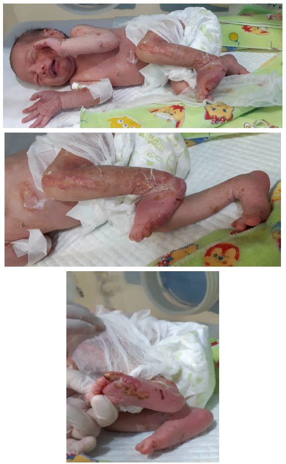
At the next day's examination, the wound was wet with pus and the left leg still swollen. From laboratory examination there was a slight increase in coagulation factors, Prothrombin time 18.1 seconds (control 15 seconds), Acquired Prothrombin Time 53.5 seconds (control 36.4 seconds) and Index Normal Ratio 1.36 seconds (control 1.11 seconds), which suspected a secondary wound infections. In accordance with the clinical picture obtained from patients additonal diagnosis of impetigo bullosa and cellulitis was added. Babies are given nutrimix infusion fluid according to the needs of the baby, antibiotics followed by topical fusidic acid. Oral milk administration is raised and the baby is consulted to the dermatovenereology division. From the dermato-venereology division given advice to continue intravenous antibiotics, topical fusidic acid and compress with NaCl 0.9% in the wound area both dry and pus. On the third day, the wounds were dried out with scaling crust, reduced leg swelling and stabilize oxygen saturation (Ficture 3). Oral and topical antibiotic administration is continued with a compress, no longer used parenteral nutrition and the baby can already drink according to the needs. During fourth day of treatment, there were improve-ment in the wound, the wound have dried up although there were still some areas with pus. Antibiotic therapy and other supportive therapies continued. Therapy is continued until the eighth day of treatment. On the eighth day of treatment, the general state of the baby appears improving, good intake, the wounds dried up and the swelling on the legs is reduced. The baby was discharged to continue treatment on the next day. The baby was adopted and continued treatment at home.
3. Discussion
This is the first case of a newborn baby with suspected multiple insect and leech bites being abused at our center. At the time the baby was found, the baby's body appeared to be full of wounds and surrounded with ants and leeches. The term "bug bite" is commonly used to denote both bites and stings inflicted by members of the phylum Arthro-poda. Arthropods make up the largest division of the animal kingdom, representing approximately 80% of all known animals. Defining characteristics include the presence of an exoskeleton, jointed appendages, and a body composed of specialized regional seg-ments. The four medically significant classes of arthropods are Chilopoda, Diplopoda, Insecta, and Arachnida [1]. Insect bites and stings maybe a minor problem or may lead to serious medical problems trough insect venom or borne illness transmission and or severe allergic reactions. Bees, wasps, and ants do not bite, but produce dermatological reactions by their stings [1, 3, 4].
Even though some arthropods are capable of injecting venom when biting, most envenomation occurs via a stinger connected to a venom gland. Notable arthropods possessing stingers include bees, wasps, hornets, fire ants, and scorpions. Both bites and stings create tissue injury which can serve as a portal of entry for secondary bacterial infection [1, 5]. Most insect bites may cause local inflammatory reactions within a few hours without complications. However, more severe local symptoms, papular urticaria, systemic allergic reactions, and transmission of a disease-causing pathogen are also possible. Uncom-monly, local reactions evolve to become vesicular, bullous or necrotic [1, 5]. Allergic responses to arthropod salivary antigens are common and contri-bute to the development of localized and systemic rashes and cutaneous pruritus. The most significant allergic response to arthropod bites and stings is the development of anaphylaxis, which can be rapidly fatal. While some arthropods can deliver toxic venom, the most significant pathophysiologic impact of arthropod bites is their potential to transmit several clinically important diseases [1, 5].
The reaction reflects an allergic response to proteins in the insect’s saliva, leading in about three-quarters of all persons to an immediate allergic reaction (wheal) and in about one-half to a delayed reaction (papule). The bites of mosquitoes and other blood-sucking insects only rarely cause serious disease [6]. IgE-mediated systemic allergic reactions are of far greater clinical significance; induced by the stings of insects belonging to the order Hymenoptera, they are associated with an immediate (anaphylactic) response that can have fatal consequences. They are most commonly caused by honeybees (Apis mellifera, here after designated simply bees) and certain species of wasp in the family Vespidae (particularly Vespula vulgaris and V. germanica, hereafter designated wasps). Anaphylaxis is occasionally caused by other species of Vespidae, such as Dolichovespula spp., hornets (Vespa crabro), and bees (mainly bumblebees [Bombus spp.]). Such reactions are rarely induced by the stings of ants (which also belong to the order Hymenoptera) or other insects, such as mosquitoes [6]. The pathogenesis of insect-associated infections, either bacterial or fungal, is unclear, as these infections are considered rare [7, 8]. In those patients who sustain insect bites, but present to the emergency department twenty four hours after the bite the differential diagnosis should be expanded. These patients could be experiencing an extended, localized allergic reaction or could be developing an infection secondary to the sting/bite or both. Some studies suggest that bacteria can be carried by insects such as Salmonella, Shigella, and E-coli [9]. Excoriation or erosion of the skin following scratching could lead to superinfections, mostly bacterial. As insects are occasionally attracted by organic waste, contami-nation of insects by a variety of fungi is possible, and this could explain the frequent coinfection with bacteria [7].
In this cases there are wounds accompanied by bullae and pus on some parts of the body, there are erythe-matous macules, erosion, excoriation, blisters and some areas are accompanied by pus on the head, chest, stomach, legs, fingers and he left leg appears erythematous, swollen, appears unable to move acti-vely, even difficult to straighten. We diagnosed with impetigo bullosa and cellulitis due to bites from insects and leeches. The infection results from inoculation of bacteria through of many means including a breakdown in the skin barrier from abrasion, laceration, puncture wound, crush injury, or burn can caused cellulitis [9]. Bacterial skin and soft tissue infections (SSTIs) often follow insect bites and can present a broad spectrum of clinical manifest-ations ranging from impetigo and ecthyma to erysipelas, abscess formation, necrotizing cellulitis, and myonecrosis [9, 10]. In addition, ongoing infection from abscesses, ulcers, and folliculitis may spread beyond a self limited capsule to surrounding skin acutely and rapidly [9]. In this case, the swelling on the baby's left leg characterized by signs of inflammation and infection which diagnosed with cellulitis strongly suspected due to the bacterial invasion of wounds that can result from insect or leech bites. The microorganisms responsible for cell-ulitis in our cases were not identified. Cellulitis, as a result of an insect bite, has been described and may initially be confused with an early allergic reaction to the insect sting or bite. Furthermore and most import-antly, it is unknown whether the cellulitis developed as a result of inoculation of pre existing bacteria or inoculated into the wound from insect or animal that served either as a reservoir or vector for pathogenic bacteria [9]. Most cellulitis seen in the emergency department is attributed to an infection from Streptococcal group A or Staphylococcus species, although other bacteria including Pasteurella, Vibrio species, Eikenella corrodens are known to cause cellulitis [9, 11].
Impetigo is a superficial vesiculopustular skin infection occurring mostly on exposed areas of the face and extremities. Affected patients usually have multiple lesions but little or no systemic toxicity. Impetigo classified as either primary or secondary. Primary impetigo involves previously normal skin affected by direct bacterial invasion. Secondary impetigo involves infection forming at a previous skin wound site. Any disturbance of the skin barrier leads to access to fibronectin receptors by GABHS and S aureus which require fibronectin for colonization. Trauma, cuts, insect bites, surgery, atopic dermatitis, burns, and varicella are common mechanisms of skin breakdown [12, 13]. This type of impetigo in patients is bullous impetigo which is adapted to the picture of lesions in infants. Bullous impetigo is often caused by Staphylococcus aureus which then when the bullae ruptures will become a brown crust according to the picture in the baby. Cellulitis generally appears following trauma, with appearance of local tenderness, pain and erythema. The area involved is red, hot and swollen. Cellulitis is usually caused by S. Auerus. Staphylococcal cell-ulitis is usually associated with purulent lesions with erythema, in accordance with the clinical picture of the baby. Cultures from wounds or blood can further help delineate the causative organism and thus can help in the administration of antibiotics in therapy. In this patient, gram examination or culture both from the wound and blood has not been done, but with the use of antibiotics adapted to the picture of lesions, the baby can quickly experience improvement [13, 14].
Leeches are invertebrates of phyllum Annelida and class Hirudinea. There are freshwater, terrestrial, and marine leeches. Some, but not all, leeches are hematophagus. Hemophagic leeches attach to their host and remain there until they become full, at which point they fall off to digest the blood [2]. Leech’s saliva contains hirudin (a potent antit-hrombin), histamine like inhibits platelet adhesion at the injured site [2]. An adult leech can ingest 1 milliliter per minute of blood, and the area of attachment can bleed for 10 hours to as long as 7 days in some instances [15, 16]. Leeches normally carry parasites in their digestive tract, which cannot survive in human and do not pose a threat. However, bacteria, viruses, and parasites from previous blood sources can survive within a leech for months, and may be transmitted to human [2, 16]. In patients there is impetigo bullosa with cellulitis which we suspect can be caused by insect bite or bite from leeches. Most leech bites will not significantly alter coag-ulation pathways, although case reports do exist where prothrombin (PT) and partial thromboplastin time (PTT) times were affected [15]. There is a lengthening of both the value of PT, APTT and INR of the patient’s, but because it is not negiable and there is no active bleeding then we do not provide therapy as specific. Antimicrobial therapy is an essential element in management of skin, soft tissue and muscle infection. Establishing the bacterial etiology and the bacterial susceptibility initially by Gram stain, and later by culture can allow for selection of proper antimicrobial therapy. Often, however, the initial therapy is empiric, based on epidemiological, historical and clinical features [13, 17, 18].
4. Conclusion
Insect or leech bites can cause bacterial infections caused by the presence of wounds that can be the focus of infection or bacteria that may be carried by insects or leeches. In this case there is indeed a deficiency with the unable to do a specific exam-ination to identify especially the bacteria that cause infection in patients. Despite the shortcomings, the baby’s conditions improved significantly from the first encounter with full of wounds. We also cannot clearly define insects or animals that may cause trauma to the baby because the baby was found abandoned, but we tried with appropriate treatment procedures.
References
- Jim Powers J, McDowell RH. Insect Bites. Stat Pearls (2020).
- Saha M, Nagi S. Intraperitoneal leech: A rare complication of leech bite. Journal of Indian Association of Pediatric Surgeons 16 (2011): 4.
- Hernandez RG, Cohen BA. Insect bite-induced hypersensitivity and the SCRATCH principles: a new approach to papular urticaria. Pediatrics 118 (2006): e189.
- Singh S, Mann BK. Insect bite reactions. Indian J Dermatol Venereol Leprol 79 (2013): 151-164.
- Moraru GM, Goddard JI. The Goddard Guide to Arthropods of Medical Importance, 7th edition, CRC Press, Boca Raton, FL (2019).
- Przybilla B, Rueff F. Insect Stings. Dtsch Arztebl Int 109 (2012): 238-248.
- Kontoyiannis PD, Koons GL, Hicklen RS, et al. Insect Bite-Associated Invasive Fungal Infections. OFID (2019).
- Truskinovsky AM, Dick JD, Hutchins GM. Fatal infection after a bee sting. Clin Infect Dis 32 (2001): E36-E8.
- Derlet RW, Richards JR. Cellulitis From Insect Bites:A case series. Callif J Emerg Med IV (2003).
- Diaz JH. Skin and Soft Skin and Soft Tissue Infections Following Marine Injuries and Exposures in Travelers. J. Travel Med 21 (2014): 207-221.
- Swartz MN. Cellulitis and subcutaneous tissue infctions. In: Mandell, Douglas, and Bennett’s Principles and Practice of Infectious Diseases 5th edition. Mandell, Bennett, Dolin Eds.;Philadelphia; Churchill Livingstone Publishers (2000): 1037-1057.
- Nardi NM, Schaefer TJ. Impetigo. StatPearls (2022): 1-11.
- Brook I. Microbiology and management of soft tissue and muscle infections. Int J Surg 6 (2008): 328-338.
- Sukumaran V. Bacterial skin and soft tissue infections.Aust Prescr 39 (2016): 159-163.
- Conley K, Jamal Z, Andrew L, et al. Leech Bite. NCBI Bookshelf (2021).
- Maetz B, Abbou R, Andreoletti JB, et al. Infections following the application of leeches: two case reports and review of the literature. Journal of Medical Case Reports 6 (2012): 364.
- McKnight AJ, Koshy JC, Xue AS, et al. Pediatric compartment syndrome following an insect bite: a case report. HAND 6 (2011): 337-339
- Clinical Immunology: Principles and Practice, 5th edition, Rich RR, Fleisher TA, Shearer WT, et al (Ed s), Elsevier (2019).


