Multimodality Imaging of Pericardial Hematoma
Article Information
Mustafa Ajam1*, Manmohan Singh2, Abdelrahman Ali3, Zachary Elder4, Rasikh Ajmal2, Ahmed Rashed5, Mohamed Shokr6
1Department of Pulmonary and Critical Care Medicine, Oregon Health and Science University, Portland, OR, USA
2Department of Cardiology, Detroit Medical Center/ Wayne state university, Detroit, MI, USA
3Department of Medicine, Mercy Hospital and Medical Center, Chicago, IL, USA
4American University of the Caribbean School of Medicine, Medical Student, USA
5Department of Cardiology, Methodist Le Bonheur Healthcare, Memphis, TN, USA
6Leon H. Charney Division of Cardiology, Cardiac Electrophysiology, NYU Langone Health, New York University School of Medicine, New York, NY, USA
*Corresponding Author: Dr. Mustafa Ajam MD, Department of Pulmonary and Critical Care Medicine Oregon Health & Science University, Portland OR 97239, USA
Received: 13 January 2020; Accepted: 25 January 2020; Published: 23 February 2020
Citation: Mustafa Ajam, Manmohan Singh, Abdelrahman Ali, Zachary Elder, Rasikh Ajmal, Ahmed Rashed, Mohamed Shokr. Multimodality Imaging of Pericardial Hematoma. Cardiology and Cardiovascular Medicine 5 (2021): 172-181.
View / Download Pdf Share at FacebookAbstract
A 66-year-old male, with a history of atrial fibrillation on chronic anticoagulation therapy, and a known pericardial hematoma, presented to the hospital with worsening dyspnea initially attributed to right sided pleural effusion. Further imaging with Computed Tomography (CT) scan of the thorax and Transthoracic Echocardiography (TTE) revealed enlarging mass-like pericardial lesion with scattered calcifications compressing the right ventricle. Contrast echocardiography demonstrated prominent contrast enhancement and vascularity raising the concern for a malignant nature of the pericardial lesion. Positron Emission Tomography (PET) scan of the myocardium did not reveal any metabolic activity of the lesion. Eventually a biopsy of the lesion was obtained, and it confirmed the diagnosis of a pericardial hematoma. This report highlights the utility of the current cardiac imaging modalities in the evaluation and diagnostic work up of pericardial hematomas.
Keywords
Pericardial diseases; Spontaneous pericardial hematomas; Multi-modality cardiac imaging; Case report
Article Details
1. Introduction
The normal pericardium is normally less than two-millimeter thickness and contains 25-50 ml of transudative fluid. TTE is a commonly used imaging modality to detect pericardial diseases and their hemodynamic effects. Cross sectional imaging such as cardiac computed tomography (cCT) and cardiac magnetic resonance imaging (cMRI) are often needed for a more detailed assessment [1]. A Pericardial hematoma is a rare entity, that can develop following blunt or penetrating chest trauma, cardiac surgery, epicardial pacing or myocardial infarction [2]. Moreover, sponta- neous pericardial hematoma formation, is very rarely reported in literature, notably seen in some cases of severe coagulopathy [3].
Coventional echocardiography may not provide detailed assessment of pericardial hematomas; however, the advents in cCT and cMRI have helped tremendously in diagnosing these subtle conditions in the recent years [4]. In this report, we demonstrate the utility of various cardiac imaging modalities in the diagnostic work up of pericardial hematomas.
2. Case Presentation
2.1 Patient’s information and clinical findings
A 66-year-old male with a history of end stage renal disease status post renal transplantation and atrial fibrillation presented with worsening dyspnea and abdominal distention over 2 months. He is known to have chronic pericardial hematoma, 3 x 3.5 cm in size, diagnosed at an outside hospital one year prior to presentation.
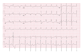
On examination, he presented with a blood pressure of 175/119 mmHg, heart rate of 84 beats per minute (bpm), dullness to percussion over the lung bases, and hepatomegaly. ECG (Figure 1) showed atrial fibrillation, right bundle branch block (RBBB) and premature ventricular contractions (PVC).
2.2. Diagnostic assessment
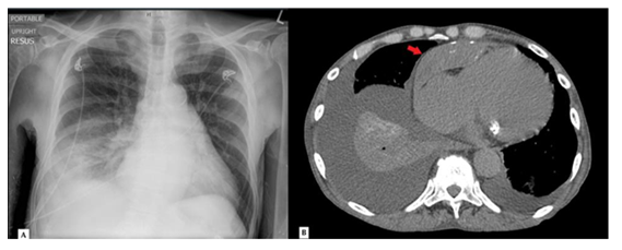
Blood work revealed kidney function at his baseline with a creatinine level of 1.83 mg/dl (normal 0.7-1.3 mg/dl), stable hemoglobin level of 11.7 gm/dl (normal 13.3-17.1 gm/dl), normal white blood cell count of 4,600 cells/µL (normal 3,500-10,600 cells/µL) and troponin level of 0.07 ng/dl (normal < 0.04 ng/dl). Chest X Ray (Figure 2A) did not show pericardial calcifica- tions; however, it revealed right lower lobe opacities and pleural effusion. CT scan of the thorax (Figure 2B) revealed cardiomegaly and mass like pericardial effusion with scattered calcifica- tions compressing the right ventricle (RV).
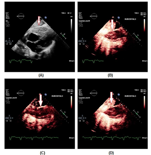
TTE showed normal left ventricle size and systolic function. It also revealed large extra-cardiac well circumscribed mass measuring 9x3 cm (compared to 3x3 cm one year prior to presentation), that appeared echogenic with heterogenous echotexture, in the vicinity of the tricuspid annulus (figure 3A). On real time, low power myocardial contrast echocardiography, the mass demonstrated prominent contrast enhancement and appeared well vascularized (Figures 3B, 3C, 3D).
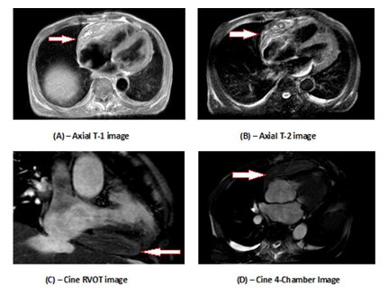
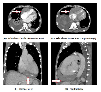
Previous images including CMRI and CCT were obtained for further assessment and comparison (Figures 4 and 5).
2.3 Intervention and follow up
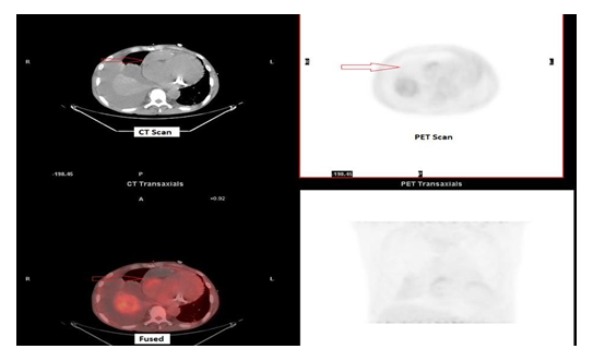
In light of the increasing size of the mass; a decision to further investigate the nature of the mass was deemed necessary to rule out possible tumor-related etiologies. PET scan of the myocardium was obtained, and it did not demonstrate any metabolic activity of the mass with absent Fluoro- deoxyglucose (FDG) uptake (Figures 6,7 and 8). In the meanwhile, coronary angiogram, done for NSTEMI, did not reveal any contribution of the coronary arteries to the blood supply of the mass.
The patient’s kidney function worsened over the hospitalization course and the patient was trans- ferred to another facility for further evaluation and management by their transplant nephrology team. At the outside hospital, a biopsy of the pericardial lesion was obtained and was suggestive of a hematoma.
3. Discussion
Clinical manifestations of a pericardial hematoma can range from asymptomatic accidental discovery to cardiac tamponade [5]. Modern multimodality imaging techniques constitute an essential aspect of the diagnostic and therapeutic approach to various pericardial pathologies including hematomas. In this case we demonstrated a stepwise approach in the evaluation of a pericardial hematoma, which is necessary to avoid false positive results and resource-exhaustion. Echocardiography continues to be the initial test to be performed in most of the cases given its wide availability. Other techniques such as cCT and cMR are considered second line choices that are used for better anatomic delineation and tissue characterization [4]. In this report we highlight the advantages of the currently available cardiac imaging modalities in the diagnostic work up of pericardial hematomas.
3.1 2D – Echocardiography
Echocardiography is highly sensitive and specific for detecting pericardial effusions. For loca- lized effusions, it can provide information regarding the tissue density, which may point the dia- gnostic work up towards exudative nature or a clot rather than simple transudative nature [4].
The presence of low acoustic reflectance echoes in an area of distended pericardial space may suggest a pericardial hematoma or clot formation [5]. Although it may not visualize the lesion itself due to size or location related difficulties, it can still identify the hemodynamic changes that may be exerted by these hematomas on cardiac chambers [6].
3.2 Contrast Echocardiography
Contrast echocardiography can provide more information regarding the vascularity of the peri- cardial lesion. Highly vascular lesions or masses can point toward a possible malignant lesion rather than a simple benign lesion [7].
3.3 Cardiac computed tomography (cCT)
Features of pericardial hematomas on CT are not described precisely in the literature. The blood is of high attenuation initially, which decreases over time, and as the hematoma progresses, it organizes and may become fibrotic and calcified. Moreover, hematomas do not enhance with intravenous contrast medium. Cardiac CT angiography is very useful in defining the boundaries of pericardial hematomas. If the attenuation value on CT scan is greater than that of water, then an effusion is more likely to be due to hemopericardium, malignancy, purulent exudates or hypothyroid-associated effusion [8, 9].
3.4 Cardiac MRI (cMRI)
cMRI is very useful in the diagnosis of pericardial hematomas. The characteristics of signal in- tensity on T-1 and T-2 weighted images depend on the chronicity of the hematoma. Acute hema- tomas are characterized by high intensity signal on T-1 weighted and T-2 weighted images [10, 11]. Hematomas 1-4 weeks old i.e. subacute, shows heterogenous signal with areas of high intensity signal on T-1 and T-2 weighted images [10, 12]. Chronic hematomas usually show dark periphery and a low signal internally that may represent calcifications, fibrosis or hemosiderin deposition [13, 14]. High intensity signal on T-1 and T-2 weighted images represent hemorrhagic fluid [15]. Delayed gadolinium enhancement images may help differentiate hematomas from coronary or ventricular pseudoaneurysms or neoplasms; as hematomas do not enhance with Gadolinium.
3.5 Cardiac PET scan
FDG-PET imaging is mainly used for tumor diagnosis because of the fact that FDG, a glucose analog, is taken up only by tumor cells. It is not of much use in diagnosing hematomas. Howe- ver, because of the fact that FDG can also be taken up by macrophages and tissue with granula- tion and inflammation [16, 17] it can sometimes help in diagnosing chronic expanding hemato-mas, where the FDG uptake is more pronounced in the peripheral rim of the mass as a result of inflammation. However, a similar pattern might be seen if a malignant tumor has a tendency of central necrosis.
4. Conclusion
A multimodality approach is usually needed in the diagnostic work up of pericardial hemato- mas. Echocardiography remains the initial desired test. Although it may not demonstrate the hematoma itself; however, it will help in delineating its hemodynamic significance. Contrast Echocardiography and Cardiac PET scan can play a role in the differential diagnosis of a pericar- dial lesion; where increased vascularity or metabolic activity can point towards the possibility of a malignant lesion. cCT scans and especially cMRI can provide a more accurate and detailed as- sessment of pericardial hematomas and their chronicity.
Acknowledgement
None.
Financial Disclosure
None to declare.
Conflict of Interest
None
Informed Consent
Informed consent was obtained from the patient.
Author Contributions
All authors discussed the case and contributed to the final manuscript. Ajam M and Shokr M approved the final version of the manuscript.
Data Availability
Data sharing is not applicable to this article.
References
- Tower-Rader A, Kwon D. Pericardial Masses, Cysts and Diverticula: A Comprehensive Review Using Multimodality Imaging. Prog Cardiovasc Dis 59 (2017): 389-397.
- Vilacosta I, Gómez J, Domínguez J, Domínguez L, Bañuelos C, Ferreirós J, et al. Massive pericardiac hematoma with severe constrictive pathophysiologic complications after insertion of an epicardial pacemaker. Am Heart J 130 (1995): 1298-1300.
- Deshmukh A, Subbiah SP, Malhotra S, Deshmukh P, Pasupuleti S, Mohiuddin S, et al. Spontaneous hemopericardium leading to cardiac tamponade in a patient with essential thrombocythemia. Cardiol Res Pract 18 (2011): 247-274.
- Klein AL, Abbara S, Agler DA. American Society of Echocardiography clinical recom- mendations for multimodality cardiovascular imaging of patients with pericardial disease: en- dorsed by the Society for Cardiovascular Magnetic Resonance and Society of Cardiovascular Computed Tomography. J Am Soc Echocardiogr 26 (2013): 965-1012.
- García-Fernández MA, Moreno M, Rossi PN, Lopez-Sendon JL, Bañuelos F. Echocardiogra- phic features of hemopericardium. Am Heart J 107 (1984): 1035-1036.
- Isobe M, Yamaoki K, Sugiyama T. Right ventricular inflow obstruction due to giant hematoma formed by chronic constrictive pericarditis. Intern Med 32 (1993): 346-349.
- Kirkpatrick JN, Wong T, Bednarz JE, Spencer KT, Sugeng L, Ward RP, et al. Differential diagnosis of cardiac masses using contrast echocardiographic perfusion imaging. J Am Coll Cardiol 43 (2004): 1412-1419.
- Tomoda H, Hoshiai M, Furuya H, Oeda Y, Matsumoto S, Tanabe T, et al. Evalution of peri- cardial effusion with computed tomography. Am Heart J 99 (1980): 701-706.
- Kamath S, Roobottom C. Hyperdense pericardial effusion in dermatomyositis and contrast induced nephropathy. Emergency Radiol 11 (2005): 177-179.
- Seelos KC, Funari M, Chang JM, Higgins CB. Magnetic resonance imaging in acute and sub- acute mediastinal bleeding. Am Heart J 123 (1992): 1269-1272.
- Vilacosta I, Gomez J, Dominguez J, et al. Massive pericardiac hematoma with severe con- strictive pathophysiologic complications after insertion of an epicardial pacemaker. Am Heart J 130 (1995): 1298-1300.
- Meleca MJ, Hoit BD. Previously unrecognized intrapericardial hematoma leading to refractory abdominal ascites. Chest 108 (1995): 1747-1748.
- Brown DL, Ivey TD. Giant organized pericardial hematoma producing constrictive pericardi- tis: a case report and review of the literature. J Trauma 41 (1996): 558-560.
- Ferguson ER, Blackwell GG, Murrah CP, Holman WL. Evaluation of complex mediastinal masses by magnetic resonance imaging. J Cardiovasc Surg (Torino) 39 (1998): 117-119.
- Isobe M, Yamaoki K, Sugiyama T. Right ventricular inflow obstruction due to giant hematoma formed by chronic constrictive pericarditis. Intern Med 32 (1993): 346-349.
- Hamada K, Myoui A, Ueda T, Higuchi I, Inoue A, Tamai N, et al. FDG-PET imaging for chronic expanding hematoma in pelvis with massive bone destruction. Skeletal Radiol 34 (2005): 807-811.
- Kwon YS, Koh WJ, Kim TS, Lee KS, Kim BT, Shim YM. Chronic expanding hematoma of the thorax. Yonsei Med J 48 (2007): 337-340.

