Methodical Review on Poxvirus Replication, Genes Responsible for the Development of Infection and Host Immune Response Against the Disease
Article Information
Takele Tesgera Hurisa, Huaijie Jia, Guohua Chen, Fang Yong Xiang, Xiao-Bing He, Xia oxia Wang, Zhizhong Jing*
State Key Laboratory of Veterinary Etiological Biology, Key Laboratory of Veterinary Public Health of Agriculture Ministry, Lanzhou Veterinary Research Institute, Chinese Academy of Agricultural Sciences, Lanzhou 730046, Gansu, China
*Corresponding Authors: Zhizhong Jing, Livestock and Poultry Zoonosis, Lanzhou Veterinary Research Institute, Chinese Academy of Agricultural Sciences, Lanzhou 730046, Gansu, China
Takele Tesgera, Livestock and Poultry Zoonosis, Lanzhou Veterinary Research Institute, Chinese Academy of Agricultural Sciences, Lanzhou 730046, Gansu, China
Received: 06 April 2019; Accepted: 15 April 2019; Published: 15 April 2019
Citation: Takele Tesgera Hurisa, Huaijie Jia, Guohua Chen, Fang Yong Xiang, Xiao-Bing He, Xia oxia Wang, Zhizhong Jing. Methodical Review on Poxvirus Replication, Genes Responsible for the Development of Infection and Host Immune Response Against the Disease. Archives of Microbiology & Immunology 3 (2019): 003-019.
View / Download Pdf Share at FacebookAbstract
Poxviruses are among the best known and most feared viruses in the world and their emergence was estimated to be around thousands of years ago. Poxviruses use the majority of their genes for intonation of their host antiviral reaction and, assumed as virulence genes. This review aims to cite information about the replication of poxvirus, genes responsible for causing the disease and host defense mechanism against the virus. Glycosaminoglycan’s (GAGs) is supposed to be the receptors of poxvirus during entrance to cells for replication. More than one enzyme is encoded by several poxviruses for production of DNA, which probably increases genome replication in inactive cells. Based on fluorescent dye visualization, the DNA synthesis of Poxvirus is detected within two hours after infection takes place in a separate juxtanuclear location known as factories. The Acquired immune response is a complex interaction of a number of cell types and the consequently lead to the generation of specific antiviral antibody of B lymphocytes and in virus-specific DTH and cytotoxic responses by T lymphocytes. In poxvirus infection, immunodeficiency syndrome reflects both a primary and secondary immunosuppression. Poxviruses infections have a varied effect on host macromolecular Syntheses and act against interferon activities.
Keywords
Genes; Immune response; Infection; Pox virus; Replication
Article Details
1. Introduction
Poxviruses are among the more known and most feared viruses in the world and are oval or brick-shaped 200-400 nm long particles and can be visualized by the best light microscopes. The emergence of Poxviruses was estimated to be around thousands of years ago [1-3]. For replication, the majority of poxviruses enter different cell types from numerous species of animals and human in a manner that is typically self-determining of species-specific receptors and uses virion proteins that are conserved in all poxviruses [1]. Handling of the host antiviral immune system, chiefly, the natural immune system is a crucial technique for virus replication and development of infection. The Acquired immune response is a complex interaction of a number of cell types which results in the generation of specific antiviral antibody by B lymphocytes and in virus-specific DTH and cytotoxic responses by T lymphocytes. A specific antibody can react directly by viral components with or without complement, or it can attach to the surface of a phagocytic cell via the Fc receptor and support the cell with specific reactivity against the poxvirus (ADCC). The complexity of the immune system is such that, it is not fully developed until after birth, and, as with other pathogens, young animals show an increased susceptibility to poxvirus infection in the absence of protective maternal antibody owing in part to this immunological immaturity. Even though few poxvirus genes are not important in others, they are significant for virus replication in the separation of cells or host animals. Studies also revealed that a majority of the genes of pox virus are only important for modulation of host antiviral reaction, due to this fact, termed as virulence genes [4, 5]. Few genes impact viral reproduction merely in a set of cell lineages that come from diverse tissues or host type. These genes proceed on poxvirus unambiguous variation in tropism, along with host range has been considered as host range genes [4-6]. More studies have revealed on the genera of orthopoxvirus and Lepori poxvirus to predict as several poxviruses encode a distinctive collection of host range genes [7]. Based on the current scientific grouping, twelve dissimilar poxvirus genes have been identified; K3L, E3L, and K1L are among the group having one gene; likewise others, including separin, C7L family and TNFRII family display numerous members which expected consequence from lineage replication actions [6]. Laboratory animals were used to functionally characterize the factors of pox virus by removal of gene out for investigations that related generally with the operation of various cellular targets, including cellular kinases and phosphatases, apoptosis and several antiviral pathways [5, 8]. In order to cause the disease to animals, these genes are very crucial for infection on cell lineage, otherwise no development of the disease. As evidenced by experiments, many studies on laboratory animals revealed that unknown factor affects viral pathogenicity; however, the laboratory animals used were still having the disease [9, 10]. Due to the fact that poxviruses are among the more known and the most feared diseases across the world, this review aims to cite information about replication, genes responsible to cause the disease and host defense mechanism against the virus.
2. Structure of Pox Viruses
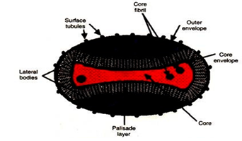
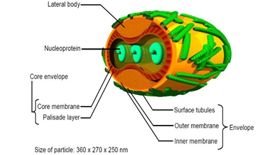
Poxviruses are oval or brick-shaped 200-400 nm long particles and can be visualized by the best light microscopes. The external surface is ridged in parallel rows, sometimes arranged helically (Figure 1). Viral particles (virions) are generally enveloped (external enveloped virion-EEV). The intracellular mature virion (IMV) form of the virus contains a different envelope and is also infectious. The internal structures of Poxviruses are shaped like flattened capsules/barrels or are lens or pill-shaped (Figure 2). On the basis of their species, they vary in their shape, but generally appear brick like or as an oval form similar to a rounded brick. The virion size is around 200 nm in diameter and 300 nm in length and carries its genome in a single, linear, double-stranded segment of DNA. The external surface of the virion is ridged in parallel rows, sometimes arranged helically. The particles are extremely complex and contain more than 100 different proteins.
Figure 2: Showing a model of poxvirus cut-away in cross-section to show the internal structures. Poxviruses are shaped like flattened capsules/barrels or are lens or pill-shaped. Their structure is complex, neither icosahedra nor helical. This model is based on Vaccinia, the smallpox virus. The structures are also highly variable and often incompletely studied. (Source: Poxvirus cronodon.com).
3. Replication of Poxvirus
3.1 Virus entry into cells
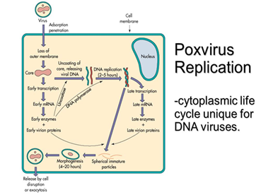
Vaccinia virus was used for the investigation of the way in which poxviruses enter into the host cell for replication. During replication of the pox virus, the pH-independent attachment with cell membrane is more vital as compared with low pH-dependent endosomal attachment system which used by influenza virus[12-15]. Likewise, the enveloped virus also fuses in a pH-independent manner with the plasma membrane [15, 16]. Poxviruses need to have 32 KDa, 29 KDa polypeptide and a domain of 54 to 58 KDa (the STE) polypeptide for attachment to the target cell. After attachment, proteins ranging from 54 to 58 KDa polypeptide, a 34KDa polypeptide and 17 KDa to 5 KDa dimer polypeptide are believed to be used for penetration of the cell for entrance to the cytoplasm where its replication takes place. During the process, proteolytic cleavage is important which is supplied by either trypsin or plasma membrane fusion [17, 18]. In addition, as described by Rodriguez et al. [19, 20] a 14 KDa polypeptide is also supposed to be involved in this process. Several stages are involved in the replication of poxviruses. First, the virus bind to the receptor on the host surface; Glycosaminoglycan’s (GAGs) is supposed to be the receptors of the pox virus. After the virus attached to the receptor on the surface of the host cell, it forced to enter to cytoplasm and removal of its coating takes place. During complete replication, two uncoating and two gene expression phases should be undertaken; during uncoating, as soon as the virus enters into the host cell, the outer membrane is removed and attached to cellular membrane then the core is released to cytoplasm immediately. In the case of gene expression, before the genome is replicated, the early genes encode the nonstructural proteins which are important for replication of the genome. Following genome replication, the late genes are expressed and encode structural proteins; finally, the complete virus is formed and assembled together in five steps of development to become the end exocytosis of the new matured virus (Figure 3).
The intracellular mature virion is formed after the immature virion assembled its proteins and the protein become aligned to make the brick shaped intracellular enveloped virion subsequently attached to the cell plasma and the cell related enveloped mature virus is created. Lastly, the cell-related enveloped virus encounters the microtubules and the virion makes ready to come out of the cell and termed as an extracellular matured virus. The assembly of the virion is taking place in the cytoplasm and presently the mechanism is under investigation for further study. Poxvirus is the largest in size which almost close to some bacteria’s and replication is fast and complex, replication takes place until host die and cellular activity stopped. Replication of poxvirus is taking place in the cytoplasm, which is rare for viruses of double-stranded DNA genome [21] (see Table 1). Also common for other large DNA viruses [22]. The unique feature of pox virus is, it uses own machinery for genome transcription, DNA dependent RNA polymerase Enzyme [23] and this enzyme is the one that makes replication of pox virus inside the cytoplasm.
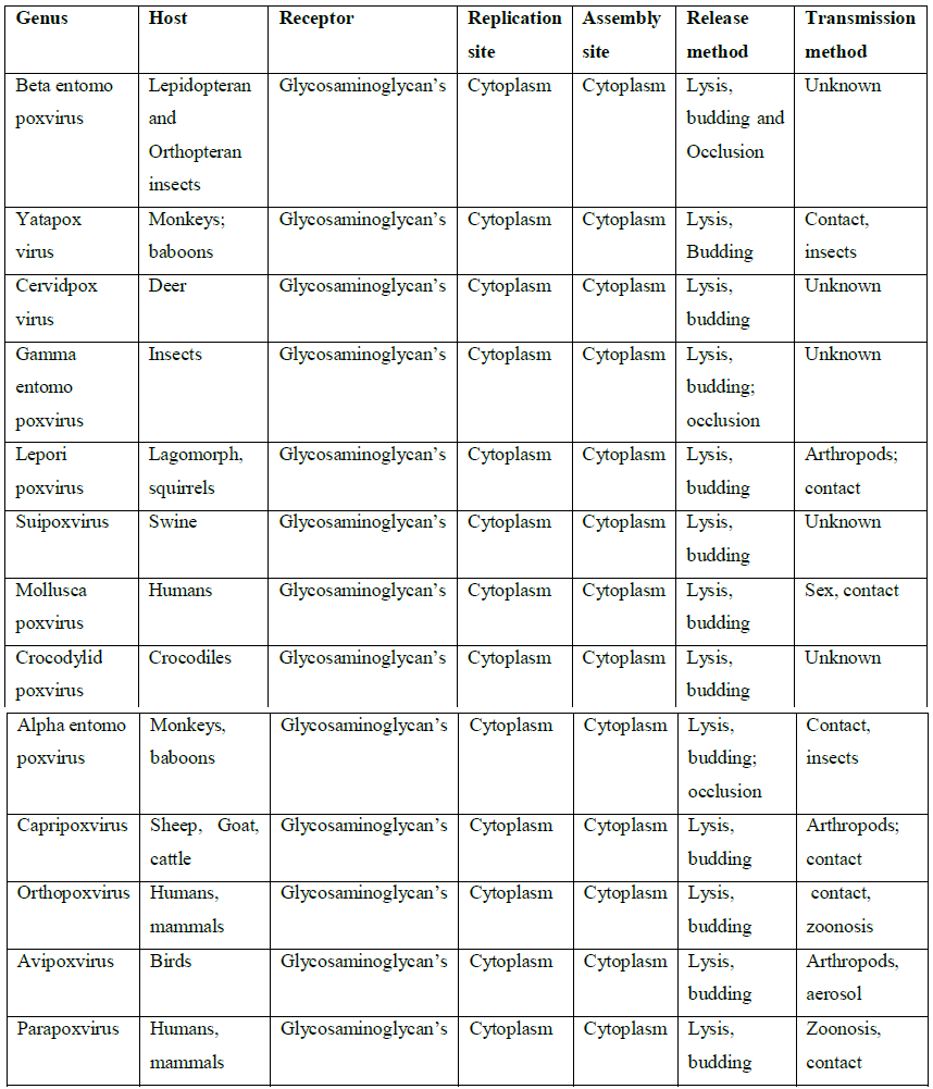
Table 1: Replication of different generas of Pox virus. Table showing host, attachment site during entry, where replication takes place, virion assembly site and after releasing how transmission to another animal is taking place for different genera of pox virus. (Source: https://en.wikipedia.org/wiki/Poxviridae).
4. Replication of Poxvirus DNA Timing and Location
Poxvirus DNA might be observed within 2 hours following entering to the target cell inside the cytoplasm, specifically at discrete juxtanuclear sites termed as factories and fluorescent dye is one of diagnostic material used for visualization (Figure 4). A single virus factory can form one virion, and many factories compared with the propagation of infection [25, 26]. Nevertheless, coalescence of individual factories often occurs with time [26, 27]. The factories are originally condensed and surrounded by endoplasmic reticulum membranes [28] and the membrane is believed to be important for replication of DNA [29]. It also mentioned that, besides virion assembly, the factories are known as the site of transcription and translation of viral mRNAs [26].
Figure 4: Reveals, an experimental study in which HeLa cells were used and infected at the same time by two recombinant vaccinia viruses, one expresses the A5 core protein fused to cyan fluorescent proteins A5-CFP (Figure B) and the rest expresses the A5 core protein fused to yellow fluorescent proteins A5-YFP (Figure B). The nuclei N and virus factories F (Figure A) were stained blue with 40, 6-diamidino-2-phenylindole (DAPI). A confocal microscopy image of a single cell in which individual factories arose from separate viruses is shown (Katsafanas and Moss, 2007). (Source: www.cshperspectives.org.) (Figure C).
5. Starting of DNA Replication
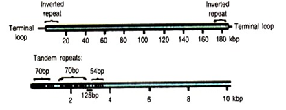
Any circular DNA molecule transfected into cells infected with VACV or Shope fibroma virus can be multiply; the replicated DNA is in the form of unbranched head to tail concatemers which could be formed by a rolling circle. And also confirmed that insertion of any DNA fragment has no effect on replication of the virus [30, 31]. Origin independent plasmid replication occurs within viral DNA factories and depends on each of the known viral proteins needed for genome replication [32]. According to Pogo et al.1984 [33]. Analysis of 3H thymidine incorporation following a shift from no permissive to a permissive temperature in cells infected with a VACV DNA negative mutant suggested that synthesis begins near the genome termini. Trials were conducted by transfecting linear DNA microchromosome indicated the availability of cis-acting replication increasing sequences within terminal 200bp. The outcomes revealed that specific replication beginning cite is within the preserved sequence between the hairpin loop and the direct repeats [34].
6. Proteins for DNA Replication
Nucleotide metabolism enzymes and replication proteins are synthesized early in poxvirus. This characteristic distinguishes proteins involved in DNA synthesis from those made at intermediate or late times that have roles in DNA processing and packaging. During DNA replication, E9L gene encodes a 117KDa DNA polymerase (Pol) [35-37] and the orthologs of these genes are common in other distinguished poxviruses. It has confirmed that the catalytic subunits of the DNA pools of another group of the virus, eukaryotic DNA pools, and Poxvirus DNA Pool has similar functions [38]. Vaccinia virus DNA Pools catalyzes primer and template-dependent DNA synthesis and possesses a range of 30 to 50 proofreading exonuclease actions [39]. Even though better one is found in the cytoplasm, the action of DNA Pool prepared from vaccinia virus is inherently distributive [40]. The DNA pool can also catalyze single-strand DNA annealing, which could generate branched molecules and thereby link DNA synthesis and recombination [41, 42].
7. Genes Responsible for Pox Virus Infection
Several genes found in poxvirus genome are not important for replication based on trials done on cell culture. However, they are mandatory for modulation of antibody produced by the host. Due to this reason, they are known as virulence genes [4, 5]. Few of these genes impact viral replication only in a set of cell lineages that came from various tissues or host species and these genes have been said to be host range genes [4-6] (Figure 4). More studies have revealed on the genera of orthopox virus and Leporipox virus to predict as several poxviruses encode a distinctive collection of host range genes [7]. According to the current scientific grouping, twelve dissimilar poxvirus genes have been identified; K3L, E3L, and K1L are among the group having one gene; likewise, others including separin, C7L family and TNFRII family display numerous members which expected consequence from lineage replication actions [6]. Laboratory animals were used to functionally characterize the factors by removal of gene out for investigations that related generally with the operation of various cellular targets, including cellular kinases and phosphatases, apoptosis and several antiviral pathways [5, 8]. These genes are incredibly crucial for infection on cell lineage, otherwise no development of the disease. Other studies on laboratory animals revealed that unknown factor affects viral pathogenicity; however, the laboratory animals used were still having the disease [9-10]. In the past, these genes were termed as host range genes when considering the cells infected without the main target animals infected by this virus [4-6]. Some experimental studies have suggested that a direct association between the diversity of host range factors and the number of host species for different poxviruses but, this association is still under argue [4, 5]. In the assessment of these fascinating questions, researchers required to establish the natural hosts for the poxviruses officially assigned to viral species and recognized by the International Committee on Taxonomy of Viruses (ICTV) [43]. Based on the available data up to now, investigators performed an extensive search for different host range genes [20].
8. Host Immune Response Against Poxvirus Infection
The Acquired antibody is a complex interaction of many cell types which results in the generation of particular antiviral antibody by B lymphocytes and in virus-specific DTH and cytotoxic responses by T lymphocytes. A particular antibody can act in response directly with viral components in presence or absence of complements, or it can bind to the surface of a phagocytic cell via the Fc receptor and support the cell with specific reactivity against the poxvirus (ADCC). The complication of the protective system is such that, it is not fully developed until late birth, and, as with other pathogens, newborn animals reveal high susceptibility to poxvirus infection in the absence of maternal immunity. For illustration, studies noticed that newborn rabbits were highly at risk to lethal infection with RPV than adult rabbits and more current trials suggested that, the undeveloped protective system was accountable for the incapacity to remove the virus [44, 45]. Associated annotations were also made by Fenner for ectromelia virus (Moscow strain) infections of suckling mice [46, 47].
9. Antibody
According to different investigations, specific antivirus antibodies can trim down virus spread by a variety of mechanisms; preventing viral attachment to the cell surface by masking viral adsorption receptors, aggregating virus, preventing virus entry, preventing virus uncoating, increasing uptake of the virus through Fc receptors by phagocytic cells, increasing virolysis or cytolysis of virus-infected cells in mixture with complement. As information’s were gathered and reviewed over years, antibody-mediated mechanisms of virus neutralization were insufficient to protect an immunologically immature host from the first infection with a powerful poxvirus [48]. Also reported that hyperimmune y globulin administered to children who had complications following vaccination for smallpox was frequently unsuccessful. Additionally, it was known that approximately all patients with Bruton's agammaglobulinemia developed defensive immunity following smallpox immunization [49], signifying that, specific neutralizing and defensive antibodies are not necessary for healing from the vaccinia virus vaccine strains. By the support of this idea, sick individuals with standard anti-vaccinia virus-immune serum can give up to progressive vaccinia [50]. Some evidence suggested that a late antibody reply may direct to progressive vaccinia in children, most likely because of the gathering of the immune system with soluble viral antigens, which accordingly prohibited neutralization of virus with antibody by competitive inhibition [51, 48].
10. T Lymphocytes
T lymphocytes are varied in phenotypic markers and functional capabilities in the population of cells and have the ability to distinguish foreign antigens that present on infected cells using MHC molecule. CD8+ T lymphocyte (TCTL or TH) and the CD4+ T lymphocyte (TH or TDTH) are among T cells which serve as effector support of the immune reaction to poxvirus infection.CD4+TH present accessory factors or help (TH) in the generation of particular antibody and CTL and CD4+TDTH cells mediate the recruitment of inflammatory cells into an injury and neutralization of the virus takes place. For example, According to Douglas and Smith [52], neutralization of vaccinia virus was increased by immune cells from blood and spleen during infection. Farther more, the significance of immune cells in defense against poxvirus infection was once more hinted by Kempe [48]. According to his report, certain children with agammaglobulinemia had no adverse reaction to vaccination with live vaccinia virus, signifying that, a further effector mechanism besides antibody, Complement, or phagocytosis of the virus by macrophages was significant in defense. Additionally, a one-year-old child with progressive vaccinia was unsuccessful to respond to administration of hyperimmune y globulin and to show a DTH reaction, while his leukocyte has the ability to destruct the virus by phagocytosis. Following the adoptive transfer of immune leukocytes and lymph node cells from donors, the child showed a local DTH reaction and weakening of the virus lesion was also observed [49, 50, 53].
11. ADCC
ADCC is a category of leukocyte (K cell), in union with a specific antibody to a virus and able to lyse the virus-infected target cells. As confirmed, the function of ADCC is not the same in all species of animals, it was more important for protection against poxvirus infections in some species than others. Based on an experimental study done on a human, in vaccinated individuals with vaccinia virus a population of Fc-R+ lymphocytes was available in a T cell down the pool of PBL, which were capable of lysing vaccinia virus-infected cells in a non-MHC restricted fashion [54-56]. The study exposed that, the addition of F ab to rabbit anti-human immunoglobulin G antisera to virus-infected targets revoked the ability of immune PBL to lyse infected cells particularly. Moreover, anti-vaccinia virus antiserum added to infected fibroblasts drastically improved the ability of non-immune Fc R PBL to lyse these cells, but not uninfected cells.
Following vaccination of footpad to adult rabbits with SFV (Madison or Patuxent strain), researchers identified ADCC in another poxvirus infections, Scott et al. [57] and increasing of T-cell and B cell proliferation in draining lymph node and spleen respectively. In addition, one more study revealed in mice that, the production of ectromelia virus-specific antibodies occurs too delayed to give defense by ADCC from a lethal primary infection in certain mouse strains [58-60], however, Upon reinfection, humoral antibody and ADCC are believed to be a significant resistant mechanism [61]. In contrast, in primary infection of Syrian hamsters rendered functionally athymic, about half of the vaccinia virus-induced cytotoxicity can be recognized as an ADCC mechanism [62].
12. Immunomodulatory Strategies of Pox Virus
To mediate host antiviral response and develop an infection, poxviruses should have the ability to invade and overcome the force full natural and acquired host immunity. Since poxviruses are categorized under viruses of large genome, they encode multiple classes of immunomodulatory proteins that have evolved specifically to inhibit such diverse process as apoptosis, the production of interferon’s, chemokines, inflammatory cytokines, the activity of cytotoxic T lymphocytes (CTLs), natural killer (NK) cells, complement and antibodies. It is not easy to trace back the evolutionary genesis of these virus-encoded immunomodulatory proteins. The obvious sequence similarity between some immunomodulatory poxvirus genes and the cDNA versions of related cellular counterparts suggests that they were once captured by ancestral retrotranscription and recombination events and then reasserted into individual virus isolates during coevolution with vertebrate hosts.
13. Immune Suppression Properties of Pox Virus
Lepari poxviruses were used to study the immunosuppressive ability of pox virus by Strayer and colleagues. Based on the investigation, MRFV and its parental strain, myxoma virus induce severe immunologic dysfunction in domestic rabbit which results in death from secondary gram-negative infection within 2 weeks of virus infection [63,65-67]. After infection by poxvirus, there are two ways of immunosuppresion; Primary suppression is mediated by virus infection of both B and T lymphocyte populations [58, 67], whereas, the secondary immunosuppression appears to result from virus induction of a soluble, nonspecific virus-induced suppressor factor from T lymphocytes [69]. Immune responsiveness improves and correlates with the production of an antagonist of virus-induced suppressor factor and anti suppressor factor [63, 70].
14. Preventing of Host Cell Metabolism
An experimental study done using Yaba virus revealed that Poxvirus infections have a diverse cause on host macromolecular syntheses including developing of tumors and infected cells grow for widespread periods after infection [71-74] and almost all of host proteins continue to be synthesized for a long time. Studies also revealed that, in Yaba virus-infected cells, remarkably, increased the synthesis of certain host proteins [75].
15. Poxvirus anti interferon property
Interferons can be categorized into three classes based on the type of cell they produced from; alpha, beta, and gamma. They are small proteins and glycoprotein’s having a molecular mass ranging from 15 to 22KDa [76]. Currently, studies suggested that an endoribonuclease, 2', 5’-A synthetase and a protein kinase are the three kinds of enzymes produced in interferon-treated cells. Also a study revealed that vaccinia virus uses early gene to prevent kiness’s activity in L929 cells [77-81] and product of 2', 5’-oligoisoadenylate synthetase activates an endoribonuclease, thus leading to the increased degradation of viral mRNAs [82, 83, 84, 85]; this progression may be inhibited by virus-induced ATPase and phosphatase or inhibition of the endoribonuclease [86-88].Another trial by Youngner et al.[89] also forwarded the supplementary supports for the existence of virus genes which modify the antiviral effects of interferon.
16. Conclusion
The main objective of this review is to cite information about replication of pox virus, genes responsible to cause the disease and host defense mechanism against the virus. Therefor Vaccinia virus was used as a model in different study areas whereas, Lepari pox virus also described for the investigation of the immunosuppressive ability of pox virus. Poxviruses are oval or brick-shaped 200-400nm long particles and can be visualized by the best light microscopes and as a group can infect a large number of animals including human being and are large double-stranded DNA viruses, which exclusively replicate in the cytoplasm of their host cells and the infections typically result in the formation of lesions, skin nodules, or disseminated rash. The emergence of Poxviruses was estimated to be around thousands of year’s ago. Nevertheless, the origin and evolution of these viruses are still unclear. Based on the studies that have been done on cell culture, many of the genes present in the poxvirus genome are not essential to viral replication. However, they are significant to the modulation of the host antiviral response and, thus are considered virulence genes. More studies have revealed on the genera of orthopoxvirus and Lepori poxvirus to predict as several poxviruses encode a distinctive collection of host range genes. A number of stages are undertaken in replication of poxviruses, first, the virus bind to the receptor on the host surface; Glycosaminoglycan’s (GAGs) is supposed to be the receptors of poxvirus. More than one enzyme is encoded by several poxviruses for production of DNA, which probably increase genome replication in inactive cells.Poxvirus DNA synthesis can usually be detected within 2hrs after infection and occurs in the cytoplasm within discrete juxtanuclear cites called factories that can easily be visualized by staining with a fluorescent dye. The Acquired antibody is a complex interaction of many cell types which results in the generation of particular antiviral antibody by B lymphocytes and in virus-specific DTH and cytotoxic responses by T lymphocytes. In poxvirus infection, there could be two kinds of immunity suppressive ways; Primary suppression is mediated by virus infection of both B and T lymphocyte populations, whereas, the secondary immunosuppression appears to result from virus induction of a soluble, nonspecific virus-induced suppressor factor from T lymphocyte. Poxvirus infections have a varied effect on host macromolecular syntheses and act against interferon activities.
References
- Hughes AL, Friedman R. Poxvirus genome evolution by gene gain and loss. Mol. Phylogenet. Evol 35 (2005): 186-195.
- Bratke KA, McLysaght A. Identification of multiple independent horizontal gene transfers into poxviruses using a comparative genomics approach. BMC Evol. Biol 8 (2008): 67.
- McLysaght A, Baldi PF, Gaut BS. Extensive gene gain associated with adaptive evolution of poxviruses.Proc. Natl. Acad. Sci. USA100 (2003): 15655-15660.
- McFadden G. Poxvirus tropism. Nat. Rev. Microbiol 3 (2005): 201-213.
- Haller SL, Peng C, McFadden G, et al. Poxviruses and the evolution of host range and virulence. Infect. Genet. Evol 21 (2014): 15-40.
- McFadden, G. Poxvirus tropism. Nat. Rev. Microbiol 3 (2005): 201-213.
- Bratke KA, McLysaght A, Rothenburg S. A survey of host range genes in poxvirus genomes. Infect. Genet. Evol 14 (2013): 406-425.
- Tulman ER, Afonso CL, Lu Z, et al. The Genome of Canarypox Virus. J. Virol 78 (2004): 353-366.
- Werden SJ, Rahman MM, McFadden G. Poxvirus Host Range Genes Chapter 3. Adv. Virus Res 71 (2008): 135-171.
- Chen W, Drillien R, Spehner D, et al. In vitro and in vivo study of the ectromelia virus homolog of the vaccinia virus K1L host range gene. Virology 196 (1993): 682-693.
- Liu J, Wennier S, Moussatche N, et al. Myxoma Virus M064 Is a Novel Member of the Poxvirus C7L Superfamily of Host Range Factors That Controls the Kinetics of Myxomatosis in European Rabbits. J. Virol 86 (2012): 5371-5375.
- Moss B. Poxvirus entry and membrane fusion. Virology 344 (2006): 48-54.
- Chang A, Metz DH. Further investigations on the mode of entry of vaccinia virus into cells. J. Gen. Virol 32 (1976): 275-282.
- Dales S. The relation between penetration of vaccinia release of viral DNA, and initiation of genetic functions. Perspect Virol 4 (1965): 47-71.
- Dales S, Kajioka R. The cycle of multiplication of vaccinia virus in Earle's strain L cells. I. Uptake and penetration.Virology 24 (1964): 278-294.
- Doms RW, Blumenthal R, Moss B. Fusion of intra and extracellular forms of vaccinia virus with the cell membrane. J. Virol 64 (1990): 4884-4892.
- Payne LG, Norrby E. Adsorption and penetration of enveloped and naked vaccinia virus particles. J. Virol 27 (1978): 19-27.
- Ichihashi Y, Oie M. Adsorption and penetration of the trypsinized vaccinia virion. Virology 101 (1980): 50-60.
- Oie M, Ichihashi Y. Modification of vaccinia virus penetration proteins analyzed by monoclonal antibodies. Virology 157 (1987): 449-459.
- Rodriguez JF, Esteban M. Mapping and nucleotide sequence of the vaccinia virus gene that encodes a 14-kDa fusion protein. J. Virol 61 (1987): 3550-3554.
- Rodriguez JF, Paez E, Esteban M. A 14,000-Mr envelope protein of vaccinia virus is involved in cell fusion and forms covalently linked trimers. J. Virol 61 (1987): 395-404.
- Mutsafi Y, Zauberman N, Sabanay I. Vaccinia-like cytoplasmic replication of the giant Mimivirus. (https://www.ncbi.nlm.nih.gov/pmc/articles/PMC2851855). Proceedings of the National Academy of Sciences USA. 107 (2010): 5978-582.
- Vincent R. Pithovirus: Bigger than Pandoravirus with a smaller genome.
- National Center for Infectious Diseases, Centers for Disease Control and Prevention, 1600 Clifton Rd., Atlanta, GA 30333, USA. DNA-dependent RNA polymerase rpo35 (Vaccinia virus)"(https://www.ncbi.nlm.nih.gov/protein/YP_233034.1). National Center for Biotechnology Information (NCBI), NIH, Bethesda, MD, USA.
- Moss B. Poxviridae: The viruses and their replication. In Fields Virology (ed. Knipe DM, Howley PM), Lippincott Williams and Wilkins, Philadelphia. (2007): 2905-2946.
- Cairns J. The initiation of vaccinia infection. Virology 11 (1960): 603-623.
- Katsafanas GC, Moss B. Colocalization of transcription and translation within cytoplasmic poxvirus factories coordinates viral expression and subjugates host functions. Cell Host Microbe 2 (2007): 221-228.
- Lin YCJ, Evans DH. Vaccinia virus particles mix inefficiently and in a way that would restrict viral recombination in coinfected cells. J Virol 84 (2010): 2432-2443.
- Tolonen N, Doglio L, Schleich S, et al. Vaccinia virus DNA replication occurs in endoplasmic reticulum-enclosed cytoplasmic mini-nuclei. Mol Biol Cell 12 (2001): 2031-2046.
- Schramm B, Krijnse Locker J. Cytoplasmic organization of poxvirus DNA replication. Traffic 6 (2005): 839-846.
- DE Lange AM, McFadden G. Sequence-nonspecific replication of transfected plasmid DNA in poxvirus-infected cells. Proc Natl Acad Sci 83 (1986): 614-618.
- Merchlinsky M, Moss B. Sequence-independent replication and sequence-specific resolution of plasmids containing the vaccinia virus concatemer junction: Requirements for early and late trans-acting factors. In Cancer cells 6: Eukaryotic DNA replication (ed. Kelly T, Stillman B), Cold Spring Harbor Laboratory Press, Cold Spring Harbor, NY (1988): 87-93.
- De Silva FS, Moss B. Origin-independent plasmid replication occurs in vaccinia virus cytoplasmic factories and requires all five known poxvirus replication factors. Virol J 2 (2005): 33.
- Pogo BG, Berkowitz EM, Dales S. Investigation of vaccinia virus DNA replication employing a conditional lethal mutant defective in DNA. Virology 132 (1984): 436-444.
- Du S, Traktman P. Vaccinia virus DNA replication: Two hundred base pairs of a telomeric sequence confer optimal replication efficiency on minichromosome templates.Proc Natl Acad Sci 93 (1996): 9693-9698.
- Jones EV, Moss B. Mapping of the vaccinia virus DNA polymerase gene by marker rescue and cell-free translation of selected mRNA. J Virol 49 (1984): 72-77.
- Traktman P, Sridhar RC, Roberts BE. Transcriptional mapping of the DNA polymerase gene of vaccinia virus.J Virol 49 (1984): 125-131.
- Earl PL, Jones EV, Moss B. Homology between DNA polymerases of poxviruses, herpesviruses, and adenoviruses: Nucleotide sequence of the vaccinia virus DNA polymerase gene. Proc Natl Acad Sci 83 (1986): 3659-3663.
- Wang TS, Wong SW, Korn D. Human DNA polymerase a: Predicted functional domains and relationships with viral DNA polymerases. FASEB J 3 (1989): 14-21.
- Challberg MD, Englund PT. Purification and properties of the deoxyribonucleic acid polymerase induced by vaccinia virus. J Biol Chem 254 (1979): 7812-7819.
- McDonald WF, Klemperer N, Traktman P. Characterization of a processive form of the vaccinia virus DNA polymerase. Virology 234 (1997): 168-175.
- Willer DO, Mann MJ, ZhangWD, Evans DH. Vaccinia virus DNA polymerase promotes DNA pairing and strand-transfer reactions. Virology 257 (1999): 511-523.
- Hamilton MD, Evans DH. Enzymatic processing of replication and recombination intermediates by the vaccinia virus DNA polymerase. Nucleic Acids Res 33 (2005): 2259-2268.
- International Committee on Taxonomy of Viruses—Taxonomy. Available online: https://talk.ictvonline.org/taxonomy/w/ictv-taxonomy (2017).
- Allison AC. Immune responses to Shope fibroma virus in adult and newborn rabbits. J. Natl. Cancer Inst. 36 (1966): 869-876.
- Duran-Reynals F. Production of degenerative inflammatory or neoplastic effects in the newborn rabbit by the Shope fibroma virus. Yale J. Biol. Med 3 (1940): 99-114.
- Fenner F. Studies in mousepox (infectious ectromelia of mice). VII. The effect of age of the host upon the response to infection. Aust. J. Exp. Biol. Med. Sci 27 (1949): 45-53.
- Fenner F, Ratcliffe FN. Myxomatosis. Cambridge University Press, Cambridge (1965).
- Kempe CH. Studies on smallpox and complications of smallpox vaccination. Pediatrics 20 (1960): 176-189.
- Fulginiti VA, Kempe CH, Hathaway WE, et al. Progressive vaccinia in immunologically deficient individuals, p. 129-145. In D. Bergsma. Immunologic deficiency diseases in man. Birth defects original article series. National Foundation March of Dimes, New York 4 (1968).
- Hansson O, Johansson SG, Vahlquist B. Vaccinia grangrenosa with normal humoral antibodies. Acta Paediatr. Scand 55 (1966) :264-272.
- Fenner F, Henderson DA, Arita I, et al. Smallpox and its eradication. World Health Organization, Geneva (1988).
- Douglas SR, Smith W. A study of vaccinal immunity in rabbits by means of in vitro methods. Br. J. Exp. Pathol 11 (1930): 96-111.
- O'Connell CJ, Karzon DT, Barron AL, et al. Progressive vaccinia with normal antibodies. A case possibly due to deficient cellular immunity. Ann.Intern. Med 60 (1964): 282-289.
- McFarland HF, Pedone CA, Mingioli ES, et al. The response of human lymphocyte subpopulations to measles, mumps, and vaccinia viral antigens. J. Immunol 125 (1980): 221-225.
- Moeller-Larsen A, Haahr S. Humoral and cell-mediated immune responses in humans before and after revaccination with vaccinia virus. Infect. Immun 19 (1978): 34-39.
- Perrin LH, Zinkernagel RM, Oldstone MBA. Immune response in humans after vaccination with vaccinia virus: generation of a virus-specific cytotoxic activity by human peripheral lymphocytes. J. Exp. Med 146 (1977): 949-969.
- Scott CB, Holdbrook R, Sell S. Cell-mediated immune response to Shope fibroma virus-induced tumors in adult rabbits. JNCI 66 (1981): 681-689.
- Blanden RV. Mechanisms of recovery from a generalized viral infection: mousepox. I. The effects of anti-thymocyte serum. J. Exp. Med 132 (1970): 1035-1053.
- Blanden RV. Mechanisms of recovery from a generalized viral infection: mousepox. III. Regression of infectious foci. J. Exp. Med 133 (1971): 1090-1104.
- Blanden RV, Gardner ID. The cell-mediated immune response to ectromelia virus infection. I. Kinetics and characteristics of the primary effector T cell response in vivo.Cell. Immunol 22 (1976): 271-282.
- Fenner F. Studies in mousepox (infectious ectromelia of mice). IV. Quantitative investigations on the spread of the virus through the host in actively and passively immunized animals.Aust. J. Exp. Biol. Med. Sci 27 (1949): 1-18.
- Nelles MJ, Duncan WR, Streilein JW. Immune response to acute virus infection in the Syrian hamster. II. Studies on the identity of virus-induced cytotoxic effector cells. J. Immunol 126 (1981): 214-218.
- Strayer DS, Dombrowski J. Immunosuppression during viral oncogenesis. V. Resistance to virus-induced immunosuppressive factor. J. Immunol 141 (1988): 347-351.
- Stitz L, Althage A, Hengartner H. Natural killer cells vs cytotoxic T cells in the peripheral blood of virus-infected mice. J. Immunol 134 (1985): 598-602.
- Strayer DS, Leibowitz JL. Virus-lymphocyte interactions during the course of immunosuppressive virus infection. J. Gen. Virol 68 (1987): 463-472.
- Strayer DS, Sell S. Immunohistology of malignant rabbit fibroma virus-a comparative study with rabbit myxoma virus. JNCI 71 (1983): 105-116.
- Strayer DS, Skaletsky E, Leibowitz JL. In vitro growth of two related leporipoxviruses in lymphoid cells.Virology 145 (1985): 330-334.
- Strayer DS, Skaletsky E, Leibowitz JL, et al. The growth of malignant rabbit fibroma virus in lymphoid cells. Virology 158 (1987): 147-157.
- Strayer DS, Korber K, Dombrowski J. Immunosuppression during viral oncogenesis. IV. Generation of soluble virus-induced immunologic suppressor molecules. J. Immunol 140 (1988): 2051-2059.
- Strayer DS, Leibowitz JL. Reversal of virus-induced immune suppression. J. Immunol 136 (1986): 2649-2653.
- Niven JSF, Armstrong JA, Andrewes CH, et al. Subcutaneous "growths" in monkeys produced by a poxvirus. J. Pathol. Bacteriol 81 (1961): 1-10.
- Sproul EE, Metzgar RS, Grace JT. The pathogenesis of Yaba virus-induced histiocytomas in primates. Cancer Res 23 (1963): 671-675.
- Yohn DS, Grace JT, Haendiges VA. A quantitative cell culture assay for Yaba tumor virus. Nature (London) 202 (1964): 881-883.
- Yohn DS, Marmol FR, Olsen RG. Growth kinetics of Yaba tumor poxvirus after in vitro adaptation to Cercopithecus kidney cells. J. Virol 5 (1970): 205-211.
- Vafai A, Rouhandeh H. Analysis of Yaba tumor poxvirus-induced proteins in infected cells by two-dimensional gel electrophoresis. Virology 120 (1982): 65-76.
- Joklik WK. Interferons. In B. N. Fields and D. M. Knipe (ed.), Virology, 2nd ed. Raven Press, New York (1990): 383-411.
- Hovanessian AG, Galabru J, Meurs E, et al. The rapid decrease in the levels of the double-stranded RNA-dependent protein kinase during (1987).
- Paez E, Esteban M. The resistance of the vaccinia virus to interferon is related to an interference phenomenon between the virus and the interferon system. Virology 134 (1984):12-28.
- Rice AP, Kerr IM. Interferon-mediated, double-stranded RNA-dependent protein kinase is inhibited in extracts from vaccinia virus-infected cells. Nature (London) 50 (1984): 229-236.
- Whitaker-Dowling P, Younger JS. Vaccinia rescue of VSV from interferon-induced resistance: reversal of translation block and inhibition of protein kinase activity. Virology 131 (1991): 128-136.
- Whitaker-Dowling P, Younger JS. Characterization of a specific kinase inhibitory factor produced by vaccinia virus which inhibits the interferon-induced protein kinase.Virology 137 (1984): 171-181.
- Baglioni C, Minks MA, Maroney PA. Interferon action may be mediated by activation of a nuclease by pppA2'p5'A2'p5'A. Nature (London) 273 (1978): 684-687.
- Brown GE, Lebleu B, Kawakita M, et al. Increased endonuclease activity in an extract from mouse Ehrlich ascites tumor cells which had been treated with a partially purified interferon preparation: dependence on double-stranded RNA. Biochem. Biophys. Res. Commun 69 (1976): 114-122.
- Kerr IM, Brown RE. pppA2'pS'A2'pS'A: an inhibitor of protein synthesis synthesized with an enzyme fraction from interferon-treated cells. Proc. Natl. Acad. Sci USA 75 (1978): 256-260.
- Ratner L, Sen GC, Brown GE, et al. Interferon, double-stranded RNA and RNA degradation. Characteristics of an endonuclease activity. Eur. J. Biochem 79 (1977): 565-577.
- Esteban M, Benavente J, Paez E. Mode of sensitivity and resistance of vaccinia virus replication to interferon. J. Gen. Virol 67 (1986): 801-808.
- Paez E, Esteban M. Nature and mode of action of vaccinia virus products that block activation of the interferon-mediated PPP(A2'p)nA-synthetase. Virology 134 (1984): 29-39.
- Rice AP, Kerr SM, Roberts WK, et al. Novel 2',5'-oligoadenylates synthesized in interferon-treated, vaccinia virus-infected cells. J. Virol 56 (1985): 1041-1044.
- Youngner JS, Thacore HR, Kelly ME. The sensitivity of ribonucleic acid and deoxyribonucleic acid viruses to different species of interferon in cell cultures. J. Virol 10 (1972): 171-178.

