Macroangiopathy in Systemic Sclerosis-Case Report and Literature Review
Article Information
Topolyanskaya SV1*, Bubman LI2
1I.M. Sechenov First Moscow State Medical University (Sechenov University), RF Health Ministry, Hospital Therapy Department No.2, Moscow, Russia
2War Veterans Hospital No.3, Moscow, Russia
*Corresponding Author: Svetlana Topolyanskaya, I.M. Sechenov First Moscow State Medical University (Sechenov University), RF Health Ministry, Hospital Therapy Department No.2, Moscow, Russia
Received: 25 June 2020; Accepted: 13 July 2020; Published: 20 July 2020
Citation:
Topolyanskaya SV, Bubman LI. Macroangiopathy in Systemic Sclerosis-Case Report and Literature Review. Fortune Journal of Rheumatology 2 (2020): 074-083.
View / Download Pdf Share at FacebookAbstract
The case report of systemic sclerosis debuting in an elderly man with critical ischemia of the lower extremities and with amputation of both legs is described. The features of this case included the large vessels pathology in the very beginning of the disease, as well as the occurrence and rapid progression of significant fibrosis and hyperpigmentation of the lower limbs skin after amputation of one of them. At the same time, the Raynaud phenomenon, sclerodactyly, and signs of visceral pathology were absent in the patient. Possible causes of the development of macroangiopathy in patients with systemic sclerosis are discussed.
Keywords
Systemic Sclerosis, Vasculopathy, Macroangiopathy, Fibrosis, Hyperpigmentation, Anticentromere Antibodies
Systemic Sclerosis articles, Vasculopathy articles, Macroangiopathy articles, Fibrosis articles, Hyperpigmentation articles, Anticentromere Antibodies articles
Systemic Sclerosis articles Systemic Sclerosis Research articles Systemic Sclerosis review articles Systemic Sclerosis PubMed articles Systemic Sclerosis PubMed Central articles Systemic Sclerosis 2023 articles Systemic Sclerosis 2024 articles Systemic Sclerosis Scopus articles Systemic Sclerosis impact factor journals Systemic Sclerosis Scopus journals Systemic Sclerosis PubMed journals Systemic Sclerosis medical journals Systemic Sclerosis free journals Systemic Sclerosis best journals Systemic Sclerosis top journals Systemic Sclerosis free medical journals Systemic Sclerosis famous journals Systemic Sclerosis Google Scholar indexed journals Vasculopathy articles Vasculopathy Research articles Vasculopathy review articles Vasculopathy PubMed articles Vasculopathy PubMed Central articles Vasculopathy 2023 articles Vasculopathy 2024 articles Vasculopathy Scopus articles Vasculopathy impact factor journals Vasculopathy Scopus journals Vasculopathy PubMed journals Vasculopathy medical journals Vasculopathy free journals Vasculopathy best journals Vasculopathy top journals Vasculopathy free medical journals Vasculopathy famous journals Vasculopathy Google Scholar indexed journals Macroangiopathy articles Macroangiopathy Research articles Macroangiopathy review articles Macroangiopathy PubMed articles Macroangiopathy PubMed Central articles Macroangiopathy 2023 articles Macroangiopathy 2024 articles Macroangiopathy Scopus articles Macroangiopathy impact factor journals Macroangiopathy Scopus journals Macroangiopathy PubMed journals Macroangiopathy medical journals Macroangiopathy free journals Macroangiopathy best journals Macroangiopathy top journals Macroangiopathy free medical journals Macroangiopathy famous journals Macroangiopathy Google Scholar indexed journals Fibrosis articles Fibrosis Research articles Fibrosis review articles Fibrosis PubMed articles Fibrosis PubMed Central articles Fibrosis 2023 articles Fibrosis 2024 articles Fibrosis Scopus articles Fibrosis impact factor journals Fibrosis Scopus journals Fibrosis PubMed journals Fibrosis medical journals Fibrosis free journals Fibrosis best journals Fibrosis top journals Fibrosis free medical journals Fibrosis famous journals Fibrosis Google Scholar indexed journals Hyperpigmentation articles Hyperpigmentation Research articles Hyperpigmentation review articles Hyperpigmentation PubMed articles Hyperpigmentation PubMed Central articles Hyperpigmentation 2023 articles Hyperpigmentation 2024 articles Hyperpigmentation Scopus articles Hyperpigmentation impact factor journals Hyperpigmentation Scopus journals Hyperpigmentation PubMed journals Hyperpigmentation medical journals Hyperpigmentation free journals Hyperpigmentation best journals Hyperpigmentation top journals Hyperpigmentation free medical journals Hyperpigmentation famous journals Hyperpigmentation Google Scholar indexed journals Anticentromere Antibodies articles Anticentromere Antibodies Research articles Anticentromere Antibodies review articles Anticentromere Antibodies PubMed articles Anticentromere Antibodies PubMed Central articles Anticentromere Antibodies 2023 articles Anticentromere Antibodies 2024 articles Anticentromere Antibodies Scopus articles Anticentromere Antibodies impact factor journals Anticentromere Antibodies Scopus journals Anticentromere Antibodies PubMed journals Anticentromere Antibodies medical journals Anticentromere Antibodies free journals Anticentromere Antibodies best journals Anticentromere Antibodies top journals Anticentromere Antibodies free medical journals Anticentromere Antibodies famous journals Anticentromere Antibodies Google Scholar indexed journals etiology articles etiology Research articles etiology review articles etiology PubMed articles etiology PubMed Central articles etiology 2023 articles etiology 2024 articles etiology Scopus articles etiology impact factor journals etiology Scopus journals etiology PubMed journals etiology medical journals etiology free journals etiology best journals etiology top journals etiology free medical journals etiology famous journals etiology Google Scholar indexed journals pro-inflammatory cytokines articles pro-inflammatory cytokines Research articles pro-inflammatory cytokines review articles pro-inflammatory cytokines PubMed articles pro-inflammatory cytokines PubMed Central articles pro-inflammatory cytokines 2023 articles pro-inflammatory cytokines 2024 articles pro-inflammatory cytokines Scopus articles pro-inflammatory cytokines impact factor journals pro-inflammatory cytokines Scopus journals pro-inflammatory cytokines PubMed journals pro-inflammatory cytokines medical journals pro-inflammatory cytokines free journals pro-inflammatory cytokines best journals pro-inflammatory cytokines top journals pro-inflammatory cytokines free medical journals pro-inflammatory cytokines famous journals pro-inflammatory cytokines Google Scholar indexed journals small arteries articles small arteries Research articles small arteries review articles small arteries PubMed articles small arteries PubMed Central articles small arteries 2023 articles small arteries 2024 articles small arteries Scopus articles small arteries impact factor journals small arteries Scopus journals small arteries PubMed journals small arteries medical journals small arteries free journals small arteries best journals small arteries top journals small arteries free medical journals small arteries famous journals small arteries Google Scholar indexed journals scleroderma vasculopathy articles scleroderma vasculopathy Research articles scleroderma vasculopathy review articles scleroderma vasculopathy PubMed articles scleroderma vasculopathy PubMed Central articles scleroderma vasculopathy 2023 articles scleroderma vasculopathy 2024 articles scleroderma vasculopathy Scopus articles scleroderma vasculopathy impact factor journals scleroderma vasculopathy Scopus journals scleroderma vasculopathy PubMed journals scleroderma vasculopathy medical journals scleroderma vasculopathy free journals scleroderma vasculopathy best journals scleroderma vasculopathy top journals scleroderma vasculopathy free medical journals scleroderma vasculopathy famous journals scleroderma vasculopathy Google Scholar indexed journals
Article Details
2. Introduction
Systemic sclerosis is a systemic autoimmune disease of unknown etiology, characterized by three main manifestations: fibrosis of the skin and internal organs, vasculopathy with a predominant involvement of the microvasculature in the pathological process and systemic inflammation with various autoantibodies and pro-inflammatory cytokines. Vasospasm, endothelial dysfunction and hemostasis disorders are considered typical for vascular pathology in case of systemic sclerosis. Pathomorphological features of scleroderma vasculopathy include intimal proliferation and fibrosis, along with thickening and fibrosis of the media of small arteries. Raynaud's phenomenon and digital ischemia of limbs are considered as classical manifestations of systemic sclerosis. According to some authors, scleroderma vasculopathy caused by complex interactions of various pathological processes, including autoimmune attacks, violation of compensatory vasculogenesis and angiogenesis, endothelial-to-mesenchymal transition, endothelial dysfunction and coagulation disorder/fibrinolysis system [1]. In severe cases of the disease, vasculitis and thrombosis of small arteries can develop, which can cause occlusion of digital arteries. Different types of vasculopathy in systemic sclerosis often contribute to the formation of digital ulcers and gangrene, ultimately leading to amputation of the fingers of the upper or lower extremities. However, the disorder of larger vessels, the more so leading to high amputation of the limbs, is described extremely rarely with systemic sclerosis.
In the case report we presented, systemic sclerosis debuted in a patient with clinical manifestations of critical ischemia of the lower extremities, which resulted in high amputation of them.
2. Case Report
Patient K, 64 years, hospitalized with complaints of long-term non-healing wounds after amputations of the 3rd and 5th fingers of the left hand, trophic ulcers of the left leg, hyperpigmentation and a noticeable tightening of the skin of the left lower limb and the anterior abdominal wall.
2.1 The history of this disease
He considers himself ill for the past 1.5 years. The disease onset with the appearance of an acute pain in his right leg. The pain syndrome was regarded as a low back pain. In a blood test, normochromic normocytic anemia, thrombocytosis and an elevation of ESR up to 45 mm/h were detected; these laboratory changes persisted throughout the follow-up. The severity of the pain syndrome gradually increased; against this background, edema and "blackening" of the fingers of the right foot arose with the subsequent development of dry gangrene of I-II fingers. In the next 10 months, the patient was hospitalized (up to 8 times) with a diagnosis of “Obliterating atherosclerosis of the lower extremities”. However, the effect of the medication therapy was not observed, the manifestations of critical ischemia of the lower limb progressed.
When describing the results of angiography, attention was drawn to the occlusion of the posterior and anterior tibial arteries, along with significant vasospasm of the arteries of the legs; there were no signs of larger arteries disorders (from the iliac to the popliteal). The angioplasty of the posterior tibial artery was performed; due to technical difficulties, angioplasty of the anterior tibial artery was not possible. Despite surgical treatment, the patient's condition continued to deteriorate, signs of critical ischemia of the right lower limb increased; pain and trophic disturbances appeared in the fingers of the left hand. During the next inpatient treatment, in addition to chronic occlusion of the right posterior and anterior tibial arteries, occlusion of the left radial and ulnar arteries was revealed. According to the results of a skin biopsy of the gluteal region significant derma fibrosis was found, but this finding was not reflected in the clinical management. In connection with increasing ischemia of the fingers and the lower limb, the amputation of the right leg was performed at the level of the middle third of the thigh, and the III and V fingers of the left hand were amputated.
After the operation, hyperpigmentation and tightening of the skin of the left lower limb appeared, with a gradual spread of these changes from the back surface of the left foot to the upper third of the thigh and with the subsequent transition to the anterior abdominal wall and stump of the right thigh. In connection with the significant skin induration, the patient felt an extremely unpleasant sensation of “tightening” and difficulty in the range of movements in the intact lower limb. Hyperpigmentation and tightening of the skin on the extensor surface of the left forearm (where the ulnar and radial artery occlusion was detected and the fingers were amputated) joined these disorders. Long-term non-healing wounds of the stumps of the III and V hand fingers were persisted, which was accompanied by intense pain. Trophic ulcers appeared in the projection of the ulnar processes and trophic ulcers on the left lower leg and foot. In connection with the above changes, the patient was admitted to the hospital.
2.2 Upon admission to the hospital
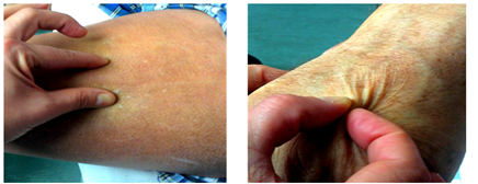
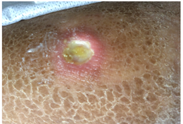
Skin and visible mucous membranes are pale. Hyperpigmentation of the skin of the left lower limb, stump of the right thigh, anterior abdominal wall and extensor surface of the left forearm. Significant induration of the skin and subcutaneous tissue of the left lower limb and anterior abdominal wall. The inability to collect a skin fold in these areas (Figure 1). The skin on the hands and face is without features. Raynaud's syndrome is not. Internal organs-without significant features. Hypotrophy of the muscles of the left forearm and hand. In the projection of the ulnar processes, there are 1 × 1 cm trophic ulcers filled with granulations and fibrin, with serous-purulent discharge, without perifocal inflammation. The stumps of the third and fifth fingers of the left hand are vicious, with protruding bone phalanges of gray color, in the absence of detachable and perifocal inflammation. In the lower third of the left leg, a 2 × 1 cm trophic ulcer performed with whitish granulations, with fibrin deposits and serous discharge, but without perifocal inflammation. On the inner surface of the lower leg, at the border of its upper and middle third, a 1 × 1 cm trophic ulcer, with callous, raised edges, with flaccid whitish granulations, with serous discharge, but without perifocal inflammation (Figure 2).
In blood tests: ESR - 35 m/h, hemoglobin - 88 g/l, platelets - 434x109/l; C-reactive protein - 24 mg/l; in a biochemistry - without clinically significant pathology. The study of limbs vessels (duplex scanning with color Doppler mapping) revealed occlusion of the left radial artery and the distal segment of the both ulnar arteries was detected; hemodynamically significant lesions of both iliac arteries, as well as arteries of the left lower limb (femoral, popliteal, anterior and posterior tibial) were not detected. The ankle-brachial index is 1.14 (normal). Mild atherosclerosis of extracranial vessels without signs of hemodynamically significant stenosis was found. When instrumental examination of internal organs - without features.
2.3 The results of a histological examination of a fragment of the skin, subcutaneous tissue and muscle-fascial flap of the anterior abdominal wall
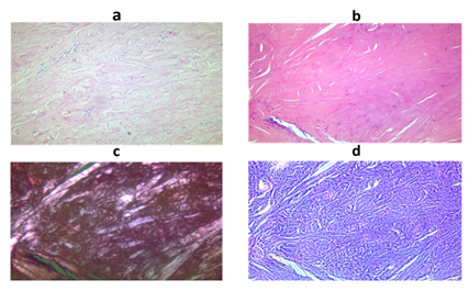
Derma thickened. The epidermis has the usual structure, small papillae, in some places vacuole dystrophy, parts of basal and spine-shaped epithelial cells are found. Collagen fibers of the papillary dermis are densified. The content of cellular elements (fibroblasts, macrophages, lymphocytes and mast cells) does not go beyond the norm. An elevated content of hemosiderophages is noted. Especially significant changes is registered in the reticular layer of the dermis. Collagen bundles are swollen, tightly connected to each other. In the papillary and reticular layers, moderate perivascular lymphohistiocytic infiltration is visible (Figure 3a). In the reticular layer of the dermis, extensive areas of hyalinosis are visible, which is characterized by the fusion of collagen bundles, a decrease in the number of fibroblasts and dystrophic changes in the remaining cells (Figure 3b). Under polarization microscopy, birefringence (anisotropy) of only part of the bundles of collagen fibers is observed, which indicates conformational changes in collagen (Figure 3c). Phase-contrast microscopy in the reticular layer of the dermis, including in the foci of hyalinosis, reveals the fibrillarity of collagen bundles (Figure 3d). Fascia and connective tissue septa are sclerotic and hyaline. There are no inflammatory infiltrates. Conclusion: the described morphological changes are characteristic of the scleroderma process.
2.4 The results of an immunological blood test
A marked increase in anticentromere antibodies in the blood serum (more than 300 units/ml, with a norm of 0-10 units/ml), antibodies to Scl-70-0.1 units/ml (normal). Taking into account the examination data, the results of immunological blood test and the skin histopathological examination, the patient was diagnosed with “Systemic sclerosis”. Taking into account macroangiopathy, which is atypical for systemic sclerosis, multiple occlusive lesions of medium and small caliber arteries, the patient’s male gender and long-term active smoking experience, it was suggested that the patient has two rare diseases in the form of systemic sclerosis and thromboangiitis obliterans. However, given the disease onset in old age, the absence of obvious signs of productive or necrotizing vasculitis and thrombotic occlusion of the vessels (with a triple morphological study), the intactness of the venous vascular bed, the diagnosis of concomitant thromboangiitis obliterans is very doubtful.
In the following months, despite immunosuppressive and vascular therapy, progression of ischemic changes in the remaining lower limb was noted, and therefore, the left lower limb was amputated at the middle third of the thigh. After discharge, ischemic changes on the right hand began to progress. Subsequently, the vicious right thigh stump was formed, so this stump was re-amputated. In the postoperative period, signs of disorganization of mental activity were observed, lack of movements in the left arm and speech disorders were also noted. However, when performing multi-spiral computed tomography of the brain, no convincing signs of a stroke were found. Subsequently, multiple organ failure progressed, due to which the patient died.
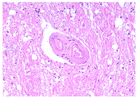
According to the autopsy results, scleroderma lesions of the skin and blood vessels were confirmed, and symmetric ischemic infarctions were detected in the subcortical nuclei of both cerebral hemispheres. Vascular pathology in the brain was mixed-a combination of scleroderma vasculopathy and atherosclerotic lesions, but without hemodynamically significant stenosis (max-20%) of the brain arteries (Figure 4).

Figure 1: Skin fold on the front of the thigh and hand.

Figure 2: Trophic ulcer on the left leg in combination with skin changes.

Figure 3: Morphological features of the skin in biopsy specimens (a) compaction and sclerosis of the reticular dermis, mild lymphohistiocytic perivascular infiltrates; stained with hematoxylin and eosin, x100; (b) a large area of hyalinosis in the reticular layer of the dermis, fusion of collagen bundles, degeneration and necrosis of fibroblasts; stained with hematoxylin and eosin, x100; (c) polarization microscopy of collagen structures in the area of hyalinosis, x100; (d) phase contrast microscopy of the hyalinosis site, revealing the fibrillarity of collagen bundles; x100.

Figure 4: Brain vascular pathology (autopsy findings).
3. Discussion
In systemic sclerosis, the involvement of large peripheral arteries in the pathological process, in contrast to the classic lesion of small digital vessels, is considered a casuistic rarity. However, in recent years, more and more often in the world literature, descriptions of cases or a series of cases of systemic sclerosis accompanied by macroangiopathy are published [2-5]. A number of studies have shown a significant increase in the risk of the formation of pathological changes in large vessels in case of systemic sclerosis, as compared with the general population [2, 6, 7]. Involvement of large vessels in the pathological process in systemic sclerosis can be due to both the sclerosis process itself and the concomitant atherosclerotic lesion, the risk of developing which in patients with scleroderma is slightly increased compared with the general population, although these data are quite contradictory [2, 8]. The coexistence of macro- and microangiopathy in systemic sclerosis significantly increases the risk of developing critical limb ischemia and the likelihood of subsequent amputation. According to the literature, in most patients with systemic sclerosis with the development of critical limb ischemia, there was a lesion of not only small, but also larger limb arteries, as in our observation.
In systemic sclerosis, the most common lesion is the tibial arteries and arteries of the forearm [9-11], as well as in our patient. So, in a recent study pathology of the forearm arteries were detected in 20% of patients with systemic sclerosis [12]. It should be noted that in this study no correlation was found between microvascular and macrovascular disorders, suggesting that different pathogenetic mechanisms can act in different vessels size [12]. In the pathological process with scleroderma, the ulnar artery is mainly involved (a significant bilateral occlusion of which was also found in our patient), although an exhaustive explanation for this fact could not be found [11]. In addition, in the case described by us, there was a lesion of the third and fifth fingers of the left hand followed by amputation, which is consistent with data from other authors, indicating frequent damage to the second and fifth finger arteries of the hand, along with the ulnar artery [3]. Unlike the data of other authors who observed damage to the ulnar and lower leg arteries many years after the onset of scleroderma, our patient suffered vascular damage with the development of critical ischemia in the onset of the disease and only then typical skin manifestations of scleroderma occurred [11].
Critical limb ischemia is a rare but dangerous complication of systemic sclerosis [13]. Descriptions of cases of critical limb ischemia in patients with systemic sclerosis are few [6, 9, 10, 14, 15]. It is significant that most of the described patients have been diagnosed with a limited form of systemic sclerosis [6, 14, 16].
The relationship between vascular pathology (up to the development of critical limb ischemia) and anticentromere antibodies found in a number of studies is quite interesting [9, 13]. It has been shown, for example, that in patients with systemic sclerosis, the presence of anticentromere antibodies is associated with the development of critical ischemia and amputation of fingers, along with an increased risk of developing occlusive lesion of large vessels. It is suggested that anticentromere antibodies can serve as one of the pathogenetic mechanisms of the development of vascular pathology in case of systemic sclerosis, including macroangiopathy [7, 9, 17, 18].
According to individual authors, it is anticentromere antibodies (in combination with an older age, smoking, and a limited form of scleroderma) that are largely responsible for the development of macroangiopathy in systemic sclerosis [17, 18]. Observations by Japanese authors suggest that gangrene of the limbs can develop with the presence of positive anticentromere antibodies even against the background of a complete absence of dermatofibrosis or with a minimal degree of its severity [18]. In the same series of cases, no visceral manifestations of systemic sclerosis were found [18].
Our data are completely consistent with the results of the above studies: in the described patient with a limited form of scleroderma, the content of anticentromere antibodies was extremely high (more than 300 IU/ml), while the level of antibodies to Scl-70 was within the normal range. In addition, in the course of numerous examinations, our patient did not reveal any visceral signs of systemic sclerosis.
Unfortunately, in cases of critical ischemia, there are frequent cases of limb amputation, as well as unsuccessful surgical treatment of this severe complication [6, 9, 10, 14, 15]. Revascularization in patients with systemic sclerosis is associated with a number of difficulties. In the pathological process with scleroderma, small arteries are primarily involved; therefore, revascularization of large vessels is not able to ensure the complete restoration of sufficient blood flow in ischemic tissues. In addition, scleroderma is characterized by severe vasospasm, hyperplasia, and thickening of the vascular intima, including at the sites of anastomosis and below, which causes shunt failure after the operation [9].
A similar situation was noted in our patient. During angiography, he showed a significant vasospasm with thrombotic changes in the arteries of the lower leg, due to which the attempt to recanalize one of the lower leg arteries was initially unsuccessful, and critical ischemia of the right lower limb progressed in the postoperative period. It has been shown that with systemic sclerosis, there is a higher risk of thrombotic and thromboembolic complications. The plasma levels of different hemostasis-related markers (such as D-dimer, von Willebrand factor, fibrinogen) were significantly elevated in patients with scleroderma compared to healthy participants [19].
The development of macroangiopathy in systemic sclerosis may be based on a number of factors and, first of all, systemic inflammation, persistent vasospasm of small vessels, endothelial dysfunction, increased levels of lipoprotein A and oxidized low-density lipoproteins [3, 8]. A number of studies in patients with scleroderma have been demonstrated correlation between the progression of pathological changes in the microvasculature vessels and the involvement of larger vessels in the pathological process [5]. The microvasculature vessels pathology precedes, as a rule, the development of macroangiopathy [8].
An important feature of the described case is the development of macronagiopathy and critical limb ischemia, which entailed amputation, before the appearance of typical manifestations of scleroderma. This is consistent with the concept of Matucci-Cerinic M. et al. according to which the initial changes in scleroderma occur precisely in the vascular bed [5]. The result of vasculopathy is ischemic and reperfusion tissue damage, along with the release of a significant number of growth factors and tissue hypoxia. All this taken together determines the development of progressive tissue fibrosis. It is significant that not only vascular pathology leads to fibrosis and organ failure, but vasculopathy itself is partially caused by progressive fibrosis with a subsequent decrease in vascular lumen [5].
An important role in systemic sclerosis, especially in the development of vasculopathy, is played by endothelin (a powerful vasoconstrictor released from endothelial cells), the content of which in patients with scleroderma is markedly increased. It is no accident that it is endothelin receptor antagonists that currently occupy one of the leading positions in the treatment of this disease. According to experimental data, endothelin is not only a powerful vasoconstrictor, but also a biologically active agent involved in the development of skin hyperpigmentation in patients with scleroderma. It is shown, for example, an increase in the synthesis of endothelin-1 by skin keratinocytes, along with a significant correlation between hyperproduction of endothelin-1 keratinocytes and the severity of hyperpigmentation of the skin, which may be due to the melanogenic potential of endothelin-1 [20]. In connection with these data, the concept of a possible correlation between the significant hyperpigmentation present in our patient and severe vasculopathy mediated by endothelin-1 is acceptable. However, it was not possible to determine the content of endothelin-1 in our patient.
General population factors such as smoking, diabetes mellitus or dyslipidemia can become additional risk factors for the development of macroangiopathy in systemic sclerosis [8]. In addition, endothelial dysfunction associated with scleroderma contributes to the onset and progression of atherosclerotic vascular lesions [8]. Thus, the close relationship between traditional risk factors for cardiovascular diseases, on the one hand, and inflammatory and autoimmune disorders, on the other, can contribute to the formation and progression of macroangiopathy in some cases of systemic sclerosis [8].
In the case described by us, traditional factors for the development of atherosclerosis are noted - age, gender and smoking. In addition, smoking can also act as an additional risk factor for the development of macroangiopathy with critical limb ischemia due to the scleroderma process [13, 14]. The effect of this risk factor cannot be excluded in our patient; his smoker index is more than 50 packs/year. However, with multiple studies of the vessels revealed only mild atherosclerotic changes in extracranial and intracranial vessels and the aorta, the complete intactness of such characteristic for atherosclerotic vascular lesions as the iliac and femoral arteries. At the same time, occlusion of the arteries of the forearm (which is not typical for the atherosclerotic process) and lower legs was noted, which to a greater extent suggests the pathology of the vessels caused precisely by scleroderma.
Along with this, autopsy results indicated multiple ischemic strokes with bilateral brain damage. It is noteworthy that changes in the intracranial arteries were of a mixed nature-both scleroderma and atherosclerotic. It should be noted that there is very little data regarding stroke in patients with scleroderma. Data of the only meta-analysis investigated the results of four retrospective studies demonstrated a statistically significant increase ischemic stroke risk among patients with systemic sclerosis [21]. However, the exact relationships between ischemic strokes and systemic sclerosis are still not completely clear.
4. Conclusion
Thus, an important feature of this clinical case is the onset of systemic sclerosis in the form of clinical manifestations of macroangiopathy with the development of critical limb ischemia, requiring surgical intervention. Unlike classical limited systemic sclerosis, our patient does not have a typical Raynaud's phenomenon. The first symptoms of skin lesions arose after amputation of the lower limb, and over the next few months there was a rapid progression of induration and hyperpigmentation of the skin. The localization of skin changes was distinguished by its originality: the primary lesion of the skin of the lower extremities and the anterior abdominal wall with complete intactness of the skin of the hands. Unlike the classic limited form of systemic sclerosis, where an advanced disease phase is formed on average 5 years after onset, this happened in our patient within a few months. In the presented case report, vascular disorders predominate in the clinical picture of the disease, there is no damage to internal organs and systems, and in the immunological analysis of blood high titers of anticentromere antibodies were detected with normal rates of antibodies to Scl-70, as in other cases of limited systemic sclerosis.
Conflict of Interests
The authors claim no conflicts of interest.
Acknowledgement
The authors express their gratitude to the professors Shehter A.B. and Radenska-Lopovok S.G., as well as to the doctor Alekperov R.T. for help in analyzing this case.
References
- Asano Y, Sato S. Vasculopathy in scleroderma. Semin Immunopathol 37 (2015): 489-500.
- Herrick A. Diagnosis and Management of Scleroderma Peripheral Vascular Disease. Rheum Dis Clin N Am 34 (2008): 89-114.
- Hettema ME, Bootsma H, Kallenberg CGM. Macrovascular disease and atherosclerosis in SSc. Rheumatology 47 (2008): 578-583.
- Ho M, Veale D, Eastmond C, et al. Macrovascular disease and systemic sclerosis. Ann Rheum Dis 59 (2000): 39-43.
- Matucci-Cerinic M, Kahaleh B, Wigley FM. Evidence That Systemic Sclerosis Is a Vascular Disease. Arthritis Rheum 65 (2013): 1953-1962.
- Youssef P, Brama T, Englert H, et al. Limited scleroderma is associated with increased prevalence of macrovascular disease. J Rheumatol 22 (1995): 469-472.
- Veale DJ, Collidge TA, Belch JJF. Increased prevalence of symptomatic macrovascular disease in systemic sclerosis. Ann Rheum Dis 54 (1995): 853-855.
- Cannarile F, Valentini V, Mirabelli G, et al. Cardiovascular disease in systemic sclerosis. Ann Transl Med 3 (2015): 8.
- Deguchi J, Shigematsu K, Ota S, et al. Surgical result of critical limb ischemia due to tibial arterial occlusion in patients with systemic scleroderma. J Vasc Surg 49 (2009): 918-923.
- Dorevitch MI, Clemens LE, Webb JB. Lower limb amputation secondary to large vessel involvement in scleroderma. Br J Rheumatol 27 (1988): 403-406.
- Stafford L, Englert H, Gover J, et al. Distribution of macrovascular disease in scleroderma. Ann Rheum Dis 57 (1998): 476-479.
- Bieber A, Dolnikov K, Chizik V, et al. Microangiopathy and forearm arterial blood flow in systemic sclerosis: a controlled study. Clinical Rheumatol (2020).
- Sharp CA, Akram Q, Hughes M, et al. Differential diagnosis of critical digital ischemia in systemic sclerosis: Report of five cases and review of the literature. Sem Arthr Rheum 46 (2016): 209-216.
- Reidy ME, Steen V, Nicholas JJ. Lower extremity amputation in scleroderma. Arch Phys Med Rehabil 73 (1992): 811-813.
- Dick EA, Aviv R, Francis I, et al. Catheter angiography and angioplasty in patients with scleroderma. Br J Radiol 74 (2001): 1091-1096.
- Hashimoto T, Satoh T, Yokozeki H. Toe gangrene associated with macroangiopathy in systemic sclerosis: a case series on the unreability of the ankle-branchial index. Acta Derm Venereol 98 (2018): 532-533.
- Wan MC, Moore T, Hollis S, et al. Ankle brachial pressure index in systemic sclerosis: in?uence of disease subtype and anticentromere antibody. Rheumatology 40 (2001): 1102-1105.
- Takahashi M, Okada J, Kondo H. Six cases positive for anti-centromere antibodies with ulcer and gangrene in the extremities. Br J Rheumatol 36 (1997): 889-893.
- Habe K, Wada H, Higashiyama A, et al. The plasma levels of ADAMTS-13, von Willebrand Factor, VWFpp, and fibrin-related markers in patients with systemic sclerosis having thrombosis. Clin Appl Thromb Hemost 24 (2018): 920-927.
- Tabata H, Hara N, Otsuka S, et al. Correlation between diffuse pigmentation and keratinocyte-derived endothelin-1 in systemic sclerosis. Int J Dermatol 39 (2000): 899-902.
- Ungprasert P, Sanguankeo A, Upala S. Risk of ischemic stroke in patients with systemic sclerosis: A systematic review and meta-analysis. Modern Rheumatology 26 (2016): 128-131.
