Expansile Lytic Lesions of Rib: Two Rare Case Reports
Article Information
Dr. Priyanka Yadav, Dr. Lokesh Singh, Dr. Uma Debi*, Dr. Vikas Bhatia, Dr. Anindita Sinha, Dr. MS Sandhu
Department of Radiodiagnosis and Imaging, Postgraduate Institute of Medical Education and Research, Chandigarh, India
*Corresponding Author: Lokesh Singh, Department of Radiodiagnosis and Imaging, Postgraduate Institute of Medical Education and Research, Chandigarh, India
Received: 23 October 2019; Accepted: 20 March 2020; Published: 15 May 2020
Citation: Priyanka Yadav, Lokesh Singh, Uma Debi, Vikas Bhatia, Anindita Sinha, MS Sandhu. Expansile Lytic Lesions of Rib: Two Rare Case Reports. Archives of Clinical and Medical Case Reports 4 (2020): 428-434.
View / Download Pdf Share at FacebookAbstract
Of all primary bony tumors, chest wall is involved in only 5 – 8 % cases. Chondroblastoma and aneurysmal bone cyst (ABC) rarely occur in chest wall. Here, we would be presenting two such rare cases .One, a 26 year old male presenting with history of chest pain for 1 ½ year and other a 37 year old male with the chief complaint of pain in left shoulder for 3 years. X –ray, CT scan and MRI was done followed by guided FNAC and diagnosis of aneurysmal bone cyst in case1 and chondroblastoma in case 2 was made.
Keywords
Expansile lytic lesion; Aneurysmal bone cyst; Chondroblastoma; Chest Wall; CT; MRI
Expansile lytic lesion articles,neurysmal bone cyst articles, Chondroblastoma articles, Chest Wall articles, CT; MRI articles
Expansile lytic lesion articles Expansile lytic lesion Research articles Expansile lytic lesion review articles Expansile lytic lesion PubMed articles Expansile lytic lesion PubMed Central articles Expansile lytic lesion 2023 articles Expansile lytic lesion 2024 articles Expansile lytic lesion Scopus articles Expansile lytic lesion impact factor journals Expansile lytic lesion Scopus journals Expansile lytic lesion PubMed journals Expansile lytic lesion medical journals Expansile lytic lesion free journals Expansile lytic lesion best journals Expansile lytic lesion top journals Expansile lytic lesion free medical journals Expansile lytic lesion famous journals Expansile lytic lesion Google Scholar indexed journals lytic lesion articles lytic lesion Research articles lytic lesion review articles lytic lesion PubMed articles lytic lesion PubMed Central articles lytic lesion 2023 articles lytic lesion 2024 articles lytic lesion Scopus articles lytic lesion impact factor journals lytic lesion Scopus journals lytic lesion PubMed journals lytic lesion medical journals lytic lesion free journals lytic lesion best journals lytic lesion top journals lytic lesion free medical journals lytic lesion famous journals lytic lesion Google Scholar indexed journals Aneurysmal bone cyst articles Aneurysmal bone cyst Research articles Aneurysmal bone cyst review articles Aneurysmal bone cyst PubMed articles Aneurysmal bone cyst PubMed Central articles Aneurysmal bone cyst 2023 articles Aneurysmal bone cyst 2024 articles Aneurysmal bone cyst Scopus articles Aneurysmal bone cyst impact factor journals Aneurysmal bone cyst Scopus journals Aneurysmal bone cyst PubMed journals Aneurysmal bone cyst medical journals Aneurysmal bone cyst free journals Aneurysmal bone cyst best journals Aneurysmal bone cyst top journals Aneurysmal bone cyst free medical journals Aneurysmal bone cyst famous journals Aneurysmal bone cyst Google Scholar indexed journals bone cyst articles bone cyst Research articles bone cyst review articles bone cyst PubMed articles bone cyst PubMed Central articles bone cyst 2023 articles bone cyst 2024 articles bone cyst Scopus articles bone cyst impact factor journals bone cyst Scopus journals bone cyst PubMed journals bone cyst medical journals bone cyst free journals bone cyst best journals bone cyst top journals bone cyst free medical journals bone cyst famous journals bone cyst Google Scholar indexed journals Chondroblastoma articles Chondroblastoma Research articles Chondroblastoma review articles Chondroblastoma PubMed articles Chondroblastoma PubMed Central articles Chondroblastoma 2023 articles Chondroblastoma 2024 articles Chondroblastoma Scopus articles Chondroblastoma impact factor journals Chondroblastoma Scopus journals Chondroblastoma PubMed journals Chondroblastoma medical journals Chondroblastoma free journals Chondroblastoma best journals Chondroblastoma top journals Chondroblastoma free medical journals Chondroblastoma famous journals Chondroblastoma Google Scholar indexed journals treatment articles treatment Research articles treatment review articles treatment PubMed articles treatment PubMed Central articles treatment 2023 articles treatment 2024 articles treatment Scopus articles treatment impact factor journals treatment Scopus journals treatment PubMed journals treatment medical journals treatment free journals treatment best journals treatment top journals treatment free medical journals treatment famous journals treatment Google Scholar indexed journals Xray articles Xray Research articles Xray review articles Xray PubMed articles Xray PubMed Central articles Xray 2023 articles Xray 2024 articles Xray Scopus articles Xray impact factor journals Xray Scopus journals Xray PubMed journals Xray medical journals Xray free journals Xray best journals Xray top journals Xray free medical journals Xray famous journals Xray Google Scholar indexed journals CT scan articles CT scan Research articles CT scan review articles CT scan PubMed articles CT scan PubMed Central articles CT scan 2023 articles CT scan 2024 articles CT scan Scopus articles CT scan impact factor journals CT scan Scopus journals CT scan PubMed journals CT scan medical journals CT scan free journals CT scan best journals CT scan top journals CT scan free medical journals CT scan famous journals CT scan Google Scholar indexed journals Chest Wall articles Chest Wall Research articles Chest Wall review articles Chest Wall PubMed articles Chest Wall PubMed Central articles Chest Wall 2023 articles Chest Wall 2024 articles Chest Wall Scopus articles Chest Wall impact factor journals Chest Wall Scopus journals Chest Wall PubMed journals Chest Wall medical journals Chest Wall free journals Chest Wall best journals Chest Wall top journals Chest Wall free medical journals Chest Wall famous journals Chest Wall Google Scholar indexed journals MRI articles MRI Research articles MRI review articles MRI PubMed articles MRI PubMed Central articles MRI 2023 articles MRI 2024 articles MRI Scopus articles MRI impact factor journals MRI Scopus journals MRI PubMed journals MRI medical journals MRI free journals MRI best journals MRI top journals MRI free medical journals MRI famous journals MRI Google Scholar indexed journals
Article Details
1. Introduction
In skeletal tumors, metastatic bone tumors have the most common occurrence [1] and primary bone tumors of chest wall accounts for only 5 - 8 % of all bony tumors [2]. Of these approximately 95% occur in ribs and remainder in the sternum [3]. Fibrous dysplasia is the most common benign rib lesion followed by osteochondroma, and enchondroma [4]. Chondroblastoma and aneurysmal bone cyst (ABC) rarely occurs in chest wall. Here, we would be presenting two rare case of expansile lytic lesion of rib, one of aneurysmal bone cyst and other of chondroblastoma.
2. Case 1
A 26 year old male presented with history of chest pain for 1 ½ year with breaking of voice for 2 years. For which chest X ray PA and lateral view followed by CT scan and MRI was done. Chest X ray (Figure 1A) PA view revealed a heterogenous well defined rounded radio-opacity in left lung field in mid zone. On lateral view (Figure 1B) the lesion was occupying posterior mediastinum. Axial CT scan Bone window (Figure 2A) revealed exophytic, expansile, multiloculated lytic lesion of ~ 8.6 × 8.5 × 5.7 cm arising from the posterior aspect of left 6th rib. The lesion was extending into ipsilateral thoracic cavity along the left lower lobe posterior segment with passive atelectasis of adjacent lung parenchyma and mild compression of descending thoracic aorta, However fat plane with aorta was maintained. Extensive rim and septal calcification was noted within the lesion. No extension into neural foramina is noted. No associated soft tissue component was seen. Axial T2W MRI (Figure 2B) also revealed multiloculated cystic lesion with fluid fluid level in few loculation with low signal intensity peripheral rim. CT guided FNAC from the lesion demonstrated hemorrhagic content.
3. Case 2
A 37 year old male came with the chief complaint of pain in left shoulder radiating to left upper arm for 3 years. On chest x- ray, PA view showed (Figure 3), a well defined heterogenous round opacity in left lung field ,occupying the upper zone. Rest of the bilateral lung field was normal. On Axial CECT chest, mediastinal and lung window (Figure 4A and 4B) revealed a well defined expansile lytic lesion involving the left first rib on anteromedial aspect ,extending from 1st costochondral junction upto the angle of rib, measuring ~ 6.2 × 4.9 × 4.5 cm ( AP × CC × TR). There is incomplete peripheral rim of thin cortex , which was interrupted at places. The lesion was projecting towards pleura into thoracic cavity and anteriorly into the chest wall. No infiltration was noted into lung parenchyma or chest wall. Findings were suggestive of benign lesion likely chondroid origin. On Axial MRI T2W image (Figure 5), the lesion showed T2 hyperintensity consistent with chondroid matrix. USG guided FNAC was done. Figure 6A (Giemsa 400X): demonstrate mononuclear chondroblastic cells, singly dispersed and in loose cohesive clusters these cells are round to polygonal, with solitary ovoid nuclei, finely granular chromatin, inconspicuous nucleoli and occasional nuclear grooves. Figure 6B (Giemsa 100X): smears were hypercellular with sheets and individually scattered mononuclear chondroblastic cells, and diagnosis of chondroblastoma was made. The lesion was surgically excised.
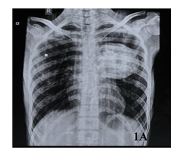
Figure 1A: (CXR PA) Well defined rounded radio-opacity in left lung field in mid zone.
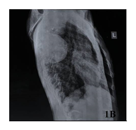
Figure 1B: (CXR LATERAL VIEW) Lesion occupying posterior mediastinum.
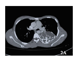
Figure 2A: (AXIAL CT BONE WINDOW) Exophytic, expansile, multiloculated, lytic lesion arising from the posterior aspect of left 6th rib.
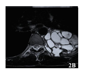
Figure 2B: (Axial T2W MRI) Multiloculated cystic lesion with fluid fluid level in few loculation with low signal intensity peripheral rim.
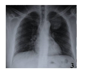
Figure 3: (CXR PA view) Well defined, round opacity in left lung field occupying the upper zone.
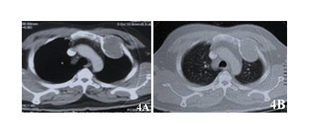
Figure 4 A-B: (Axial CECT chest) Mediastinal and lung window shows well defined, expansile, lytic lesion involving the left first rib on anteromedial aspect, extending from 1st costochondral junction upto the angle of rib, with incomplete peripheral rim of thin cortex, which is interrupted at places.
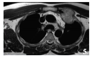
Figure 5: (Axial MRI T2W) Lesion showed T2 hyperintensity consistent with chondroid matrix.
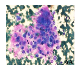
Figure 6A: (Giemsa 400X) Mononuclear chondroblastic cells, singly dispersed and in loose cohesive clusters these cells are round to polygonal, with solitary ovoid nuclei, finely granular chromatin, inconspicuous nucleoli and occasional nuclear grooves.
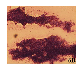
Figure 6B: (Giemsa 600X) Smears are hypercellular with sheets and individually scattered mono nuclear chondroblastic cells.
4. Discussion
Rib lesions can be traumatic, metabolic, inflammatory, neoplastic or related to congenital disorders. Among neoplastic lesions, metastasis is the most common malignant rib lesion, mostly with primary from breast, prostate gland, lung, or kidney. Chondrosarcoma is the most common primary malignant rib lesion, usually occurring at or near the costochondral junction [12]. Most common differential for expansile lytic lesions of bone are –fibrous dysplasia, unicameral bone cyst, aneurysmal bone cyst, chondroblastoma, osteoblastoma, chondromyxoid fibroma and metastasis (renal, thyroid and breast), lymphoma(diffuse large B cell), leukaemia, langerhans cell histiocytosis [5]. Fibrous dysplasia is most common benign tumour of rib where as metastasis and myeloma are most common malignant tumours of rib. Plasmacytoma is another differential diagnosis of expansile lytic lesion of rib which occurs as a result of destruction of the cortex of bone with extension into surrounding soft tissue [6]. Although percutaneous biopsies of rib lesions under image guidance has high diagnostic yield ,lesions with no associated extra-osseous component have a lower biopsy success rate [13]. Unless proven otherwise, all rib lesions must be considered as potentially malignant.Prompt intervention is required .Wide resection with tumor-free margins provide the best chance for cure in both benign and malignant lesions [14].
4.1 Aneurysmal Bone Cyst
Aneurysmal bone cyst can occur as primary as well as secondary tumors. Primary lesion occur commonly under 30 years of age. Clinically they present with mild pain, swelling and restricted movement [11]. On imaging appear as a well defined solitary expansile osteolytic bone lesion, filled with blood. It is because of its expansile nature that it had been named as aneurysmal bone cyst. Aneurysmal bone cyst is composed of numerous blood-filled arteriovenous communications [11]. Expansion of these cysts is due to increased venous pressure or secondary to trauma. It is a metaphyseal or diaphyseal tumor involving usually the long bones. ABC involving the ribs has rare occurance. On MR imaging, it shows multi-lobulated mass lesion with thin peripheral rim of low signal intensity [7]. Presence of fluid levels indicating hemorrhage, is not pathognomonic as it can occur in primary as well as secondary tumors [8].
4.2 Chondroblastoma
Chondroblastoma classically involves the epiphyseal region of long bones and occurrence in chest wall is rare. It is located at an ossification site and is therefore usually found at the costo-chondral or costo-vertebral junction. The lesion may look aggressive on a CT. MRI shows considerable edema in the bone marrow and adjacent soft tissues. [9]. In chest wall it usually involves ribs and scapulae. If found in ribs, it is encountered at later age as compared to its usual occurrence in 2nd decade. As it arises from ossification centre costo-vertebral or costo-chondral junctions are typical site of occurance. These tumors can show aggressive nature and are difficult to differentiate from malignant tumors [10]. Aneurysmal bone cysts can occur secondarily in these tumors. Associated soft tissue and bone marrow edema can be seen.
5. Conclusion
Aneurysmal bone cysts (ABC) and Chondroblastoma have rare occurrence in chest wall. ABC can be primary or secondary with multiloculated appearance. Fluid levels are seen in both primary as well as secondary tumors. Chondroblastoma typically involves costo-vertebral or costo-chondral junctions with later age of occurrence as compared to classical tumors which occur in 2nd decade.
References
- Greenspan A, Jundt G, Remagen W. Differential diagnosis in orthopaedic oncology. 2nd ed. Phila-delphia, Pa: Lippincott Williams & Wilkins (2007): 458-480.
- Teitelbaum SL. Twenty years experience with intrinsic tumors of the bony thorax at a large institution. J Thorac Cardiovasc Surg 63 (1972): 776-782.
- Waller DA, Newman RJ. Primary bone tumours of the thoracic skeleton: an audit of the Leeds regional bone tumour registry.Thorax 45 (1990): 850-855.
- Incarbone M, Pastorino U. Surgical treatment of chest wall tumors. World J Surg 25 (2001): 218-230.
- Hartenstine J, Jackson H, Mortman K. A 38-year-old woman with an osteolytic rib lesion. Chest 149 (2016): e79-e85.
- Sharma D, Rawat V, Yadav R. A rare case of multiple myeloma presenting as lytic lesion of the rib. J Clin Diagn Res 10 (2016): 20-21.
- Beltran J, Simon DC, Levy M, et al. Aneurysmal bone cysts: MR imaging at 1.5 T. Radiol 158 (1986): 689-690.
- Hudson TM, Hamlin DJ, Fitzsimmons JR. Magnetic resonance imaging of fluid levels in an aneurysmal bone cyst and in anticoagulated human blood. Skeletal Radiol 13 (1985): 267-270.
- Zarqane H, Viala P, Dallaudière B, et al. Tumors of the rib., Diagn Interv Imaging 94 (2013): 1095-1108.
- Mayo-Smith W, Rosenberg AE, Khurana JS, et al. Chondroblastoma of the rib: a case report and review of the literature. Clin Orthop Relat Res 251 (1990): 230-234.
- Eisenberg RL. Bubbly Lesions of Bone. Am J Roentgenol 193 (2009): W79-W94.
- Levine BD, Motamedi K, Chow K, et al. CT of rib lesions. Am J Roentegenol 193 (2009): 5-13.
- Sakellaridis T, Gaitanakis S, Piyis A. Rib tumors: a 15-year experience. Gen Thorac Cardiovasc Surg 62 (2014): 434-40.
- Jakanani GC, Saifuddin A. Percutaneous image-guided needle biopsy of rib lesions: a retrospective study of diagnostic outcome in 51 cases. Skeletal Radiol 42 (2013): 85-90.
