Endomyocardial Biopsy-Induced Coronary-Cameral Fistula
Article Information
Sander Trenson, MD 1,2, Daniel Hofer, MD1, Mateusz Sokolski, MD, PhD 1,3, Fran Mikulicic MD 1, Frank Ruschitzka MD, PhD 1, Andreas J. Flammer MD 1, Stephan Winnik MD, PhD1*
1Heart Center Zurich, Department of Cardiology, Zurich, Switzerland
2Department of Cardiovascular Sciences, University of Leuven, Leuven, Belgium
3Department of Heart Diseases, Wroclaw Medical University, Wroclaw, Poland
*Corresponding Authors:Stephan Winnik, Heart Center Zurich, Department of Cardiology, Zurich, Switzerland
Received: 13 October 2020; Accepted: 22 October 2020; Published: 26 October 2020
Citation: Sander Trenson, Daniel Hofer, Mateusz Sokolski, Fran Mikulicic, Frank Ruschitzka, Andreas J. Flammer, Stephan Winnik. Endomyocardial Biopsy-Induced Coronary-Cameral Fistula. Cardiology and Cardiovascular Medicine 4 (2020): 620-622.
View / Download Pdf Share at FacebookAbstract
A 45-year-old patient presented for coronary allograft vasculopathy screening 7 years after heart transplantation. Coronary angiography revealed a fistula between the left anterior descending artery and the right ventricle.
Keywords
Biopsy; Cameral Fistula
Article Details
Case report description
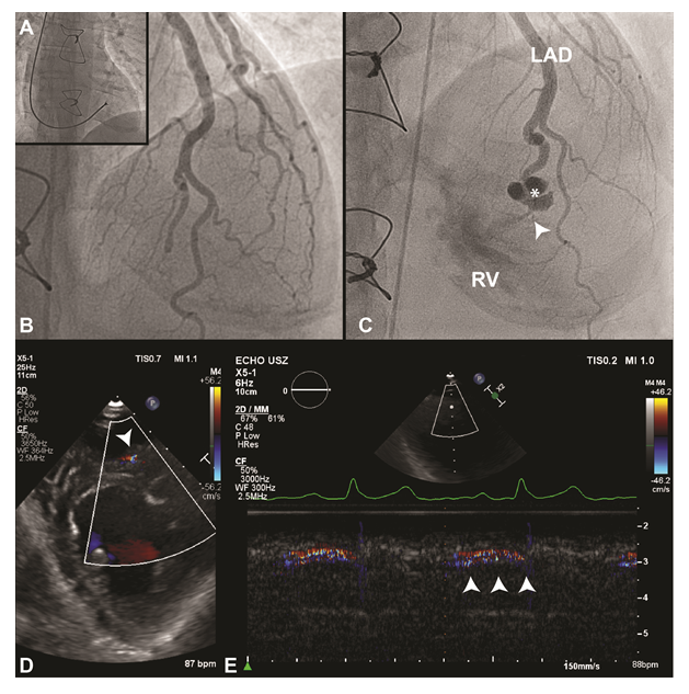
A 45-year-old patient presented for coronary allograft vasculopathy screening 7 years after heart transplantation for a non-compaction cardiomyopathy (Figure 1A, Video S1). He denied symptoms of heart failure. Immunosuppression was within therapeutical range. Coronary angiography revealed a fistula arising from an aneurysmatically dilated septal perforator, originating from the left anterior descending artery, and draining into the right ventricle (Figure 1C, Video S2). Oxygen saturation abruptly increased by 8.5% from mixed-venous to the pulmonary artery, suggesting a hemodynamically non-relevant left-to-right shunt (Qp/Qs 1.35). The fistula was not evident during a coronary angiogram one year post-transplantation (Figure 1B, Video S3). Transthoracic echocardiography confirmed a small left-to-right shunt during diastole at mid-papillary level (Figure 1D-E). A relevant steal phenomenon was excluded by stress echocardiography. Coronary artery fistulas are a not uncommon finding in heart transplant patients with a reported prevalence from 2.8 to 23.2%. These iatrogenic complications of endomyocardial biopsies, mostly originate from the right coronary artery or left anterior descending artery and drain into the right ventricle. In the absence of symptoms, the prognosis is good as long as the shunts are small. In symptomatic patients, closure of the fistula may be achieved through various percutaneous or surgical techniques. In this patient, the aneurysmatic deformation is probably due to a high pressure-gradient, suggestive of a small shunt. Thus, regular clinical evaluation and yearly echocardiographic follow-up for surveillance of right ventricular volume overload and/or apical ischemia during exercise, which are the two most important complications, should be performed.
A- Periprocedural fluoroscopy image of the biopsy localization. B – First coronary angiogram after heart transplantation, showing no fistula. C – Coronary angiogram 7 years after transplantation showing a fistula (white arrow) arising from an aneurysmatically dilated septal perforator (white
asterisk), originating from the left anterior descending artery, and draining into the right ventricle (RV). D - Transthoracic echocardiography confirmed a small left-to-right shunt at mid-papillary level. E – Color doppler M-mode confirmed flow during diastole (white arrows).
Ethics
This report complies with the declaration of Helsinki. Informed consent has been given by the subject.
Disclosure
Sander Trenson reports a grant from the Frans Van de Werf Fund. The other authors report no conflicts of interest to this case report.

