Emphysematous Pyelonephritis and Cystitis Complicated by Pyonephrosis
Article Information
Hassane Moussa Diongole1, Maazou Halidou1, Doutchi Mahamadou1, Abdoul Razack Hamidou Zakou2, Abdoul Aziz Garba1, Kodo Abdoulaye2, Chaibou Laouali2‚ Abdoul Aziz Seribah2
1Faculty of Health Sciences, University/ of Zinder National Hospital, Niger
2National Hospital of Zinder, Niger
*Corresponding Author: Hassane Moussa Diongole, Faculty of Health Sciences, University/ of Zinder National Hospital, Niger.
Received: 30 April 2022; Accepted: 18 May 2022; Published: 24 May 2022
Citation: Hassane Moussa Diongole, Maazou Halidou, Doutchi Mahamadou, Abdoul Razack Hamidou Zakou, Abdoul Aziz Garba, Kodo Abdoulaye, Chaibou Laouali‚ Abdoul Aziz Seribah. Emphysematous Pyelonephritis and Cystitis Complicated by Pyonephrosis. Archives of Nephrology and Urology 5 (2022): 52-56.
View / Download Pdf Share at FacebookKeywords
Cystitis; Pathology; Pyonephrosis; Uroscanner; Emphysematous pyelonephritis cystitis
Cystitis articles; Pathology articles; Pyonephrosis articles; Uroscanner articles; Emphysematous pyelonephritis cystitis articles
Cystitis articles Cystitis Research articles Cystitis review articles Cystitis PubMed articles Cystitis PubMed Central articles Cystitis 2023 articles Cystitis 2024 articles Cystitis Scopus articles Cystitis impact factor journals Cystitis Scopus journals Cystitis PubMed journals Cystitis medical journals Cystitis free journals Cystitis best journals Cystitis top journals Cystitis free medical journals Cystitis famous journals Cystitis Google Scholar indexed journals Pathology articles Pathology Research articles Pathology review articles Pathology PubMed articles Pathology PubMed Central articles Pathology 2023 articles Pathology 2024 articles Pathology Scopus articles Pathology impact factor journals Pathology Scopus journals Pathology PubMed journals Pathology medical journals Pathology free journals Pathology best journals Pathology top journals Pathology free medical journals Pathology famous journals Pathology Google Scholar indexed journals Pyonephrosis articles Pyonephrosis Research articles Pyonephrosis review articles Pyonephrosis PubMed articles Pyonephrosis PubMed Central articles Pyonephrosis 2023 articles Pyonephrosis 2024 articles Pyonephrosis Scopus articles Pyonephrosis impact factor journals Pyonephrosis Scopus journals Pyonephrosis PubMed journals Pyonephrosis medical journals Pyonephrosis free journals Pyonephrosis best journals Pyonephrosis top journals Pyonephrosis free medical journals Pyonephrosis famous journals Pyonephrosis Google Scholar indexed journals Uroscanner articles Uroscanner Research articles Uroscanner review articles Uroscanner PubMed articles Uroscanner PubMed Central articles Uroscanner 2023 articles Uroscanner 2024 articles Uroscanner Scopus articles Uroscanner impact factor journals Uroscanner Scopus journals Uroscanner PubMed journals Uroscanner medical journals Uroscanner free journals Uroscanner best journals Uroscanner top journals Uroscanner free medical journals Uroscanner famous journals Uroscanner Google Scholar indexed journals Emphysematous pyelonephritis cystitis articles Emphysematous pyelonephritis cystitis Research articles Emphysematous pyelonephritis cystitis review articles Emphysematous pyelonephritis cystitis PubMed articles Emphysematous pyelonephritis cystitis PubMed Central articles Emphysematous pyelonephritis cystitis 2023 articles Emphysematous pyelonephritis cystitis 2024 articles Emphysematous pyelonephritis cystitis Scopus articles Emphysematous pyelonephritis cystitis impact factor journals Emphysematous pyelonephritis cystitis Scopus journals Emphysematous pyelonephritis cystitis PubMed journals Emphysematous pyelonephritis cystitis medical journals Emphysematous pyelonephritis cystitis free journals Emphysematous pyelonephritis cystitis best journals Emphysematous pyelonephritis cystitis top journals Emphysematous pyelonephritis cystitis free medical journals Emphysematous pyelonephritis cystitis famous journals Emphysematous pyelonephritis cystitis Google Scholar indexed journals red blood cells articles red blood cells Research articles red blood cells review articles red blood cells PubMed articles red blood cells PubMed Central articles red blood cells 2023 articles red blood cells 2024 articles red blood cells Scopus articles red blood cells impact factor journals red blood cells Scopus journals red blood cells PubMed journals red blood cells medical journals red blood cells free journals red blood cells best journals red blood cells top journals red blood cells free medical journals red blood cells famous journals red blood cells Google Scholar indexed journals diabetic nephropathy articles diabetic nephropathy Research articles diabetic nephropathy review articles diabetic nephropathy PubMed articles diabetic nephropathy PubMed Central articles diabetic nephropathy 2023 articles diabetic nephropathy 2024 articles diabetic nephropathy Scopus articles diabetic nephropathy impact factor journals diabetic nephropathy Scopus journals diabetic nephropathy PubMed journals diabetic nephropathy medical journals diabetic nephropathy free journals diabetic nephropathy best journals diabetic nephropathy top journals diabetic nephropathy free medical journals diabetic nephropathy famous journals diabetic nephropathy Google Scholar indexed journals nephrectomy articles nephrectomy Research articles nephrectomy review articles nephrectomy PubMed articles nephrectomy PubMed Central articles nephrectomy 2023 articles nephrectomy 2024 articles nephrectomy Scopus articles nephrectomy impact factor journals nephrectomy Scopus journals nephrectomy PubMed journals nephrectomy medical journals nephrectomy free journals nephrectomy best journals nephrectomy top journals nephrectomy free medical journals nephrectomy famous journals nephrectomy Google Scholar indexed journals
Article Details
1. Introduction
Emphysematous pyelonephritis is an infection of the renal parenchyma characterized by the presence of air in the cavities and perirenal spaces most often of bacterial origin. Emphysematous pyelonephritis is a rare disease, first described by Kelly and Mac Callum in 1898 [1] and cystitis in 1961 by H. Bailey [2]. It is a medico-surgical emergency because it can threaten the vital prognosis by the occurrence of septic shock. The diagnosis is made by imaging in front of urinary signs. The management is complex and in this situation a nephrectomy is most often necessary. The authors report a case of pyelonephritis and emphysematous cystitis in the same patient treated at Zinder National Hospital.
Observation
Harouna Mayaou is 63 years old, received as an outpatient for impaired kidney function discovered in a context of lumbar pain.
No medical and surgical history was reported. As for the history of the disease, the symptomatology dates back to about 10 days with lumbar pain, reason for which the patient consults in a clinic in the city of Zinder. He received antibiotic therapy with ceftriaxone. Biological explorations showed impaired renal function (creatinine level = 339 micromole / l and urea = 1.09 g / l), hyperleukocytosis at 20,000 / mm3 on the blood count and an Hb level at 14 g / dl. Faced with this AKI, the patient was referred and then hospitalized in the nephrology department. On admission, the general condition was satisfactory, there was good mucocutaneous discoloration and moderate dehydration. Blood pressure = 110/80 mmHg, heart rate = 109 /min and respiratory rate = 21 / min. He presented with an irritative syndrome made up of pollakiuria, micturition burning and urgency. The physical examination noted tenderness of the left lumbar fossa with positive lumbar contact and a positive Jordaneau sign.
The explorations noted:
- The urine dipstick: proteinuria = ++, hematuria = +++; Nitrites =0 and Leucocytes =++
- Cytobacteriological examination of urine = turbid urine with numerous leukocytes but the culture was sterile.
- CRP = 19.8 mg/l
- Kidney ultrasound= the right kidney measures 117 mm for the long axis and the left kidney 189 mm with dilation of the pyelocaliceal cavities
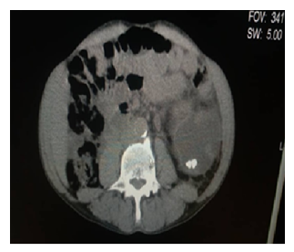
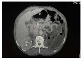
The patient is hospitalized to correct the dehydration with isotonic dirty serum 2 liters per day and the antibiotic therapy started with ceftriaxone 2 g per day, we added ofloxacin 200 mg at a rate of 200 mg morning and evening. The evolution is marked after 4 days of treatment by the disappearance of the clinical signs and the creatinine has returned to 82 micromol/l. Faced with this evolution, we requested a uroscanner which showed a left kidney with bumpy contours. Its pyelocaliceal cavities are dilated of liquid and aeric density. Its cortex, which is enhanced after injection of contrast product, is thinned and measures 3 mm. There is the presence of a 14 mm lower caliceal radiopaque stone. Absence of left renal secretion and excretion. We sent the patient to the urology department, and he received a ureteral stent. A reassessment will be made in 4 weeks for a possible nephrectomy or preservation of the kidney.

Figure 1: CT UROS: axial section without injection of contrast product, passing through the lower pole of the left kidney, showing major pyelocaliceal dilation, associated with lower caliceal lithiasis.

Figure 2: CT UROS: axial slice after injection of contrast product in the late phase showing left nephromegaly, seat of air bubble, without sign of excretion, while pyelocaliceal opacification of the right kidney is observed.

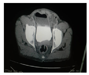
Figure 3: CT UROS: axial section after injection in the late phase, passing through the bladder showing the presence of air in the bladder.
2. Discussion
Emphysematous urinary tract infections are rare and very serious (in some cases). Non-aerobic gas-forming bacteria are the basis of these infections [3]. These are mainly emphysematous pyelonephritis and cystitis. They generally occur in the cases described on fragile ground such as diabetes and bladder neuropathy [2]. The particularity of our patient is the fact that he is not diabetic and that he was not in septic shock. This situation can be explained by the fact that he received early antibiotic therapy. This antibiotic therapy would also explain the absence of germs identified in the culture. In the series of the literature, in more than half of the cases the germ is found the occurrence of renal failure is a criterion of severity, in our context it is multifactorial (infectious and functional). The diagnosis is made by imaging, in particular the uroscanner, which explains the delay in diagnosis. For our observation we requested the uroscanner for the exploration of hydronephrosis in search of a ureteral obstacle in front of the clinical signs of pyelonephritis. In our patient there is no obstacle that could justify this hydronephrosis. But in the series published generally a ureteral lithiasis is found [1]. Management is based on dual antibiotic therapy guided by antibiogram. The urine must also be drained generally by placing ureteral stent [4]. As was the case with our patient. Conservative treatment is based on evaluation of the renal parenchyma. This evaluation allows in the case of non-functionality of the kidney to perform a nephrectomy [5].
3. Conclusion
Emphysematous infections of the urinary tract are rare and, in some cases, life-threatening. The diagnosis must be evoked in front of any severe urinary tract infection with gasogenic germs and confirmed by a uroscanner.
References
- Ouyahia M, Rais A, Gasmi S. et al. Pyélonéphrite emphysémateuse. Ann. Fr. Med. Urgence 3 (2013): 327-330.
- Clémenc A, Meyrieux A, Schneider T. Cystite emphysémateuse. Journal Européen des Urgences et de Réanimation 31 (2019): 57-59.
- Motih Hamade A, Baddouch A, Elalaoui H, et al. Pyélonéphrite emphysémateuse associée à une cystite emphysémateuse.
- Yannick Dimi, Messian gallouo. Pyélonéphrite bilatérale et cystite emphysémateuses compliquant une anurie obstructive: a propos d’un cas et revue de littérature. URO ‘ANDRO 2 (2020).
- Derouiche A, Ouni A, Agreni A, et al. The Management of Emphysematous Pyelonephritis: About 21 Cases. The Journal of Urology 39 (2008): 49-56.
