Electric Phenomenon in Bones as a Result of Piezoelectricity of Hydroxyapatite
Article Information
Maciej Pawlikowski
AGH University Science and Technology, Cath. Mineralogy, Petrography and Geochemistry, al. Mickiewicza 30,
30-049 Kraków, Poland
*Corresponding Author: AGH University Science and Technology, Cath. Mineralogy, Petrography and
Geochemistry, al. Mickiewicza 30, 30-049 Kraków, Poland
Received: 08 May 2017; Accepted: 12 May 2017; Published: 15 May 2017
View / Download Pdf Share at FacebookAbstract
Conducted biomineralogical studies indicate piezoelectric properties of apatite, which is one of the major components of bone. This publication presents hypothetical correlations between piezoelectric properties of bone apatite and the accompanying electromagnetic phenomena in bones and their environment. It notes the correlations between electrical properties of bones and changes in the electromagnetic field around bones, as well as connections between external pressure changes etc. and electrical phenomena in the bones.
Keywords
Apatite Piezoelectricity; Bone; Electromagnetic Phenomena
Article Details
1. Introduction
The first research data regarding piezoelectricity was the result of many years of work by the Curie brothers [1-10]. It applied to electrical phenomena occurring in quartz crystals under mechanical pressure. These studies have been extended to other minerals and synthetics, showing the presence of similar phenomena in them.
Particularly interesting was the study of electrical phenomena in the bones. Those piezoelectric phenomena have long been of interest both in biochemical and electrical terms [11-23] as well as in a practical sense, mainly in relation to bone healing and acceleration of fracture unions [24-28]. The influence of electrical phenomena in the bones on the functioning of bone elements, including bone cell morphology, has also been observed [29-30].
Recent studies have shown that bone piezoelectricity may be associated with the piezoelectric properties of bone hydroxyapatite [18, 22-23]. Those studies have become the basis for the considerations presented in this publication. It attempts to explain the relationship between the piezoelectricity of bone hydroxyapatite and the piezoelectricity of the entire bone. The interpretation of the described phenomena has also been discussed in the context of the potential relationship of bone piezoelectricity to other phenomena. Research on the piezoelectric properties of bones opens new prospects for understanding many biochemical phenomena in bones and other organs [31].
2. Study Conducted with the Author's Own Resources
Considering the impact of bones on the functioning of the body, we need to take into account their huge surface: after converting the surface of all bone trabeculae from all human bones to one surface, we get the value of 380 000 to 420 000 m2 [18]. On that surface, elements of bone such as bone marrow, body fluids, cells and others get in contact with bone collagen containing micro-crystals of hydroxyapatite. On this contact point, there is constant physicochemical equilibrium between the "biological world" and the "mineral world" of bone. On such a vast surface, elements are passed both ways throughout a human’s life, and in the course of life, this system of minerals vs. "biology" is evolving.
All biological processes occurring in the bones are accompanied by electrical phenomena connected, among others, with their piezoelectric properties. Their relevance seems to be difficult to overestimate.
Piezoelectric testing of bone hydroxyapatite is not possible at the present apparatus level due to the nano-size of its crystals. As a result, piezoelectricity of this mineral was studied on large crystals (Figure 1A) and the obtained results can be transferred to nano-crystals in bones. Likewise, electrical phenomena observed in large crystals of apatite can be transferred to nano-crystals found in bones (Figure 1 B).
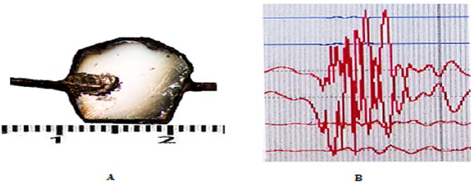
Figure 1: A - Example of a hydroxyapatite plate cut perpendicularly to the optical axis Z after dusting with gold. The dark edge is a paraffin border; after dusting with gold and removal of the border, upper and lower surfaces of the plates were electrically separated. B - Example of the currents chart recorded during a hand movement [22].
When discussing piezoelectricity, we need to pay attention to the details of the relation between collagen and bone hydroxyapatite, as well as electrical phenomena manifesting outside the bone.
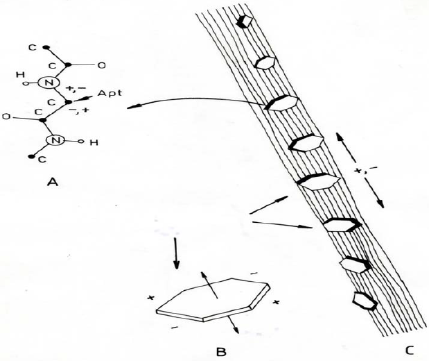
Figure 1: A- Potential site for the transfer of electrical charges from the mechanically excited apatite crystal to the collagen fiber found in the bone structure. B - visualization of the induction of electrical charges in a mechanically deformed plate crystal of apatite. C- schematic of induction of electrical charges in mechanically deformed collagen-apatite fiber (trabecula).
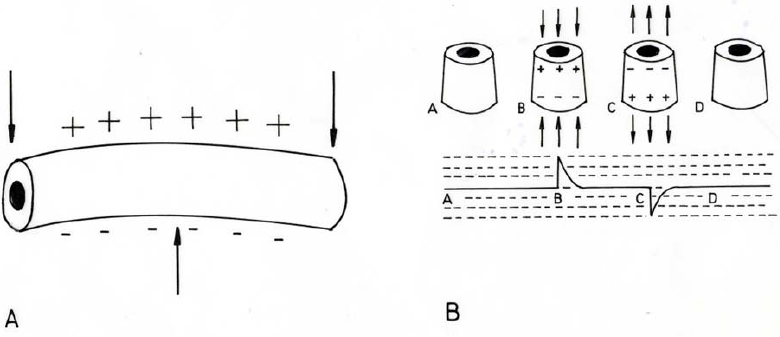
Figure 2: Distribution of electrical charges in bones [19] A - long bone, bent; B - long bone; A, D - at rest; B –weight-bearing; C – not weight-bearing.
While discussing the above phenomena, we should address some of the problems that may be related to them, such as:
- Mechanical bone stress and osteoporosis,
- Influence on the functioning of certain tissues and organs
- External relations between pressure, gravity and electromagnetic field, and electrical phenomena occurring in bones
- Mechanical phenomena occurring in bones vs. external electromagnetic field.
- Influence of piezoelectric phenomena on fracture healing.
3. Mechanical Bone Stress and Osteoporosis
Generated "bone" currents can also affect bone collagen itself, especially the binding between collagen and hydroxyapatite. It appears that currents generated during stress (during walking) promote stability of these bonds. Absence of these currents can probably cause the bindings (ionic: apatite - collagen) to weaken or be broken. The effect is loosening and weakening of the bone structure.
This situation takes place in bone unaffected by gravity, eg. in astronauts in space. Studies show that osteoporosis in conditions where gravity exists is an effect of clogging of micro-arteries in the bones. Crystallizing substances in those arteries are both phosphates and cholesterol [21,23].
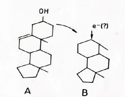
Piezoelectrically induced currents in bone apatite may affect various bone components, including arteries in which endothelial cells have a polar structure (Figure 3).
This issue is complex and not fully explained, and it requires further experimental research. It seems that treating astronauts’ bones in space with weak alternating currents could probably minimize the osteoporosis process.
Figure 3: Sketch of the theoretical influence of currents generated in bone on the structure of destroyed phospholipid with polar structure of endothelial cell membrane. A- phospholipid with polar structure of endothelial
cell membrane. B- structure of partially destroyed phospholipid with endothelial cell membrane (eg. due to inflammation in the body). The site of destruction may be deprived of electrical charges by flowing "bone" currents. This way, the possibility of using damage site as the center of calcification or cholesterol crystallization is reduced.
4. Influence on the Functioning of Certain Tissue and Organs
The bone system: trabeculae – bone marrow and other elements (vessels, nerves, etc.) is a system of physicochemical equilibrium. It changes throughout life, from youth to old age. Its function is to transfer elements from the "biological part" to the "mineral part" and vice versa [18]. So far, electrical phenomena generated in bones in the "biological part" have not been studied.
Their influence seems unquestionable as the vast majority of chemical processes that take place in bones, including their "biological part", are ionic. It is also likely that the microcurrents that form in the bones stimulate the synthesis of bone-generated blood components.
5. External Relations Between Pressure, Gravity and Electromagnetic Field, and Electrical
Phenomena Occurring in Bones
Mechanically bent apatite plates generate differences of potentials at the ends of the crystals [22]. Undoubtedly, they do not cover all the problems that may be associated with piezoelectric phenomena observed in bones. Thus, walking that exerts pressure on the (leg) bones and on the hydroxyapatite microcrystals in trabeculae causes the creation of currents [12]. They are alternating currents in the up-down - up-down direction [11, 19].
It is assumed that generating currents in bone may also occur as a result of changes in external atmospheric pressure, as well as changes in gravity (eg. in astronauts). It can not be ruled out that changes in the electromagnetic field also cause micro-vibrations of bone apatite crystals and generation of currents (Figure 4). The significance of these theoretically discussed phenomena is difficult to overestimate.
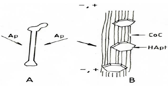
Figure 4: Schematic diagram of the influence of external pressure, gravitation on electrical phenomena in bones. A - example of pressure changes (ζp) on the human femur. B - schematic of generating alternating currents in collagen fiber by hydroxyapatite crystals subjected to pressure by external forces
6. Mechanical Phenomena Occurring in Bones Vs. External Electromagnetic Field
Although the currents generated in a single micro-crystal of bone hydroxyapatite are negligible, the combination of currents generated by moist collagen fibers may probably add up [14]. A result of this phenomenon may be stronger currents and higher voltages at the ends of the collagen fibers, in whole trabeculae, and even on the surface of bone [28].
Because stressing and unstressing of bones leads to the development of alternating currents, walking causes the same flow of currents [29, 19]. Currents generated in this way undoubtedly influence the creation of a weak, alternating electromagnetic field around the bone that generates these currents. It is also an alternating field, and its value depends not only on the value of the generated currents, but also on the enormous surface area of the bone (the sum of the surfaces of all the trabeculae generating currents). It may be too daring to say that the bone that generates alternating currents can be treated like a great transceiver antenna, but it may actually be so.
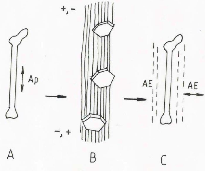
Figure 5: Diagram of the mechanism of generating an alternating electromagnetic field by the femur stressed during walking. A- femur under alternating pressure (ζp) during walking. B- image of a collagen fiber generating alternating currents through deformed hydroxyapatite crystals. C- hypothetical image of electromagnetic field
changes around (AE) under the influence of alternating currents generated in the bone while walking.
7. Influence of Piezoelectric Phenomena on Fracture Healing
Complexity of the bone healing process is obvious. Among its elements are the generation of collagen with centers of apatite crystallization, the phenomena of angiogenesis (Pawlikowski - Biomineralogy of angiogenesis - in print), the re-launch of alkaline phosphatase synthesis, and others.
After a bone fracture, various components of bone are destroyed, including collagen fibers mineralized with apatite in damaged trabeculae. For this reason, as well as through the stabilization of the fracture (plaster fixation, AO plates, etc.), electrical systems occurring in the unbroken, stressed bone get damaged and disturbed. Hence, the use of artificial - external alternating electromagnetic field may stimulate bone healing by approximating the "electrical conditions" occurring in bones to natural conditions. This alternating field may also probably stimulate angiogenesis and other processes. Although some publications suggest the toxicity of the alternating electromagnetic field used to accelerate bone healing [27], it seems very important in this case to match the generated field to the field produced by the natural bone. It requires further research; however, at the current stage of knowledge it is purely theoretical.
Summary
Despite the fact that the currents in bones are very weak (as they are in the brain), they are absolutely elementary. Their incompletely recognized role for the functioning of the whole body is difficult to overestimate.
References
- Curie J, Curie P. Développement, par pression, de l’électricité polaire dans les cristaux hémièdres à faces inclinées. Comptes rendus 91 (1980): 294-295.
- Curie J, Curie P. Sur l’électricité polaire dans les cristaux hémièdre à faces inclinées. présentée par m. desains. Comptes rendus de lAcadémie des sciences 91 (1880): 383-386.
- Curie J, Curie P. Lois du dégagement de l’électricité par pression, dans la tourmaline. Présentée par M. Friedel. Comptes rendus de l’Académie des sciences 92 (1881): 186-188.
- Curie J, Curie P. Les cristaux hémièdres à faces inclinées, comme sources constantes d'électricité. Comptes rendus de l’Académie des sciences 93 (1881): 204-207.
- Curie J, Curie P. Sur les phénomènes électriques de la tourmaline et des cristaux hémièdre à faces inclinées. Présentée par M. Friedel. Comptes rendus de l’Académie des sciences 92 (1881): 350-353.
- Curie J, Curie P. Contractions et dilatations produites par des tensions électriques dans les cristaux hémièdres à faces inclines. Comptes rendus de l’Académie des sciences 93 (1881): 1137-1140.
- Curie J, Curie P. Déformations électriques du quartz. Présentée par. Comptes rendus de l’Académie des sciences 95 (1882): 914-917.
- Curie J, Curie P. Phénomènes électriques des cristaux hémièdre à faces inclinées. J de Physique 1 (1882): 245- 251.
- Curie J, Curie P. Quartz piézo-électrique. Phil Mag 36 (1883): 340-342.
- Curie J, Curie P. Dilatation électrique du quartz. J. de Physique 8 (1889): 149-170.
- Fukada E, Yasuda I. On the piezoelectric effect of bone. J Phys Soc Jpn 10 (1957): 1158-1162.
- Basset CA, Becker OR. Generation of electric potentials by bone in response to mechanical stress. Science 137 (1962): 1063-1064.
- Cerquiglini S, Cignitti M, Salleo A. On the origin of electrical effects produced by stress in the hard tissues of living organisms. Life Science 6 (1967): 2651-2560.
- Anderson JC, Eriksson C. Electrical properties of wet collagen. Nature 21 (1968): 166-168.
- Korostoff E. A Linear piezoelectric model for characterizing stress generated potentials in bone. J Biomech 12 (1979): 335-347
- Johnson MW, Chakkalakal DA, Harper RA, Katz JL. Comparizon of the electromechanical effects in wet and dry bone. J Biomechanics 13 (1980): 437-442.
- Salzstein RA, Pllack SR, Mak AFT, Petrov N. Electromechanical potentials in cortical bone. Journal of biomechanics 20 (1987): 261–270.
- Pawlikowski M, Niedwiedzki T. Mineralogia koci (Mineralogy of bones – book). Polish Acad Sci Kraków 111.
- Szewczenko J. Zjawiska elektryczne w kociach d?ugich. Elektrotechn 81 (2005): 94-97.
- Ahn AC, Grodzinsky AJ. Relevance of collagen piezoelecticity to Wolf’s law: a critical review. Med Eng Phys 31 (2009): 733–741.
- Pawlikowski M. Osteoporosis as a source of tissue mineralization research on osteoporosis therapy and dissolution of arterial mineralization. Journal of Life Sci 8 (2014): 610-625.
- Pawlikowski M. Biomineralogical investigation of apatite piezoelectricity. Traumatologija i ortopedija Rossji T 22 (2016): 57-62.
- Pawlikowski M. Biomineralogy of osteoporosis. Acad Jour Biotechnology 4 (2016): 138-144.
- Weigert M, Wehahn C. The influence of electric potentials on plated bones. Clin Orthop 124 (1977): 20-30.
- Starkebaum W, Pollack SR, Korostoff E J. Microelectrode studies of stress-generated potentials in four-point bending of bone. J Biomed Mater Res 13 (1979): 729-751.
- Pienkowski D, Pollack SR. The origin of stress-generated potentials in fluid-saturated bone. J Orthop Res 1 (1983): 30-41.
- Jacobson-Kram D, Tepper J, Kuo P. Evaluation of potential genotoxicity of pulsed electric end electromagnetic fields used for bone growth stimulation. Mutation Research 388 (1997): 45-57.
- Yasuda I. Fundamental aspects of fracture treatment J Kyoto Med Soc 4 (1953): 395-406.
- Williams WS, Breger L. Analysis of stress distribution and piezoelectric response in cantilever bending of bone and tendon. Annals of the New York Academy of Sciences 238 (1974): 121-130.
- Weinbaum S, Cowin SC, Zeng Yu. A model for the excitation of osteocytes by mechanical loading-induced bone fluid shear stresses. J Biomech 27 (1994): 339–360.
- Wilson ERM, Dowker SEP. Apatite structure. Advances in X-ray Analysis 45 (2002): 172-181
