Effects of Non-Invasive Right Prefrontal Stimulation on Cognitive Performance of ADHD Patients
Article Information
Lihi Bokovza2, Guy Baz2, Hadar Shalev1,2 *
1Department of Psychiatry, Soroka University Medical Center
2Department of Life Sciences and the Zlotowski Centre of Neuroscience, Ben-Gurion University of the Negev, Beer-Sheva, Israel
*Corresponding Author: Dr. Hadar shalev, Department of Psychiatry, Soroka University Medical Center; Department of Life Sciences and the Zlotowski Centre of Neuroscience, Ben-Gurion University of the Negev, Beer-Sheva, Israel
Received: 19 November 2020; Accepted: 25 November 2020; Published: 16 December 2020
Citation:
Lihi Bokovza, Guy Baz, Hadar Shalev. Effects of Non-Invasive Right Prefrontal Stimulation on Cognitive Performance of ADHD Patients. Journal of Psychiatry and Psychiatric Disorders 4 (2020): 400-414.
View / Download Pdf Share at FacebookAbstract
Aims: Attention deficit hyperactivity disorder (ADHD) is a prevalent neuropsychiatric disorder. Recent study from our lab has shown lasting electrophysiological alterations and clinical improvement following 15 daily high-frequency (HF) deep transcranial magnetic stimulation (dTMS) sessions directed to the right prefrontal cortex (rPFC). This paper focuses on the cognitive performance of ADHD patients before and after treatment and compared to non-ADHD controls.
Methods: Ninety-six ADHD and 57 non-ADHD subjects underwent cognitive assessment using computerized battery. Forty-nine ADHD patients completed HF dTMS or sham treatment (n=26 real and 23 sham). Completers underwent a second Mindstreams assessment.
Results: Reaction times (RT) and their standard deviations (RT SD) were significantly higher in the ADHD group compared to non-ADHD. Reduced accuracy was evident within the ADHD group in tasks with high cognitive load and also in a verbal memory and a problem-solving task. HF dTMS resulted in a significant improvement in two tasks, as well as higher effect sizes in other tasks, compared to sham treatment.
Conclusions: Cognitive deficits related to ADHD are manifested in the Mindstreams battery mostly in terms of RT, and in some cases in accuracy. HF dTMS may improve cognitive abilities but an increase of sample sizes is required.
Keywords
Neuropsychiatric disorder; Attention deficit hyperactivity disorder; Deep transcranial magnetic stimulation; ADHD
Neuropsychiatric disorder articles; Attention deficit hyperactivity disorder articles; Deep transcranial magnetic stimulation articles; ADHD articles
Neuropsychiatric disorder articles Neuropsychiatric disorder Research articles Neuropsychiatric disorder review articles Neuropsychiatric disorder PubMed articles Neuropsychiatric disorder PubMed Central articles Neuropsychiatric disorder 2023 articles Neuropsychiatric disorder 2024 articles Neuropsychiatric disorder Scopus articles Neuropsychiatric disorder impact factor journals Neuropsychiatric disorder Scopus journals Neuropsychiatric disorder PubMed journals Neuropsychiatric disorder medical journals Neuropsychiatric disorder free journals Neuropsychiatric disorder best journals Neuropsychiatric disorder top journals Neuropsychiatric disorder free medical journals Neuropsychiatric disorder famous journals Neuropsychiatric disorder Google Scholar indexed journals Attention deficit hyperactivity disorder articles Attention deficit hyperactivity disorder Research articles Attention deficit hyperactivity disorder review articles Attention deficit hyperactivity disorder PubMed articles Attention deficit hyperactivity disorder PubMed Central articles Attention deficit hyperactivity disorder 2023 articles Attention deficit hyperactivity disorder 2024 articles Attention deficit hyperactivity disorder Scopus articles Attention deficit hyperactivity disorder impact factor journals Attention deficit hyperactivity disorder Scopus journals Attention deficit hyperactivity disorder PubMed journals Attention deficit hyperactivity disorder medical journals Attention deficit hyperactivity disorder free journals Attention deficit hyperactivity disorder best journals Attention deficit hyperactivity disorder top journals Attention deficit hyperactivity disorder free medical journals Attention deficit hyperactivity disorder famous journals Attention deficit hyperactivity disorder Google Scholar indexed journals Deep transcranial magnetic stimulation articles Deep transcranial magnetic stimulation Research articles Deep transcranial magnetic stimulation review articles Deep transcranial magnetic stimulation PubMed articles Deep transcranial magnetic stimulation PubMed Central articles Deep transcranial magnetic stimulation 2023 articles Deep transcranial magnetic stimulation 2024 articles Deep transcranial magnetic stimulation Scopus articles Deep transcranial magnetic stimulation impact factor journals Deep transcranial magnetic stimulation Scopus journals Deep transcranial magnetic stimulation PubMed journals Deep transcranial magnetic stimulation medical journals Deep transcranial magnetic stimulation free journals Deep transcranial magnetic stimulation best journals Deep transcranial magnetic stimulation top journals Deep transcranial magnetic stimulation free medical journals Deep transcranial magnetic stimulation famous journals Deep transcranial magnetic stimulation Google Scholar indexed journals ADHD articles ADHD Research articles ADHD review articles ADHD PubMed articles ADHD PubMed Central articles ADHD 2023 articles ADHD 2024 articles ADHD Scopus articles ADHD impact factor journals ADHD Scopus journals ADHD PubMed journals ADHD medical journals ADHD free journals ADHD best journals ADHD top journals ADHD free medical journals ADHD famous journals ADHD Google Scholar indexed journals Reaction times articles Reaction times Research articles Reaction times review articles Reaction times PubMed articles Reaction times PubMed Central articles Reaction times 2023 articles Reaction times 2024 articles Reaction times Scopus articles Reaction times impact factor journals Reaction times Scopus journals Reaction times PubMed journals Reaction times medical journals Reaction times free journals Reaction times best journals Reaction times top journals Reaction times free medical journals Reaction times famous journals Reaction times Google Scholar indexed journals standard deviations articles standard deviations Research articles standard deviations review articles standard deviations PubMed articles standard deviations PubMed Central articles standard deviations 2023 articles standard deviations 2024 articles standard deviations Scopus articles standard deviations impact factor journals standard deviations Scopus journals standard deviations PubMed journals standard deviations medical journals standard deviations free journals standard deviations best journals standard deviations top journals standard deviations free medical journals standard deviations famous journals standard deviations Google Scholar indexed journals High frequency articles High frequency Research articles High frequency review articles High frequency PubMed articles High frequency PubMed Central articles High frequency 2023 articles High frequency 2024 articles High frequency Scopus articles High frequency impact factor journals High frequency Scopus journals High frequency PubMed journals High frequency medical journals High frequency free journals High frequency best journals High frequency top journals High frequency free medical journals High frequency famous journals High frequency Google Scholar indexed journals transcranial magnetic stimulation articles transcranial magnetic stimulation Research articles transcranial magnetic stimulation review articles transcranial magnetic stimulation PubMed articles transcranial magnetic stimulation PubMed Central articles transcranial magnetic stimulation 2023 articles transcranial magnetic stimulation 2024 articles transcranial magnetic stimulation Scopus articles transcranial magnetic stimulation impact factor journals transcranial magnetic stimulation Scopus journals transcranial magnetic stimulation PubMed journals transcranial magnetic stimulation medical journals transcranial magnetic stimulation free journals transcranial magnetic stimulation best journals transcranial magnetic stimulation top journals transcranial magnetic stimulation free medical journals transcranial magnetic stimulation famous journals transcranial magnetic stimulation Google Scholar indexed journals right prefrontal cortex articles right prefrontal cortex Research articles right prefrontal cortex review articles right prefrontal cortex PubMed articles right prefrontal cortex PubMed Central articles right prefrontal cortex 2023 articles right prefrontal cortex 2024 articles right prefrontal cortex Scopus articles right prefrontal cortex impact factor journals right prefrontal cortex Scopus journals right prefrontal cortex PubMed journals right prefrontal cortex medical journals right prefrontal cortex free journals right prefrontal cortex best journals right prefrontal cortex top journals right prefrontal cortex free medical journals right prefrontal cortex famous journals right prefrontal cortex Google Scholar indexed journals semi structured interview articles semi structured interview Research articles semi structured interview review articles semi structured interview PubMed articles semi structured interview PubMed Central articles semi structured interview 2023 articles semi structured interview 2024 articles semi structured interview Scopus articles semi structured interview impact factor journals semi structured interview Scopus journals semi structured interview PubMed journals semi structured interview medical journals semi structured interview free journals semi structured interview best journals semi structured interview top journals semi structured interview free medical journals semi structured interview famous journals semi structured interview Google Scholar indexed journals
Article Details
1. Introduction
Attention-deficit/hyperactivity disorder (ADHD) is a complex, chronic, and potentially debilitating disorder of brain, behavior, and development. ADHD is amongst the most common neurobehavioral problems afflicting children between 6 and 17 years of age, with an estimated prevalence of 7.2% worldwide in this age group [1-3]. Accumulating body of evidence indicates that in the majority of cases ADHD persists into adult life, where it is associated with a range of clinical and psychosocial impairments. Numerous follow-up studies of children with ADHD show that the disorder persists during adolescence and adulthood in up to two-thirds of individuals [refs 3-11] from [4].
Stimulant medications such as methylphenidate and amphetamines have been used to treat the core ADHD symptoms of over activity, impulsivity, and inattention since 1937 [5, 6]. Stimulants are effective in approximately 70% of children with ADHD and are first-line treatments for this disorder [7-10]. However, it is estimated that approximately 30% of a?ected individuals do not adequately respond or cannot tolerate stimulant treatment [8-10] as it causes a variety of adverse events [11]. Moreover, these drugs increase extracellular dopamine in the brain, and thus hold a potential for abuse [12]. Non-stimulant medications are available (atomoxetine, α-2 agonists), however, they are generally not preferred because of lower efficacy compared to stimulants [13-15]. Except for their various side effects, the pharmacological treatments available for treatment of ADHD lack an effect on the core neuropathology of the disorder. Indeed, except for sustained attention, growing body of evidence indicates that medications do not necessarily normalize neuropsychological outcomes of ADHD [16]. Therefore, drug treatment alone may not be sufficient to remediate the deficits associated with ADHD, and development of novel treatments for ADHD can be of great benefit; preferably, treatments that will be designed to target the specific neuropathology of this condition.
One such alternative is non-invasive brain stimulation using transcranial magnetic stimulation (TMS). When applied in sessions of repeated stimulation, TMS can lead to changes in neuronal activity/excitability that outlast the stimulation itself [17]. Repetitive TMS (rTMS) influences neural activity in the short and long term by mechanisms involving neuroplasticity both locally, under the stimulating coil, and at the network level, throughout the brain [18]. Multiple sessions of rTMS are gradually becoming a viable intervention for clinical neuromodulation of various conditions [19]. High frequency (HF) rTMS directed to the right prefrontal cortex (rPFC) was studied in our lab as a possible treatment for adult ADHD and proven effective by Alyagon et al. 2020 [20].
This study was conducted in continuation to Alyagon et al. 2020 [20] in order to elaborate discrimination techniques between ADHD and non-ADHD subjects and to test the effect of rTMS treatment directed to the rPFC on specific cognitive performances of ADHD patients. For that end we used the Mindstreams battery. Mindstreams by NeuroTrax is a validated battery of tasks that can be used to assess a range of cognitive domains. It includes tasks that require attention, memory, executive functions, response inhibition, continuous performance and information processing. All of the above are well established as deficient in adult subjects with ADHD [21, 22]. For each task, accuracy, reaction time and standard deviation of reaction time are calculated. Mindstreams’ construct validity as a tool for assessing ADHD was confirmed in 2007, based on a sample of 28 ADHD and 49 healthy men [21].
2. Methods
2.1 Participants
Between February 2013 to March 2019, 96 ADHD patients and 57 healthy controls were recruited using ads or mass university email. All participants provided written informed consent. ADHD subjects were assigned to either active or sham treatment (n=33, 28 respectively). Forty-nine ADHD subjects completed the treatment phase (n=26 real and 23 sham). Participants received information concerning the study requirements over the phone and were further screened by a psychiatrist using a semi structured interview (SCID) based on DSM-V criteria to verify ADHD diagnosis and to rule out psychiatric comorbidities. No minimum score of CAARS or other questionnaire was required. Detailed inclusion and exclusion criteria are listed below. ADHD participants were required to refrain from taking any psychostimulant medication for a week before, and during the three-week treatment phase. ADHD participants did not receive financial compensation, and healthy controls received a symbolic payment for their participation. The experimental protocol was approved by the ethics committee of the Soroka University Medical Center and registered at the NIH (NCT01737476).
2.2 Inclusion criteria
(1) 18-65 years old; (2) ADHD diagnosis according to DSM-V; (3) consent to abstain from other ADHD treatment for 4 weeks, starting one week prior to the first treatment and ending after the last treatment; (4) satisfactory responses on the TASS questionnaire; (5) gave informed consent for participation in the study.
2.3 Exclusion criteria
(1) additional axis 1 psychiatric disorder according to DSM-V; (2) treatment by antipsychotics, antidepressants or mood stabilizers; (3) known intolerance to TMS; (4) Cognitive or functional disability, diagnosed according to DSM-V criteria; (5) high risk for suicidality as assessed during the screening interview; (6) subjects who suffer from an unstable physical disease such as high blood pressure (>150 mmHg systolic / diastolic > 110 mmHg) or acute, unstable cardiac disease; (7) history of epilepsy or seizure – for the patient or for first degree relatives; (8) history of head injury or cerebral infraction that caused irreversible damage; (9) history of any metal in the head (outside the mouth); (10) metallic particles in the eye, implanted cardiac pacemaker or any intracardiac lines, implanted neurostimulators, intracranial implant (e.g., aneurysm clips, shunts, stimulators, cochlear implants, or electrodes) or implanted medical pumps; (11) alcohol or other substance abuse or dependence during the last 12 months before recruitment; (12) subject cannot communicate reliably with the investigator; (13) participation in a clinical trial during this clinical trial, or within the past 3 months before the beginning of this clinical trial; (14) subject cannot sign an informed consent form; (15) known or suspected pregnancy.
2.4 Study design
Healthy controls performed one session of cognitive assessment. ADHD patients were randomly assigned to real or sham deep TMS treatment which was composed of 15 treatments in a course of 3 weeks. Cognitive assessment for the ADHD group was performed on the days of the first and last treatments.
2.5 Cognitive assessment
Cognition was assessed by using the Mindstreams [23, 24] computerized battery of tests, which includes nine tasks that assess performance across an array of cognitive domains including: memory, executive function, visual spatial perception, verbal function, attention, information processing speed, and motor skills. The tasks are described on Schweiger et al., 2007 [21]. Healthy controls completed Mindstreams testing once, and ADHD participants completed it twice (before and after treatment).
2.6 TMS coils and stimulation
TMS was delivered using a Magstim Rapid2 stimulator (Magstim, UK) inducing biphasic pulses. The active and sham H-coils were built together into a single helmet, and coil selection was performed by magnetic cards, which were individual per patient. Thus, neither the patient nor the operator knew which coil was activated, i.e. to which treatment group the patient was assigned. The Real stimulation was delivered using an H6-coil which was specially designed to unilaterally stimulate the rPFC, based on the principles of H-coil family design [25-27]. Placebo stimulation was performed with a sham coil placed in the same helmet encasing the active TMS coil. The sham coil produces a similar acoustic artifact and scalp sensation as the active coil. The sham coil induces only a negligible electric field inside the brain itself due to a very rapid reduction of the field as a function of distance insured by the non-tangential orientation of the sham coil relative to the scalp and by elements producing significant field cancellation [28].
Individual left hand resting motor threshold (RMT) was measured at the beginning of each treatment, and the helmet was then moved 5 cm anteriorly and 2 cm laterally from the motor hot spot to target the rPFC. The stimulation included 40 trains, each comprised of 2sec stimulation at 120% of RMT at 18 Hz, followed by a 20sec inter train interval (ITI). Maximal stimulation intensity was set to 80% of stimulator output. According to subject’s need, stimulation intensity during the first treatments was lowered to a minimum of 100% of RMT to improve treatment tolerability.
2.7 Treatment procedure
Individual left hand resting motor threshold (RMT) was measured at the beginning of each treatment [29]. The TMS coil was then moved 5 cm anteriorly and 2 cm laterally from the motor hot spot to target the rPFC. These placement parameters were set according to the H-coil design and are fitted to stimulate the DLPFC [30] and the VLPFC [31] (which are the two most implicated pre-frontal targets in ADHD related network). Prior to each stimulation session, participants completed a short computerized cognitive training [32] in order to provoke their attention. This was done in accordance with previous publication suggesting that pre-treatment provocation may increase clinical response to rTMS [33-35].
2.8 Statistical analysis
Statistical analyses were conducted using STATISTICA software [36]. ADHD vs non-ADHD comparison was performed using one tailed Student’s T-test. In cases of violation of the equal variance assumption, Welch's T-test was performed instead of Student’s T-test. For all T-tests, the alternative hypothesis specifies that the ADHD group is greater than the non-ADHD group. Real vs sham-treatment comparison was performed using 2-way mixed model ANOVA with time (pre vs post treatment) as within-subjects factor and group (real vs sham rTMS stimulation) as between-subjects factor.
3. Results
3.1 ADHD vs non-ADHD
The overall differences between ADHD and non-ADHD subjects are detailed in the supplementary results. The major differences between these groups were assessed according to their performance on the Go-NoGo response inhibition task, Stroop task and Staged Information Processing Speed task, since these tasks assess executive functions, attention and information processing speed. Throughout all these tasks, reaction times and their standard deviations were significantly higher in the ADHD group, as can be seen in table 1. Differences in accuracy were mostly negligible, probably due to ceiling effect. For tasks where ceiling effect was not reached, differences in accuracy were indeed evident: accuracy was significantly lower in the ADHD group on the more demanding levels of the SIPS task (Figure 1), i.e. the fast speed two-digit arithmetics (level 2.3 of SIPS, t148=-1.979, p=0.025, Cohen’s d=0.33) and the medium speed three-digit arithmetics (level 3.2 of SIPS, t143=-3.708, p<0.001, Cohen’s d=0.63). On the surface, it seems like there is no difference in accuracy between the groups in the most demanding level (3.3). In practice, although both groups reached a floor effect, the difference lies in the number of subjects who performed well enough to ahcieve a score on this level (89% of non-ADHD compared to only 72% of ADHD subjects).
In the verbal memory task, accuracy of immediate recognition was significantly poorer in the ADHD group in the first and second repetitions (Rep1: t148.9=-3.482, p<0.001a, Cohen’s d=0.51; Rep2: t130.9=-3.819, p<0.001, Cohen’s d=0.53), but no significant difference was evident in recognition accuracy after a delay (Figure 2a). However, no difference was found between the groups in the non-verbal memory task, on both immediate and delayed recognition (Figure 2b).
Poorer performance was also evident in the problem-solving task, where accuracy of ADHD patients was significantly lower compared to healthy subjects (t143.1=-3.992, p<0.001a, Cohen’s d=0.58, Figure 3).
|
ADHD |
Non ADHD |
Statistics |
|||
|
RT |
GNG |
366.3 |
348.1 |
T148=2.255, p=0.013, Cohen’s d=0.38 |
|
|
SI |
Letter Color Color vs. Meaning |
418.8 436.9 |
395.4 362.7 |
T143.9=1.983, p=0.025a, Cohen’s d=0.31 T117.4=3.894, p<0.001a, Cohen’s d=0.55 |
|
|
SIPS |
All levels |
807.4 |
718.4 |
T151=5.304, p<0.001a, Cohen’s d=0.89 |
|
|
RT SD |
GNG |
87.8 |
71.2 |
T147.8=3.851, p<0.001a, Cohen’s d=0.58 |
|
|
SI |
Letter Color Color vs. Meaning |
119.1 166.5 |
87.4 79.9 |
T145.6=3.358, p<0.001a, Cohen’s d=0.51 T127.8=4.504, p<0.001a, Cohen’s d=0.65 |
|
|
SIPS |
All levels |
200 |
168.1 |
T151=4.687, p<0.001, Cohen’s d=0.78 |
|
Table 1: Statistical analysis for ADHD vs non-ADHD subjects’ reaction times (RT) and their standard deviations (RT SD) in the Go-NoGo response inhibition task (GNG), Stroop interference task (SI) and Staged Information Processing Speed task (SIPS). The SIPS is comprised of three levels of information processing load: single digit, two-digit arithmetic problems (e.g., 5-1), and three-digit arithmetic problems (e.g., 3+2-1). For each of these three levels, stimuli are presented at three different rates, incrementally increasing as testing continues. Since RT and RT SD were significantly higher in the ADHD group in all nine levels, the results presented in this table are an average of all nine levels of this task.
For all tests, the alternative hypothesis specifies that group ADHD is greater than group non-ADHD.
a Welch's T-test was performed instead of Student's T-test in cases where Levene's test was significant, suggesting a violation of the equal variance assumption.
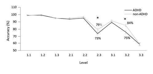
Figure 1: The staged information processing speed task (SIPS) is comprised of three levels of information processing load: single digit, two-digit arithmetic problems (e.g., 5-1), and three-digit arithmetic problems (e.g., 3+2-1). For each of these three levels, stimuli are presented at three different rates, incrementally increasing as testing continues. Accuracy in the SIPS task was significantly lower in the ADHD group compared to non-ADHD only on the more demanding levels of the task, i.e. the fast speed two-digit arithmetics (level 2.3, t148=-1.979, p=0.025, Cohen’s d=0.33) and the medium speed three-digit arithmetics (level 3.2, t143=-3.708, p<0.001, Cohen’s d=0.63).
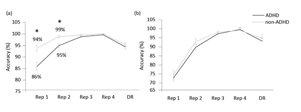
Figure 2: Accuracy in verbal and non-verbal memory tasks. In the verbal memory task (a), accuracy of immediate recognition was significantly poorer in the ADHD group in the first and second repetitions (Rep1: t148.9=-3.482, p<0.001a, Cohen’s d=0.51; Rep2: t130.9=-3.819, p<0.001, Cohen’s d=0.53), but no significant difference was evident in recognition accuracy after a delay (delayed recognition, DR). However, no difference was found between the groups in the non-verbal memory task (b), on both immediate and delayed recognition.
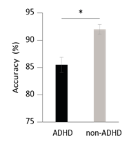
Figure 3: Problem solving task. Accuracy of ADHD patients was significantly lower compared to non-ADHD subjects (t143.1= -3.992, p<0.001a).
3.2 Real vs sham HF rTMS treatment
Performance has significantly improved in the second testing session (post treatment) in both groups, as indicated by a significant effect for time in a substantial number of parameters.
|
HF dTMS |
Sham |
|||||||
|
PRE |
POST |
Cohen's d |
PRE |
POST |
Cohen's d |
|||
|
RT |
GNG |
366.7 |
345 |
0.80 |
377.1 |
356.9 |
0.51 |
|
|
SI |
Letter Color Color vs. Meaning |
431.2 449.9 |
377.4 356.2 |
0.62 0.85 |
414.8 420.9 |
392.3 348.1 |
0.26 0.67 |
|
|
SIPS |
All levels |
821.6 |
716.4 |
1.47 |
808.4 |
727.1 |
0.91 |
|
|
RT SD |
GNG |
90.4 |
78.8 |
0.42 |
90.8 |
74.4 |
0.66 |
|
|
SI |
Letter Color Color vs. Meaning |
134.2 193.3 |
82.4 90 |
0.63 0.78 |
93.8 141.2 |
93.9 71.6 |
-0.08 0.53 |
|
|
SIPS |
All levels |
203.4 |
166 |
0.84 |
200.1 |
176.1 |
0.61 |
|
Table 2: Statistical analysis for the effect of real vs sham HF rTMS treatment on performance in the Go-NoGo response inhibition task (GNG), Stroop interference task (SI) and Staged Information Processing Speed task (SIPS), as measured by reaction times (RT) and their standard deviations (RT SD). Although not statistically significant, for some parameters, effect size (measured by Cohen’s d) was higher in the real treatment, compared to sham.
Notably, significant time X group interaction was observed in SD of RT under Stroop interference (F1,433.005 p=0.045 one tailed, Cohen’s d=0.78 for real and 0.53 for sham, Figure 4) and in accuracy at level 3.2 (medium speed, three-digit arithmetic) of SIPS (F1,45=2.991, p=0.045, Cohen’s d=0.51 for real and 0.03 for sham, Figure 5). Although not statistically significant, for some parameters, effect size (measured by Cohen’s d) was higher in the real treatment, compared to sham (Table 2), suggesting that an increase of sample sizes would result in a significantly greater improvement as a result of real, but not sham, stimulation.
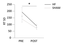
Figure 4: Stroop Interference. Decrease in standard deviation of reaction time during the interference stage of the Stroop task was more pronounced after Real high frequency (HF) rTMS treatment, compared to Sham. F1, 43=3.005, p=0.045 one tailed, Cohen’s d=0.78 for real and 0.53 for sham.
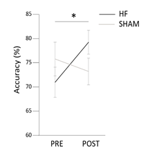
Figure 5: Staged Information Processing Speed (SIPS). Group X Time interaction for accuracy in level 3.2 (medium speed, three-digit arithmetics) of SIPS (F1, 45=2.991, p=0.045, Cohen’s d=0.51 for real and 0.03 for sham) indicates improvement in performance in the Stroop task after real high frequency (HF), but not sham, rTMS.
4. Discussion
The results of the present study support the claim that Mindstreams' version of cognitive tests is valid for assessment of cognitive performance, despite its methodological modifications compared to the known versions of these tests. As expected, in a large sample size of ADHD and non-ADHD participants, ADHD patients performed poorer compared to non-ADHD subjects in tasks that required sustained attention, response inhibition and information processing. ADHD patients also displayed a slower learning curve in a verbal memory test. Lastly, ADHD patients performed poorer on the Problem-Solving task. These findings are in line with several meta-analyses that show that adults with ADHD generally do not perform as well as healthy controls on neuropsychological tests, particularly those related to sustained and focused attention, inhibition, verbal fluency, and verbal memory [37]. Nevertheless, these findings are insufficient to determine that the Mindstreams battery can satisfactorily distinguish between individuals with ADHD and healthy controls. Actually, it seems that neuropsychological tests in general have a poor ability to discriminate between patients diagnosed with ADHD and patients not diagnosed with ADHD, as Pettersson et al. 2018 also reported [37]. One possible reason for this lack of discriminant validity may lay in the heterogeneity of this disorder: symptom manifestation varies from one patient to another in terms of attention deficit, hyperactivity and impulsivity. Perhaps different subtypes of ADHD will lead to different profiles of performance in neuropsychological tests. Therefore, characterization of ADHD subtypes, based on questionnaires and brain activity, followed by cross validation with cognitive performance (measured using neuropsychological test), may pave the way towards better diagnosis of this disorder.
Throughout most of the tasks, reaction time variability was higher for ADHD patients compared to non-ADHD subjects. Indeed, intraindividual variability is considered by many to be a core and stable feature of the disorder, and referred to frequently as a ubiquitous and etiologically important characteristic of ADHD [38]. For this reason, it was proposed as a potential endophenotype of the disorder [39, 40]. Accordingly, neurological correlates of intraindividual variability include regions implicated consistently in ADHD, including dorsolateral prefrontal, orbital frontal, and anterior cingulate cortices [41, 42]. In line with that, our results demonstrate that rTMS directed to the right dorsolateral prefrontal cortex resulted in significant decrease in RT variability under Stroop interference - a task that requires behavioral inhibition [43], a core deficit in ADHD [44, 45] which is strongly associated with rPFC dysfunction [46, 47]. Although RT variability was not reduced significantly in other tasks, effect sizes (measured by Cohen’s d) were higher in the Real group, compared to Sham (Table 2), suggesting that an increase of sample sizes would result in a significantly greater improvement as a result of real, but not sham, stimulation.
Another well-established feature of ADHD is slowed processing speed, as manifested across a wide variety of tasks including (1) naming speed, as measured by tasks such as Stroop color naming or word reading and (2) reaction time on Go-NoGo tasks [refs from [48]. This “slowing” in ADHD was associated with deficits in fundamental components of executive function underlying processing speed [48]. Our results show that this slowing down is not resolved in adulthood: ADHD patients’ RTs were significantly longer, compared to non-ADHD participants, in Go-NoGo, Stroop, and SIPS tasks. Moreover, performance was affected by the required processing load, as evident in poorer performance as the SIPS task became more demanding (levels 2.3, 3.2, 3.3). Performance in those tasks requires attention, information processing, working memory and inhibitory control, which are all affected by the PFC [49, 50]. rTMS directed to the rPFC indeed resulted in significant improvement of accuracy in level 3.2 of the SIPS, in the Real, but not Sham, group.
Our study also revealed impairments in the verbal memory of adults with ADHD. Namely, significantly lower accuracies in the first two repetitions of immediate recognition. Notably, no difference between the groups was observed in the third and fourth immediate repetitions, as well as in the delayed recognition. This observation is consistent with previous publications, both in children [51, 52] and in adults, as reviewed by Woods et al. 2002 [22]. It has been argued that word list learning tasks are difficult for individuals with ADHD because they lack inherent semantic structure that might aid with the active organization of material during learning [22]. It appears that the memory deficits exhibited by adults with ADHD reflect encoding and retrieval deficits, rather than consolidation and/or storage problems; a profile often associated with frontal-subcortical impairment [22]. The PFC regulates attention and behavior through its widespread connections to subcortical structures, as well as sensory and motor cortices [53]. Therefore, we hypothesized that rTMS directed to the rPFC would have an impact on this circuitry, and that this impact would manifest in the task. Unfortunately, we could not evaluate treatment effect on verbal memory, since on the second testing session (post treatment), both Real and Shame groups exhibited a ceiling effect with accuracies above 97% starting from the first repetition. It’s important to note that the word pairs were similar on both sessions. Thus, we can infer that the cognitive process that was taking place on the second testing session was somewhat reduced compared to the first session; i.e. the learning process that is required for formation of new associations between words did not take place in the second session, as the patients already formed these associations three weeks earlier. This finding stresses the importance of amending the task by changing the word pairs that are being used in this task each session.
The results of the present study would go further, suggesting that nonverbal memory is unimpaired in individuals with ADHD. In particular, the ADHD group did not underperform on the non-verbal memory task, which assesses memory of spatial orientation of geometric patterns and symbols. Negative findings on various other visual memory tasks have been previously reported, both in children [52] and in adults, as reviewed by Woods et al. 2002 [22]. In summary, we show that cognitive deficits related to ADHD are manifested in the Mindstreams battery of computerized cognitive tests mostly in terms of reaction time, and that under high processing load, cognitive deficits manifest also in accuracy. We also show that fifteen sessions of deep rTMS directed to the rPFC seem to improve cognitive abilities.
Acknowledgment
We are grateful to all the participants who took part in this study. Liran Fridman deserves special thanks for developing a program for data extraction of the Mindstreams data from PDF to Excel.
Funding
This research was supported by the Israeli Ministry of Science, Technology and Space.
References
- Berger I. Diagnosis of attention deficit hyperactivity disorder: much ado about something. Isr Med Assoc J IMAJ 13 (2011): 571-574.
- Thomas R, Sanders S, Doust J, et al. Prevalence of Attention-Deficit/Hyperactivity Disorder: A Systematic Review and Meta-analysis | American Academy of Pediatrics. Pediatrics 135 (2015): e994-e1001.
- Fayyad J, De Graaf R, Kessler R, et al. Cross-national prevalence and correlates of adult attention-deficit hyperactivity disorder | The British Journal of Psychiatry | Cambridge Core. Br J Psychiatry 190 (2007): 402-409.
- Kooij SJ, Bejerot S, Blackwell A, et al. European consensus statement on diagnosis and treatment of adult ADHD: The European Network Adult ADHD. BMC Psychiatry 3 (2010): 67.
- Bradley C. The behavior of children receiving benzedrine. Am J Psychiatry 94 (1937): 577-585.
- Swanson JM, McBurnett K, Wigal T, et al. Effect of stimulant medication on children with attention deficit disorder: A “review of reviews.” Except Child Rest 60 (1993): 154.
- The MTA Cooperative Group. A 14-Month Randomized Clinical Trial of Treatment Strategies for Attention-Deficit/Hyperactivity Disorder. Arch Gen Psychiatry 56 (1999): 1073-1086.
- Barkley RA. A Review of Stimulant Drug Research with Hyperactive Children. J Child Psychol Psychiatry 18 (1977): 137-165.
- Spencer T, Biederman J, Wilens T, et al. Pharmacotherapy of Attention-Deficit Hyperactivity Disorder across the Life Cycle. J Am Acad Child Adolesc Psychiatry 35 (1996): 409-432.
- Faraone SV, Buitelaar J. Comparing the efficacy of stimulants for ADHD in children and adolescents using meta-analysis. Eur Child Adolesc Psychiatry 19 (2010): 353-364.
- Biederman J, Spencer T, Wilens T. Evidence-based pharmacotherapy for attention-deficit hyperactivity disorder. Int J Neuropsychopharmacol 7 (2004): 77-97.
- Biederman J. Attention-Deficit/Hyperactivity Disorder: A Selective Overview. Biol Psychiatry 57 (2005): 1215-1220.
- Sharma A, Couture J. A Review of the Pathophysiology, Etiology, and Treatment of Attention-Deficit Hyperactivity Disorder (ADHD). Ann Pharmacother 48 (2014): 209-225.
- Dopheide JA, Pharm D, Pliszka SR. Attention-Deficit-Hyperactivity Disorder: An Update. Pharmacother J Hum Pharmacol Drug Ther 29 (2009): 656-679.
- Pliszka S. Practice Parameter for the Assessment and Treatment of Children and Adolescents With Attention-Deficit/Hyperactivity Disorder. J Am Acad Child Adolesc Psychiatry 46 (2007): 894-921.
- Advokat C. What are the cognitive effects of stimulant medications? Emphasis on adults with attention-deficit/hyperactivity disorder (ADHD). Neurosci Biobehav Rev 34 (2010): 1256-1266.
- Thut G, Pascual-Leone A. A Review of Combined TMS-EEG Studies to Characterize Lasting Effects of Repetitive TMS and Assess Their Usefulness in Cognitive and Clinical Neuroscience. Brain Topogr 22 (2009): 219.
- Diana M, Raij T, Melis M, et al. Rehabilitating the addicted brain with transcranial magnetic stimulation. Nat Rev Neurosci 18 (2017): 685-693.
- Lefaucheur J-P, André-Obadia N, Antal A, et al. Evidence-based guidelines on the therapeutic use of repetitive transcranial magnetic stimulation (rTMS). Clin Neurophysiol 125 (2014): 2150-2206.
- Alyagon U, Shahar H, Hadar A, et al. Alleviation of ADHD symptoms by non-invasive right prefrontal stimulation is correlated with EEG activity. NeuroImage Clin 26 (2020): 102206.
- Schweiger A, Abramovitch A, Doniger GM, et al. A clinical construct validity study of a novel computerized battery for the diagnosis of ADHD in young adults. J Clin Exp Neuropsychol 29 (2007): 100-111.
- Woods SP, Lovejoy DW, Ball JD. Neuropsychological Characteristics of Adults with ADHD: A Comprehensive Review of Initial Studies. Clin Neuropsychol 16 (2002): 12-34.
- Dwolatzky T, Whitehead V, Doniger GM, et al. Validity of a novel computerized cognitive battery for mild cognitive impairment. BMC Geriatr 3 (2003): 4.
- Schweiger A, Doniger G, Dwolatzky T, et al. Reliability of a novel computerized neuropsychological battery for mild cognitive impairment. Acta Neuropsychol 1 (2003): 407-413.
- Roth Y, Zangen A. Reaching Deep Brain Structures: The H-Coils. In: Rotenberg A, Horvath JC, Pascual-Leone A, editors. Transcranial Magnetic Stimulation [Internet]. New York, NY: (2014): 57-65. (Neuromethods).
- Roth Y, Zangen A, Hallett M. A Coil Design for Transcranial Magnetic Stimulation of Deep Brain Regions. J Clin Neurophysiol 19 (2002): 361.
- Zangen A, Roth Y, Voller B, et al. Transcranial magnetic stimulation of deep brain regions: evidence for efficacy of the H-Coil. Clin Neurophysiol 116 (2005): 775-779.
- Isserles M, Shalev AY, Roth Y, et al. Effectiveness of Deep Transcranial Magnetic Stimulation Combined with a Brief Exposure Procedure in Post-Traumatic Stress Disorder – A Pilot Study. Brain Stimulat 6 (2013): 377-383.
- Levkovitz Y, Isserles M, Padberg F, et al. Efficacy and safety of deep transcranial magnetic stimulation for major depression: a prospective multicenter randomized controlled trial. World Psychiatry 14 (2015): 64-73.
- Fitzgerald PB, Maller JJ, Hoy KE, et al. Exploring the optimal site for the localization of dorsolateral prefrontal cortex in brain stimulation experiments. Brain Stimulat 2 (2009): 234-237.
- Vanneste S, Ridder DD. The involvement of the left ventrolateral prefrontal cortex in tinnitus: a TMS study. Exp Brain Res 221 (2012): 345-350.
- Stern A, Malik E, Pollak Y, et al. The Efficacy of Computerized Cognitive Training in Adults With ADHD: A Randomized Controlled Trial. J Atten Disord 20 (2016): 991-1003.
- Isserles M, Shalev AY, Roth Y, et al. Effectiveness of Deep Transcranial Magnetic Stimulation Combined with a Brief Exposure Procedure in Post-Traumatic Stress Disorder – A Pilot Study. Brain Stimulat 6 (2013): 377-383.
- Carmi L, Alyagon U, Barnea-Ygael N, et al. Clinical and electrophysiological outcomes of deep TMS over the medial prefrontal and anterior cingulate cortices in OCD patients. Brain Stimulat [Internet] (2017).
- Dinur-Klein L, Dannon P, Hadar A, et al. Smoking Cessation Induced by Deep Repetitive Transcranial Magnetic Stimulation of the Prefrontal and Insular Cortices: A Prospective, Randomized Controlled Trial. Biol Psychiatry 76 (2014): 742-749.
- STATISTICA (data analysis software system), version 13. Dell Inc. (2016).
- Pettersson R, Söderström S, Nilsson KW. Diagnosing ADHD in Adults: An Examination of the Discriminative Validity of Neuropsychological Tests and Diagnostic Assessment Instruments. J Atten Disord 22 (2018): 1019-1031.
- Kofler MJ, Rapport MD, Sarver DE, et al. Reaction time variability in ADHD: A meta-analytic review of 319 studies. Clin Psychol Rev 33 (2013): 795-811.
- Castellanos FX, Sonuga-Barke EJS, Scheres A, et al. Varieties of Attention-Deficit/Hyperactivity Disorder-Related Intra-Individual Variability. Biol Psychiatry 57 (2005): 1416-1423.
- Sonuga-Barke EJS, Castellanos FX. Spontaneous attentional fluctuations in impaired states and pathological conditions: A neurobiological hypothesis. Neurosci Biobehav Rev 31 (2007): 977-986.
- Bellgrove MA, Hester R, Garavan H. The functional neuroanatomical correlates of response variability: evidence from a response inhibition task. Neuropsychologia 42 (2004): 1910–1916.
- MacDonald SWS, Nyberg L, Bäckman L. Intra-individual variability in behavior: links to brain structure, neurotransmission and neuronal activity. Trends Neurosci 29 (2006): 474-480.
- Miyake A, Friedman NP, Emerson MJ, et al. The Unity and Diversity of Executive Functions and Their Contributions to Complex, Frontal Lobe, Tasks: A Latent Variable Analysis. Cognit Psychol 41 (2000): 49-100.
- Huang-Pollock CL, Nigg JT, Carr TH. Deficient attention is hard to find: applying the perceptual load model of selective attention to attention deficit hyperactivity disorder subtypes. J Child Psychol Psychiatry 46 (2005):1211-1218.
- Sonuga-Barke EJS. Causal Models of Attention-Deficit/Hyperactivity Disorder: From Common Simple Deficits to Multiple Developmental Pathways. Biol Psychiatry 57 (2005): 1231-1238.
- Aron AR, Robbins TW, Poldrack RA. Inhibition and the right inferior frontal cortex. Trends Cogn Sci 8 (2004): 170-177.
- Aron AR, Robbins TW, Poldrack RA. Inhibition and the right inferior frontal cortex: one decade on. Trends Cogn Sci 18 (2014): 177-185.
- Jacobson LA, Ryan M, Martin RB, et al. Working memory influences processing speed and reading fluency in ADHD. Child Neuropsychol 17 (2011): 209-224.
- The prefrontal cortex: Executive and cognitive functions. New York, NY, US: Oxford University Press; (Roberts AC, Robbins TW, Weiskrantz L, editors. The prefrontal cortex: Executive and cognitive functions) 8 (1998): 248.
- Munakata Y, Herd SA, Chatham CH, et al. A unified framework for inhibitory control. Trends Cogn Sci 15 (2011): 453-459.
- Cohen MJ. Children’s Memory Scale. In: Kreutzer JS, DeLuca J, Caplan B, editors. Encyclopedia of Clinical Neuropsychology [Internet]. New York, NY: Springer New York (1997): 556-559.
- West J, Houghton S, Douglas G, et al. Response Inhibition, Memory, and Attention in Boys with Attention-Deficit/Hyperactivity Disorder. Educ Psychol 22 (2002): 533-551.
- Arnsten AFT. The Emerging Neurobiology of Attention Deficit Hyperactivity Disorder: The Key Role of the Prefrontal Association Cortex. J Pediatr 154 (2009): I-S43.
