Do Not Test The Safety of Diagnostic Ultrasound Attaching The Probe to Pregnant Small Animals
Article Information
Kazuo Maeda*
Obstetrics and Gynaecology, Tottori University Medical School, Yonago, Japan
*Corresponding Author: Prof. Kazuo Maeda, Obstetrics and Gynaecology, Tottori University Medical School, Yonago, Japan
Received: 08 February 2019; Accepted: 20 February 2019; Published: 13 March 2019
Citation: Kazuo Maeda. Do Not Test The Safety of Diagnostic Ultrasound Attaching The Probe to Pregnant Small Animals. Archives of Clinical and Medical Case Reports 3 (2019): 55-58.
View / Download Pdf Share at FacebookAbstract
A research reported abnormal fetal neuronal migration after irradiation of diagnostic ultrasound attaching the probe to the abdomen of pregnant small animal. Another irradiation of low intensity ultrasound resulted infantile brain damage followed by reduced learning ability. Fetal hepatic cellular apoptosis increased after short irradiation of Doppler ultrasound. Fetal ultrasound examination was restricted after the report.
Keywords
Fetus, Diagnostic ultrasound, Brain damage, Apoptosis, Direct probe attachment, Insulation of probe heat
Fetus articles Fetus Research articles Fetus review articles Fetus PubMed articles Fetus PubMed Central articles Fetus 2023 articles Fetus 2024 articles Fetus Scopus articles Fetus impact factor journals Fetus Scopus journals Fetus PubMed journals Fetus medical journals Fetus free journals Fetus best journals Fetus top journals Fetus free medical journals Fetus famous journals Fetus Google Scholar indexed journals Diagnostic ultrasound articles Diagnostic ultrasound Research articles Diagnostic ultrasound review articles Diagnostic ultrasound PubMed articles Diagnostic ultrasound PubMed Central articles Diagnostic ultrasound 2023 articles Diagnostic ultrasound 2024 articles Diagnostic ultrasound Scopus articles Diagnostic ultrasound impact factor journals Diagnostic ultrasound Scopus journals Diagnostic ultrasound PubMed journals Diagnostic ultrasound medical journals Diagnostic ultrasound free journals Diagnostic ultrasound best journals Diagnostic ultrasound top journals Diagnostic ultrasound free medical journals Diagnostic ultrasound famous journals Diagnostic ultrasound Google Scholar indexed journals Brain damage articles Brain damage Research articles Brain damage review articles Brain damage PubMed articles Brain damage PubMed Central articles Brain damage 2023 articles Brain damage 2024 articles Brain damage Scopus articles Brain damage impact factor journals Brain damage Scopus journals Brain damage PubMed journals Brain damage medical journals Brain damage free journals Brain damage best journals Brain damage top journals Brain damage free medical journals Brain damage famous journals Brain damage Google Scholar indexed journals health articles health Research articles health review articles health PubMed articles health PubMed Central articles health 2023 articles health 2024 articles health Scopus articles health impact factor journals health Scopus journals health PubMed journals health medical journals health free journals health best journals health top journals health free medical journals health famous journals health Google Scholar indexed journals Apoptosis articles Apoptosis Research articles Apoptosis review articles Apoptosis PubMed articles Apoptosis PubMed Central articles Apoptosis 2023 articles Apoptosis 2024 articles Apoptosis Scopus articles Apoptosis impact factor journals Apoptosis Scopus journals Apoptosis PubMed journals Apoptosis medical journals Apoptosis free journals Apoptosis best journals Apoptosis top journals Apoptosis free medical journals Apoptosis famous journals Apoptosis Google Scholar indexed journals Direct probe attachment articles Direct probe attachment Research articles Direct probe attachment review articles Direct probe attachment PubMed articles Direct probe attachment PubMed Central articles Direct probe attachment 2023 articles Direct probe attachment 2024 articles Direct probe attachment Scopus articles Direct probe attachment impact factor journals Direct probe attachment Scopus journals Direct probe attachment PubMed journals Direct probe attachment medical journals Direct probe attachment free journals Direct probe attachment best journals Direct probe attachment top journals Direct probe attachment free medical journals Direct probe attachment famous journals Direct probe attachment Google Scholar indexed journals Insulation of probe heat articles Insulation of probe heat Research articles Insulation of probe heat review articles Insulation of probe heat PubMed articles Insulation of probe heat PubMed Central articles Insulation of probe heat 2023 articles Insulation of probe heat 2024 articles Insulation of probe heat Scopus articles Insulation of probe heat impact factor journals Insulation of probe heat Scopus journals Insulation of probe heat PubMed journals Insulation of probe heat medical journals Insulation of probe heat free journals Insulation of probe heat best journals Insulation of probe heat top journals Insulation of probe heat free medical journals Insulation of probe heat famous journals Insulation of probe heat Google Scholar indexed journals patient articles patient Research articles patient review articles patient PubMed articles patient PubMed Central articles patient 2023 articles patient 2024 articles patient Scopus articles patient impact factor journals patient Scopus journals patient PubMed journals patient medical journals patient free journals patient best journals patient top journals patient free medical journals patient famous journals patient Google Scholar indexed journals medicine articles medicine Research articles medicine review articles medicine PubMed articles medicine PubMed Central articles medicine 2023 articles medicine 2024 articles medicine Scopus articles medicine impact factor journals medicine Scopus journals medicine PubMed journals medicine medical journals medicine free journals medicine best journals medicine top journals medicine free medical journals medicine famous journals medicine Google Scholar indexed journals Doppler ultrasound articles Doppler ultrasound Research articles Doppler ultrasound review articles Doppler ultrasound PubMed articles Doppler ultrasound PubMed Central articles Doppler ultrasound 2023 articles Doppler ultrasound 2024 articles Doppler ultrasound Scopus articles Doppler ultrasound impact factor journals Doppler ultrasound Scopus journals Doppler ultrasound PubMed journals Doppler ultrasound medical journals Doppler ultrasound free journals Doppler ultrasound best journals Doppler ultrasound top journals Doppler ultrasound free medical journals Doppler ultrasound famous journals Doppler ultrasound Google Scholar indexed journals
Article Details
1. Introduction
The safety of widely utilized B-mode ultrasound was studied to pregnant small animals Some experimental low intensity or short Doppler examination developed fetal abnormality. Early pregnancy Doppler fetal study was
restricted. How shall we do? Heating artifact is analyzed in this report as an answer.
2. Methods and Results
The temperature of transvaginal ultrasound probe was limited below 41?, Abdominal diagnostic ultrasound probe may be heated 41°C because its structure is the same as a vaginal probe. Animal fetus will be heated until 41°C via mate?nal abdominal wall, if the probe is directly attached maternal animal abdomen, while there is limitation of time to be heated without hazardous effect.The heat exposure time was limited by following equation by by National Council on Radiation Protection and Measurement (NCRP);
t<4(43-T) [1]
T is the maximum anticipated temperature (T in °C), t is the duration of the exposure time (min).
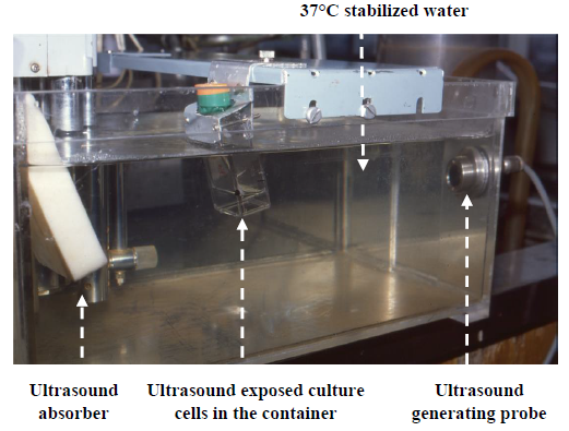
As T is 41°C, t should be shorter than 4(43-41)=42=16 min, namely, duration of 41°C temperature exposure should be less than 16 min to obtain normal outcome [1]. Thus, the small pregnant animal should not touch the ultrasound probe for 16 or more min, where the migration of neuronal cell was delayed, when the probe was attached to the animal for 30 min [2], where heating time was definitely longer than 16 min. It is natural to develop fetal abnormality due to heating artifact. Although the equation 1 was presented to study the thermal index of diagnostic ultrasound,?the?same?temperature?rise?time?is?discussed?also?in?the?estimation?of heating artifact in this report (Figure 1).

Figure 1: Experimental ultrasound was exposed to the subject, but avoiding the heat of ultrasound probe with 37? stabilized water in Japanese study group of diagnostic ultrasound safety in 1970s. No anomaly developed after exposure of pregnant animal with intense ultrasound using the system.
Ultrasound probe attachment was unknown in another ultrasound exposure experiment [3], The abnormality after short exposure of Doppler ultrasound to fetal animal was transient increase of hepatic cell apoptosis, however, the phenomenon can appear by other reasons, thus, it is difficult to decide to be caused limitedly only by ultrasound exposure [4]. It is natural to achieve abnormal outcome after thermal artifact exposure longer than the equation of NCRP report [1].?It is hoped to avoid heat artifact longer than the limit of NCRP. The radical method to avoid probe heat artifact is to insulate the probe heat with 37°C stabilized water, then experimental subject is exposed only to ultrasound, but not to heating artifact of the ultrasound generating probe. Small pregnant animals and growing cultured cells were exposed to ultrasound without heating of ultrasound probe in the Japanese national ultrasound safety study group in 1970s. Anomalous animal fetus did not develop, and the threshold of cultured cell growth suppression was 240 mW/cm2 , which was the thermal index (TI)=1 intensity level which is the threshold of clinical ultrasound exposure [5]. As human pregnant uterus is large in size filled with amniotic fluid, thus, no abnormality is expected in human case with diagnostic ultrasound exposure.
3. Discussion
A researcher to test the safety of diagnostic ultrasound safety should not attach the probe to pregnant small animal abdomen, but should test the experimental radiation within safe temperature limit, or totally shut off the heat with 37°C stabilized water, purely radiating the ultrasound without heating experimental animal with ultrasound probe. The limit of ultrasound exposure was tested by the growth curve suppression of cultured cells where the threshold was 240 mW/cm2 under the heat insulation with 37°C stabilized water [5], and threshold ultrasound intensity was the level of Thermal Index = 1. Fetal animal exposure was also done in the same radiation environment developing no anomaly in the 1970s experimental ultrasound group.
Present trend of ultrasound safety is “As low as reasonably achievable (ALARA) principle” by which further ultrasound safety is expected, e. g. our actocardogram ultrasound is 1 mW/cm2, which separates physiologic sinusoidal from pathologic one, that was unable by the CTG. Undisturbed pulse Doppler flow wave was recorded by 0.1 Thermal Index intensity [6], which was around 1/10 of 240 mW/cm2=20mW/cm2, which was CW ultrasound intensity, by which the author recorded world first CW Doppler fetal arterial blood flow wave by using own frequency demodulation system at the sound output of Doppler fetal heart beat listener Doptone, where the CW ultrasound intensity was 20mW/cm2, by which CW Doppler fetal arterial flow wave was traced too[7].
4. Conclusion
The author who reported neurological damage of small animal fetus after direct attachment of diagnostic B-mode ultrasound probe for 30 minutes to pregnant small animal would be recommended to perform the experiment excluding direct attachment of the probe by separating the probe by 37°C water to avoid the heating artifact of the probe, because of the doubt of artifact of heating the animal fetus. A pregnant small animal should be exposed to experimental diagnostic ultrasound separating 41°C warmed ultrasound probe heat with 37°C water, where the animal is purely exposed to ultrasound without heating artifact, namely, according to NCRP equation of t<4(43-T), small animal fetus tolerates 16 or less min of 41°C probe heat [1], Please attach ultrasound probe less than 16 min in the safety experiment to avoid heat artifact, if directly attach the probe to the animal abdomen. Furthermore, it is recommended to expose experimental ultrasound, insulating the heat of ultrasound probe with 37°C water,. which is inserted between the animal and ultrasound probe.
References
- National Council on Radiation Protection and Measurements. Exposure Criteria for Medical Diagnostic ultrasound: Criteteria Bases on Thermal Mechanisms. NCRP REPORT No. 113 (1992).
- Ang ES Jr, Gluncic V, Duque A, et al. Prenatal exposure to ultrasound waves impacts neuronal migration in mice. PNAS 103 (2006): 12909.
- Ping LI, Wang PJ, Zang W. Prenatal exposure to ultrasound affects learning and memory in young rats. Ultrasound Med Biol 41 (2015): 644-653.
- Pellicer B, Herraiz S, Táboas E, et al. Ultrasound bioeffects in rats: quantification of cellular damage in the fetal liver after pulsed Doppler imaging. Ultrasound in Obstet Gynecol 3 (2011): 643-648.
- Maeda K, Murao F, Tsuzaki T, et al. Experimental studies on the suppression of cultured cell growth curves after irradiation with CW and pulsed ultrasound. IEEE Trans Ultrasonics, Ferroelectrics, Freq control 33 (1986): 186-193.
- Sande RK, Matre K, Kisserad T, et al. Ultrasound safety in early pregnancy: reduced energy setting does not compromise obstetric Doppler measurements.Ultrasound Obstet Gynecol 39 (2012): 438-443.
- Maeda K, Kimura S, Nakano H, et al. Pathophysio- logy of Fetus. Fukuoka Printing, Fukuoka (1969).
