Diagnosis and Treatment of an Infected Supraorbital Ethmoid Cell Cyst
Article Information
Kento Koda1,2*, Kazuo Yasuhara1
1Department of Otolaryngology and Head and Neck Surgery, Takeda General Hospital, Japan
2Department of Otolaryngology and Head and Neck Surgery, the University of Tokyo, Japan
*Corresponding Author: Kento Koda, Department of Otolaryngology and Head and Neck Surgery, Takeda General Hospital, Japan
Received: 24 February 2022; Accepted: 07 March 2022; Published: 14 March 2022
Citation: Kento Koda, Kazuo Yasuhara. Diagnosis and Treatment of an Infected Supraorbital Ethmoid Cell Cyst. Archives of Clinical and Medical Case Reports 6 (2022): 199-202
View / Download Pdf Share at FacebookKeywords
Supraorbital Ethmoid-Cell; Computed tomography; Endoscopic surgery
Supraorbital Ethmoid-Cell articles; Computed tomography articles; Endoscopic surgery articles
Covid-19 articles Covid-19 Research articles Covid-19 review articles Covid-19 PubMed articles Covid-19 PubMed Central articles Covid-19 2023 articles Covid-19 2024 articles Covid-19 Scopus articles Covid-19 impact factor journals Covid-19 Scopus journals Covid-19 PubMed journals Covid-19 medical journals Covid-19 free journals Covid-19 best journals Covid-19 top journals Covid-19 free medical journals Covid-19 famous journals Covid-19 Google Scholar indexed journals Supraorbital Ethmoid-Cell articles Supraorbital Ethmoid-Cell Research articles Supraorbital Ethmoid-Cell review articles Supraorbital Ethmoid-Cell PubMed articles Supraorbital Ethmoid-Cell PubMed Central articles Supraorbital Ethmoid-Cell 2023 articles Supraorbital Ethmoid-Cell 2024 articles Supraorbital Ethmoid-Cell Scopus articles Supraorbital Ethmoid-Cell impact factor journals Supraorbital Ethmoid-Cell Scopus journals Supraorbital Ethmoid-Cell PubMed journals Supraorbital Ethmoid-Cell medical journals Supraorbital Ethmoid-Cell free journals Supraorbital Ethmoid-Cell best journals Supraorbital Ethmoid-Cell top journals Supraorbital Ethmoid-Cell free medical journals Supraorbital Ethmoid-Cell famous journals Supraorbital Ethmoid-Cell Google Scholar indexed journals cancer articles cancer Research articles cancer review articles cancer PubMed articles cancer PubMed Central articles cancer 2023 articles cancer 2024 articles cancer Scopus articles cancer impact factor journals cancer Scopus journals cancer PubMed journals cancer medical journals cancer free journals cancer best journals cancer top journals cancer free medical journals cancer famous journals cancer Google Scholar indexed journals Computed tomography articles Computed tomography Research articles Computed tomography review articles Computed tomography PubMed articles Computed tomography PubMed Central articles Computed tomography 2023 articles Computed tomography 2024 articles Computed tomography Scopus articles Computed tomography impact factor journals Computed tomography Scopus journals Computed tomography PubMed journals Computed tomography medical journals Computed tomography free journals Computed tomography best journals Computed tomography top journals Computed tomography free medical journals Computed tomography famous journals Computed tomography Google Scholar indexed journals Ultra Sound articles Ultra Sound Research articles Ultra Sound review articles Ultra Sound PubMed articles Ultra Sound PubMed Central articles Ultra Sound 2023 articles Ultra Sound 2024 articles Ultra Sound Scopus articles Ultra Sound impact factor journals Ultra Sound Scopus journals Ultra Sound PubMed journals Ultra Sound medical journals Ultra Sound free journals Ultra Sound best journals Ultra Sound top journals Ultra Sound free medical journals Ultra Sound famous journals Ultra Sound Google Scholar indexed journals treatment articles treatment Research articles treatment review articles treatment PubMed articles treatment PubMed Central articles treatment 2023 articles treatment 2024 articles treatment Scopus articles treatment impact factor journals treatment Scopus journals treatment PubMed journals treatment medical journals treatment free journals treatment best journals treatment top journals treatment free medical journals treatment famous journals treatment Google Scholar indexed journals CT articles CT Research articles CT review articles CT PubMed articles CT PubMed Central articles CT 2023 articles CT 2024 articles CT Scopus articles CT impact factor journals CT Scopus journals CT PubMed journals CT medical journals CT free journals CT best journals CT top journals CT free medical journals CT famous journals CT Google Scholar indexed journals Radiology articles Radiology Research articles Radiology review articles Radiology PubMed articles Radiology PubMed Central articles Radiology 2023 articles Radiology 2024 articles Radiology Scopus articles Radiology impact factor journals Radiology Scopus journals Radiology PubMed journals Radiology medical journals Radiology free journals Radiology best journals Radiology top journals Radiology free medical journals Radiology famous journals Radiology Google Scholar indexed journals Endoscopic surgery articles Endoscopic surgery Research articles Endoscopic surgery review articles Endoscopic surgery PubMed articles Endoscopic surgery PubMed Central articles Endoscopic surgery 2023 articles Endoscopic surgery 2024 articles Endoscopic surgery Scopus articles Endoscopic surgery impact factor journals Endoscopic surgery Scopus journals Endoscopic surgery PubMed journals Endoscopic surgery medical journals Endoscopic surgery free journals Endoscopic surgery best journals Endoscopic surgery top journals Endoscopic surgery free medical journals Endoscopic surgery famous journals Endoscopic surgery Google Scholar indexed journals surgery articles surgery Research articles surgery review articles surgery PubMed articles surgery PubMed Central articles surgery 2023 articles surgery 2024 articles surgery Scopus articles surgery impact factor journals surgery Scopus journals surgery PubMed journals surgery medical journals surgery free journals surgery best journals surgery top journals surgery free medical journals surgery famous journals surgery Google Scholar indexed journals
Article Details
1. Case Report
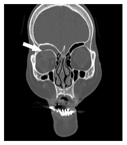
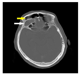
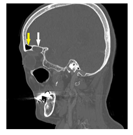
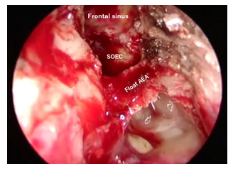
A 79-year-old woman with no significant medical history visited an emergency room with a complaint of diplopia. Ocular motility disorder was noted, and a central lesion was suspected. Contrast-enhanced computed tomography (CT) revealed a cystic lesion, and cyst infection was suspected based on the rim enhancement. Coronal images (Figure 1) suggested a frontal sinus cyst, but horizontal (Figure 2) and sagittal (Figure 3) sections revealed a Supraorbital Ethmoid-Cell (SOEC) cyst extending into the orbit. Diplopia disappeared immediately after endoscopic surgery for cyst enucleation. Anatomically, SOECs are associated with the anterior ethmoidal artery (AEA), which runs within or in continuity with the posterior border of the SOEC opening [1] (Figure 4). Therefore, in endoscopic surgery, the risk of AEA damage increases with a posterior approach, and an approach from the front is recommended. If an axillary flap [2] is not created and the nasal ridge is not sufficiently excised, it is difficult to operate using an endoscope.
Abbreviations:
AEA: Anterior ethmoidal artery; CT: Computed tomography; SOEC: Supraorbital ethmoid-cell cyst

Figure 1: Coronal section: a frontal sinus cyst invading the orbit.

Figure 2: horizontal section: cyst is posterior to the frontal sinus.

Figure 3: sagittal section: the cyst originates from the SOECs.

Figure 4: The AEA runs along the posterior border of the SOEC opening.
White arrows, cyst; yellow arrows, frontal sinus
2.1. Teaching point
Sinus lesions should be localized using horizontal, sagittal, and coronal CT images. Regarding the surgical technique, it is recommended to create a flap, remove the nasal ridge sufficiently, and approach from the front.
Acknowledgement
We would like to thank Editage (www.editage.com) for English language editing.
References
- Jang DW, Lachanas VA, White LC, et al. Supraorbital ethmoid cell: a consistent landmark for endoscopic identification of the anterior ethmoidal artery. Otolaryngol Head Neck Surg 151 (2014): 1073-1077.
- Wormald PJ. The axillary flap approach to the frontal recess. Laryngoscope 112 (2002): 494-499.
