Diagnosis and Restorative Management of Proximal Carious Lesions of Permanent Teeth using Elastic Separators. Description of the Technique and 2 Case Reports
Article Information
Berdouses EB1*, Sifakaki M2, Lagouvardos P3, Oulis CJ3
1Paediatric Dentist, Athens, Greece
2Paediatric Dentist, Clinical Instructor, Department of Paediatric Dentistry, National and Kapodistrian University of Athens, Greece
3Professor Emeritus, Department of Operative Dentistry, National and Kapodistrian University of Athens, Athens, Greece
*Corresponding Author: Berdouses EB, Paediatric Dentist, 22 Kodrou str. 15231 Halandri, Athens, Greece
Received: 11 November 2020; Accepted: 23 November 2020; Published: 04 December 2020
Citation: Berdouses EB, Sifakaki M, Lagouvardos P, Oulis CJ. Diagnosis and Restorative Management of Proximal Carious Lesions of Permanent Teeth using Elastic Separators. Description of the Technique and 2 Case Reports. Dental Research and Oral Health 3 (2020): 207-213.
View / Download Pdf Share at FacebookAbstract
Introduction and Aim: The management of the proximal cavitated carious lesions in permanent molars has been a challenge for the clinician in terms of integrity of the tooth, aesthetics and longevity of the restoration. The purpose of these case reports is to present a new technique for the management of proximal cavitated carious lesions of permanent teeth and to discuss the indications and benefits along with the problems and contraindications encountered in the clinical practice.
Case Reports: Two representative cases are presented in this paper among many others managed with this new approach. Case 1 refers to a 12 year old patient radiographically diagnosed with ICCMS stage 2 lesion on the right maxillary first permanent molar. Case 2 refers to a 15 year old radiographically diagnosed with ICCMS stage 3 and 4 lesion on the right maxillary and mandibular first permanent molars, respectively. The steps of the technique are described in detail and indications and benefits are presented along with contraindications and disadvantages of the technique. These two cases are part of a bigger study of 177 restorations followed for a median time of 4.3 years showing very similar results with class II restorations.
Conclusions: It is a technique that can correctly answer the question “cavitation or not” supporting the clinician’s decision to apply prevention or surgical intervention for the interproximal surface. It produces interdental space that can facilitate the restoration of the interproximal surface only leaving the occlusal intact. It is a technique that complies with the concept of minimal invasive dentistry showing very similar results with the class II amalgam restorations and betted results of composite ones.
Keywords
Dental Caries, Amalgam, Resin, Elastic Separators
Dental Caries articles; Amalgam articles; Resin articles; Elastic Separators articles
Dental Caries articles Dental Caries Research articles Dental Caries review articles Dental Caries PubMed articles Dental Caries PubMed Central articles Dental Caries 2023 articles Dental Caries 2024 articles Dental Caries Scopus articles Dental Caries impact factor journals Dental Caries Scopus journals Dental Caries PubMed journals Dental Caries medical journals Dental Caries free journals Dental Caries best journals Dental Caries top journals Dental Caries free medical journals Dental Caries famous journals Dental Caries Google Scholar indexed journals Amalgam articles Amalgam Research articles Amalgam review articles Amalgam PubMed articles Amalgam PubMed Central articles Amalgam 2023 articles Amalgam 2024 articles Amalgam Scopus articles Amalgam impact factor journals Amalgam Scopus journals Amalgam PubMed journals Amalgam medical journals Amalgam free journals Amalgam best journals Amalgam top journals Amalgam free medical journals Amalgam famous journals Amalgam Google Scholar indexed journals Resin articles Resin Research articles Resin review articles Resin PubMed articles Resin PubMed Central articles Resin 2023 articles Resin 2024 articles Resin Scopus articles Resin impact factor journals Resin Scopus journals Resin PubMed journals Resin medical journals Resin free journals Resin best journals Resin top journals Resin free medical journals Resin famous journals Resin Google Scholar indexed journals Elastic Separators articles Elastic Separators Research articles Elastic Separators review articles Elastic Separators PubMed articles Elastic Separators PubMed Central articles Elastic Separators 2023 articles Elastic Separators 2024 articles Elastic Separators Scopus articles Elastic Separators impact factor journals Elastic Separators Scopus journals Elastic Separators PubMed journals Elastic Separators medical journals Elastic Separators free journals Elastic Separators best journals Elastic Separators top journals Elastic Separators free medical journals Elastic Separators famous journals Elastic Separators Google Scholar indexed journals tooth articles tooth Research articles tooth review articles tooth PubMed articles tooth PubMed Central articles tooth 2023 articles tooth 2024 articles tooth Scopus articles tooth impact factor journals tooth Scopus journals tooth PubMed journals tooth medical journals tooth free journals tooth best journals tooth top journals tooth free medical journals tooth famous journals tooth Google Scholar indexed journals
Article Details
1. Introduction
The management of the proximal cavitated carious lesions in permanent molars has been a challenge for the clinician in terms of integrity of the tooth, aesthetics and longevity of the restoration. Restoring a tooth with an adequate contact point and preserving sound enamel and dentin, complying with the principles of Minimum Intervention Dentistry, are within the aims of modern dentistry. Treatment options for interproximal lesions including ICCMS stage 4 (Table 1) include the typical Class II cavity preparation and several others less popular minimal invasive techniques. In the literature minimal invasive techniques such as the tunnel, the saucer shaped and the box-only technique, introduced in an attempt to preserve more sound tooth structure and overcome the problem of durability of the restoration, have shown inferior results compared to conventional class II restorations [1-3]. Temporary elective separation of the teeth with orthodontic elastic separators as a complementary tool for diagnosing proximal carious lesion gives direct visual access to the proximal tooth surface [4]. It is the only tool we have today that unequivocally answer the question on whether the lesion is cavitated or not and it is well known since many years [5]. It also plays an important role in the decision whether a lesion will be treated with preventive measures or it will be restored (Figure 1).
|
Class |
Description |
|
|
0 |
0 |
No radiolucency |
|
RA: Initial stages |
1 |
radiolucency in the outer ½of enamel |
|
2 |
radiolucency in the inner ½ of enamel ± EDJ (enamel-dentine junction) |
|
|
3 |
radiolucency limited to the outer ?of dentin |
|
|
RB: Moderate stage |
4 |
radiolucency reaching the middle ? of dentin |
|
RC: Extensive stages |
5 |
Radiolucency reaching the inner ? of dentin, clinically cavitated |
|
6 |
Radiolucency into the pulp, clinically cavitated |
Table 1: ICCMS classification of the depth of interproximal caries lesion [6].
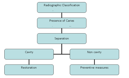
Figure 1: Decision sequence for interproximal caries detection and restoration
The possibility to take advantage of the created space and prepare, in certain cases, a direct class I restoration with better aesthetics and durability gives a new option in restoring interproximal caries. Hence, the purpose of this case report is to present in detail this new technique for the management of proximal cavitated carious lesions of permanent teeth and to discuss the indications and benefits along with the problems and contraindications encountered in clinical practice.
2. Case Report
Two representative cases are presented in this paper among many others managed with this new approach. Case 1 refers to a 12 year old patient radiographically diagnosed with ICCMS stage 2 lesion on the right maxillary first permanent molar. Case 2 refers to a 15 year old radiographically diagnosed with ICCMS stage 3 and 4 lesion on the right maxillary and mandibular first permanent molars, respectively.
2.1 Procedure Phase I
At first, guardians were informed about the benefits and the risks of the procedure and they signed the written consent form. An elastic separator of double thickness (DynaFlex® Reseps separators, DynaFlex, Missouri, USA) was inserted between the contacting surfaces for 4-5 days (Figure 2) and a space of about 2 mm was created. The process of separating the teeth usually, involves the stretching of the elastic ring between orthodontic separator placing plier ends (Figure 3). Then the stretched ring is pushed interdentally until the lower part of the ring is located under the contact point of the teeth. In our technique, the classic method is modified by using an elastic ring of double thickness (size # 2).
Sometimes, depending on the crowding of the teeth, a size #1 separating ring can be used to start opening the interdental space, followed by a #2 elastic ring. During the application of the elastic the patient feels some pressure on the teeth but overall the process is painless. Few hours after the placement of the separator the patient may start to feel pain from the pressure exerted on the teeth depending on how crowded the teeth are. If the arch is especially crowded, the separator may cause pain upon chewing due to the teeth movement. This pain begins few hours after placement and usually lasts for a day or two. Use of a pain reliever 20 min before the meals controls the pain. Usually all these symptoms will disappear one day after the placement of the separators. Oral hygiene instructions and information about any discomfort were given to the patient and was re- scheduled after 4 or 5 days.
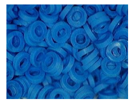
Figure 2: DynaFlex® Reseps separators double Thicknes.
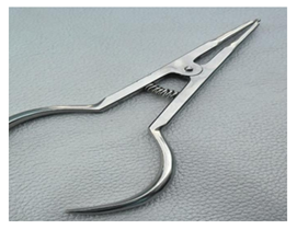
Figure 3: Orthodontic separator placing plier.
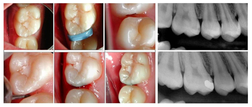
Figure 4: Clinical Procedure/Radiographic images, pre and postoperatively (Case Report 1).
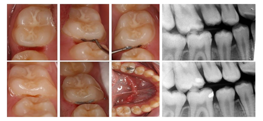
Figure 5: Clinical Procedure/Radiographic images, pre and postoperatively (Case Report 2).
2.2 Phase II
At the following appointment 4-5 days later the elastic separator was removed and 2-3 mm interproximal space could be observed (Figure 4 & 5). Direct visual examination and tactile evaluation by touching the mesial surface of the tooth with an explorer is possible to evaluate the integrity of the surface and whether it was cavitated or not. The protocol then follows two alternatives:
2.2.1 Enamel surface intact (no cavitation) - preventive management: In this case and if the carious lesion was not arrested, a protocol [7, 8] for fluoride application with 5% Duraphat varnish was applied once and reapplied 3 more times once every week, every 6 months for 2 years, taking an xray for reassessment every year.
2.2.2 Carious lesion, cavitation - operative management: Following anaesthesia and placement of a rubber dam, an Ivory matrix retainer was inserted on the adjacent tooth to protect it from accidental drilling. A high-speed turbine with a #330 carbide bur was used in a horizontal direction buccolingually to remove caries and open the cavity. With a #2 or 3 low speed bur the cavity was appropriately prepared for amalgam or composite resin. Provided that the thickness of the marginal ridge is at least 2 mm the restorative material was inserted according to the manufacturer instructions. Removal of excess material was accomplished using a finishing bur for composites and an explorer along with a dental floss for amalgam before setting. An x-ray was taken to assess the margins of the restoration sub- gingivally and the patient was dismissed, with the appropriate instructions for cleaning the area and a recall examination after one year.
3. Discussion and Comments
The described modified technique creates a diastema of 2 mm with the elastic separator of double thickness and presents several advantages compared to other techniques. First of all, it gives a better access to examine the proximal surface and give an unequivocal decision on whether the lesion is cavitated. In cases of not cavitated lesions preventive measures can be applied safely. Second, due to the created space it gives the possibility to proceed operatively and prepare a small class I cavity, maintaining the marginal ridge and saving healthy tooth structure. In fact, this new technique not only fulfils the principles of the Minimal Invasion Dentistry but also it appears to be very promising in terms of performance and durability. In a prospective study we published in 2020, [9] 177 restorations were placed on proximal surfaces of permanent molars in 140 patients (median age 12.2 years) and were followed for several years (median time 4.3 years). The results showed very similar success with the literature for the class II amalgam restorations (91.6%) and better performance (93.5%) for the composite resin restorations.
As for the choice of material, both materials showed similar performance with estimated median time to failure being 17.8 years for amalgam restorations and 21.7 years for the composite resin ones. It was estimated that the cumulative probability failure for 5 and 10 years were 6.71%, 4.75% and 21.59%, 15.69% for the amalgam and composite restorations respectively. In comparison, the survival rate of posterior class II restorations, range from 92.5% to 92.8 % for amalgam and 86.2% to 85.8% for composite resins, with the main reasons of the failures being the secondary caries and fractures of the restorations and/or the tooth [10]. At the same time, the suggested alternative techniques for restoration of proximal carious lesions such as tunnel and the saucer shaped only box techniques, were not satisfactory in their attempt to replace the classical class II cavity preparation [11]. Results regarding the failure rates of these types of restorations show that they present inferior longevity compared to conventional class II composite resin restorations. Main reasons of failure are marginal ridge fracture for the tunnel and recurrent or progressive caries for both configurations [1]. Another advantage of this technique is the low cost and easiness to perform without the need to use special training and equipment. Our experience shows that the teeth are usually separated easily within 4-5 days in all children up to 16-18 years of age. It is still a question, if it works successfully to adults in regards to the interdental space that is created and the discomfort the patients might experience. Careful selection of the cases for this new approach is very important, avoiding very deep cavities, or cavities where the marginal ridge is less than 2 mm.
Among the disadvantages of the technique, we can refer to some pressure throughout the separating process of the teeth expressed by some patients. Crowding of the arch seems to exacerbate the symptoms causing pain almost immediately after the insertion of the elastics. But overall, the application process is quite painless and all these symptoms disappear after a while. The two-appointment procedure along with the earlier loss (before 4 days) of the elastic separators in very young patients might be considered for some clinicians another disadvantage. However, by explaining to the parents or patients the advantages and the additive value of the technique, both problems can be easily managed and overcome. By using this technique, clinicians can reduce enamel microcracks and secondary caries. As a consequence, marginal discoloration, recurrent caries and postoperative sensitivity can be reduced. It is a very effective alternative to class II restorations for selected cases, but it cannot replace them. It can save though substantial tooth structure, improve the aesthetics of the area and be at least equally successful with them.
4. Conclusions
Τhe main advantages of this technique are: a) the feasibility to correctly answer the question “cavitation or not” supporting the clinician’s decision to apply prevention or surgical intervention b) the preservation of healthy tooth substance complying to modern dentistry and the MID’s principles, resulting in c) prolongation of the survival of the tooth for the benefit of the patient.
Competing Interests
The authors declare that they have no competing interests.
References
- Kopperud SE, Tveit AB, Gaarden T, et al. Longevity of posterior dental restorations and reasons for failure. Eur J Oral Sci 120 (2012): 539-548.
- McComb D. Systematic review of conservative operative caries management strategies. J Dent Educ 65 (2001): 1154-1161.
- Peters MC, McLean ME. Minimally invasive operative care. J Adhes Dent 3 (2001): 7-16.
- Pitts NB, Longbottom C. Temporary tooth separation with special reference to the diagnosis and preventive management of equivocal approximal carious lesions. Quintessence Int 18 (1987): 563-573.
- Baelum V, Hintze H, Wenzel A, et al. Implications of caries diagnostic strategies for clinical management decisions. Comm Dent Oral Epidemiol 40 (2012): 257-266.
- Pitts NB, Ismail ΑI, Martignon S, et al. ICCMSTM Guide for Practitioners and Educators. ICDAS Found (2014): 20-21.
- Petersson LG, Arthursson L, Ostberg C, et al. Caries-Inhibiting Effects of Different Modes of Duraphat Varnish Reapplication: A 3-Year Radiographic Study. Caries Res 25 (1991): 70-73.
- Urquhart O, Tampi MP, Pilcher L, et al. Nonrestorative Treatments for Caries: Systematic Review and Network Meta- analysis. J. Dent. Res 98 (2019): 14-26.
- Berdouses ED, Agouropoulos A, Sifakaki M, et al. Restorative direct management of cavitated proximal carious lesions of permanent molars using elastic separators in children and adolescents. Dent Res Oral Heal 3 (2020): 183-193.
- Moraschini V, Fai CK, Alto RM, et al. Amalgam and resin composite longevity of posterior restorations: A systematic review and meta-analysis. J Dent 43 (2015): 1043-1050.
- Mount GJ. Minimal intervention dentistry: rationale of cavity design. Oper Dent 28 (2003): 92-99.
