Control of Expression Level in Human Genes: Observations with Apoptosis Genes and Genes Involved in B cell Development
Article Information
Jay C. Brown
Department of Microbiology, Immunology and Cancer Biology, University of Virginia School of Medicine, Charlottesville, Virginia, 22908
*Corresponding author: Jay C. Brown, Department of Microbiology, Immunology and Cancer Biology, University of Virginia School of Medicine Box 800734, Charlottesville, Virginia 22908, USA
Received: 11 July 2022; Accepted: 20 July 2022; Published: 28 July 2022
Citation: Jay C. Brown. Control of Expression Level in Human Genes: Observations with Apoptosis Genes and Genes Involved in B cell Development. Journal of Bioinformatics and Systems Biology 5 (2022): 108-115.
View / Download Pdf Share at FacebookAbstract
To understand the way a gene functions in development, one needs to know about the gene product’s functional capabilities, the tissues where it is located, and the level of its expression. It is now widely accepted that transcription factors can affect the level of gene expression, but the results emphasize the need for further clarification. The study described here was carried out to determine whether the amount of a transcription factor bound in the promoter region might be directly related to the level of the gene’s expression. The study was focused on a population of human genes involved in apoptosis, a pathway known to be affected by the transcription factor Ikaros (IKZF1 gene). For each apoptosis gene, information was accumulated about its expression level and about the level of IKZF1 binding in the promoter. The two measurements were then compared and interpreted to identify instances where the amount of IKZF1 binding is related to the level of gene expression. A similar analysis was carried out with genes involved in B cell development, also a gene population influenced by IKZF1. The results identified gene groups, each containing 3-8 genes, in which the expression level was related to IKZF1 binding in the promoter, a result that supports the idea that promoter bound IKZF1 can affect the level of gene expression. A further study was performed to examine the secondary, non-IKZF1 transcription factors bound in the promoters of apoptosis and B cell development genes. Prominent amounts of RBFOX2, ASH2L and TAF1 were observed in both populations suggesting IKZF1-rich promoters may resemble each other in their content of other transcription factor binding sites as well.
Keywords
gene expression; transcription factor; promoter; IKZF1; apoptosis; B cell development
gene expression articles, transcription factor articles, promoter, IKZF1 articles, apoptosis articles, B cell development articles
gene expression articles gene expression Research articles gene expression review articles gene expression PubMed articles gene expression PubMed Central articles gene expression 2023 articles gene expression 2024 articles gene expression Scopus articles gene expression impact factor journals gene expression Scopus journals gene expression PubMed journals gene expression medical journals gene expression free journals gene expression best journals gene expression top journals gene expression free medical journals gene expression famous journals gene expression Google Scholar indexed journals transcription factor articles transcription factor Research articles transcription factor review articles transcription factor PubMed articles transcription factor PubMed Central articles transcription factor 2023 articles transcription factor 2024 articles transcription factor Scopus articles transcription factor impact factor journals transcription factor Scopus journals transcription factor PubMed journals transcription factor medical journals transcription factor free journals transcription factor best journals transcription factor top journals transcription factor free medical journals transcription factor famous journals transcription factor Google Scholar indexed journals promoter articles promoter Research articles promoter review articles promoter PubMed articles promoter PubMed Central articles promoter 2023 articles promoter 2024 articles promoter Scopus articles promoter impact factor journals promoter Scopus journals promoter PubMed journals promoter medical journals promoter free journals promoter best journals promoter top journals promoter free medical journals promoter famous journals promoter Google Scholar indexed journals IKZF1 articles IKZF1 Research articles IKZF1 review articles IKZF1 PubMed articles IKZF1 PubMed Central articles IKZF1 2023 articles IKZF1 2024 articles IKZF1 Scopus articles IKZF1 impact factor journals IKZF1 Scopus journals IKZF1 PubMed journals IKZF1 medical journals IKZF1 free journals IKZF1 best journals IKZF1 top journals IKZF1 free medical journals IKZF1 famous journals IKZF1 Google Scholar indexed journals apoptosis articles apoptosis Research articles apoptosis review articles apoptosis PubMed articles apoptosis PubMed Central articles apoptosis 2023 articles apoptosis 2024 articles apoptosis Scopus articles apoptosis impact factor journals apoptosis Scopus journals apoptosis PubMed journals apoptosis medical journals apoptosis free journals apoptosis best journals apoptosis top journals apoptosis free medical journals apoptosis famous journals apoptosis Google Scholar indexed journals B cell development articles B cell development Research articles B cell development review articles B cell development PubMed articles B cell development PubMed Central articles B cell development 2023 articles B cell development 2024 articles B cell development Scopus articles B cell development impact factor journals B cell development Scopus journals B cell development PubMed journals B cell development medical journals B cell development free journals B cell development best journals B cell development top journals B cell development free medical journals B cell development famous journals B cell development Google Scholar indexed journals ENCODE articles ENCODE Research articles ENCODE review articles ENCODE PubMed articles ENCODE PubMed Central articles ENCODE 2023 articles ENCODE 2024 articles ENCODE Scopus articles ENCODE impact factor journals ENCODE Scopus journals ENCODE PubMed journals ENCODE medical journals ENCODE free journals ENCODE best journals ENCODE top journals ENCODE free medical journals ENCODE famous journals ENCODE Google Scholar indexed journals RNA-seq articles RNA-seq Research articles RNA-seq review articles RNA-seq PubMed articles RNA-seq PubMed Central articles RNA-seq 2023 articles RNA-seq 2024 articles RNA-seq Scopus articles RNA-seq impact factor journals RNA-seq Scopus journals RNA-seq PubMed journals RNA-seq medical journals RNA-seq free journals RNA-seq best journals RNA-seq top journals RNA-seq free medical journals RNA-seq famous journals RNA-seq Google Scholar indexed journals chromosome articles chromosome Research articles chromosome review articles chromosome PubMed articles chromosome PubMed Central articles chromosome 2023 articles chromosome 2024 articles chromosome Scopus articles chromosome impact factor journals chromosome Scopus journals chromosome PubMed journals chromosome medical journals chromosome free journals chromosome best journals chromosome top journals chromosome free medical journals chromosome famous journals chromosome Google Scholar indexed journals
Article Details
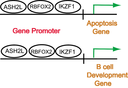
Graphical Abstract

Graphical Abstrac
1. Introduction
Control of gene expression is one of the most actively studied areas of molecular biology today, and this has been the case for more than half a century. The effort has been highly productive. As a result, we now have an advanced understanding of regulatory control as it occurs in a wide variety of organisms including humans. Relevant features uncovered include the role of transcription factors, enhancers, epigenetic modifications, and the way chromatin architecture can affect gene expression [1-4]. A neglected area, however, has been an identification of factors that affect the quantitative level of expression. Studies have addressed whether a gene is on or off but avoided the issue of whether expression is high, medium, or low. It is expected that the same factors that affect expression will also affect the level of expression, but one would like to have additional information about how such functions operate.
The study described here was designed to confront the issue of expression level directly. The goal was to test the role of a promoter bound transcription factor with the level of gene expression. Analysis was focused on a single transcription factor, Ikaros (IKZF1) and a population of 56 human genes involved in apoptosis, a system whose function is known to be affected by IKZF1 [5]. For each apoptosis gene, two values were identified, (1) the level of expression and (2) the amount of IKZF1 bound at the promoter. The values were then plotted, and the plots were interpreted to indicate whether IKZF1 was affecting expression of a gene and if so then whether the effect was to potentiate or repress the level of expression. A similar analysis was carried out with 64 human genes involved in B cell development, a function also known to be regulated by IKZF1 [6-8]. The results identify several groups of genes in which promoter bound IKZF1 is correlated with the level of gene expression.
2. Materials and Methods
2.1 Gene databases
The study was performed beginning with two human gene populations, one of genes involved in apoptosis (Supplementary Table 1) and the other of B cell development genes (Supplementary Table 2). Apoptosis and B cell development genes were derived beginning with those reported by Jourdan et al. [5] and Calonga-Solis et al. [9], respectively. Transcription levels were derived from the GTEx Portal of RNA-seq results as reported in the UCSC Genome Browser (version hg38; https://genome.ucsc.edu/ ). ChIP-seq results for IKZF1 and control transcription factor binding to the gene promoter were obtained for GM12878 cells by way of the Integrated Genome Browser (https://igv.org/app/). Secondary, non-IKZF1 transcription factor binding sites were retrieved from the ENCODE Transcription Factor ChIP Clusters data base by way of the UCSC Genome Browser. For each gene reported, the list of TF binding sites from ENCODE was examined visually, and the number of binding sites was counted. The counts were used to identify the three most prevalent binding sites which were reported as TF rank 1-TF rank 3. Gene functions were retrieved from GeneCards (https://www.genecards.org/).
2.2 Data analysis
Data were recorded with RStudio and Excel. Results were analyzed and rendered graphically with SigmaPlot 14.5.
3. Results
3.1 Experimental strategy
The goal of the project described here was to test the idea that binding of a transcription factor in the promoter region of a gene might affect the quantitative level of the gene’s expression. It was expected this might be the case as transcription factors (TF) are known to affect whether a gene is expressed or not. It is reasonable therefore to consider that the same factors that affect on vs. off might also affect the degree of on. Also, the resources needed to carry out the test are readily available. The results of RNA-seq studies yield the required information about gene expression levels and ChIP-seq results provide promoter binding information. After accumulating the above information, the study involved plotting the level of gene expression (RNA-seq results) against the amount of promoter bound TF (ChIP-seq results) to determine if the plot indicates a relationship between the two values.
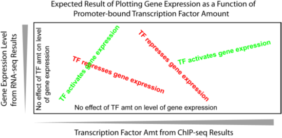
Fig. 1 shows a graphic representation of the expected results as described above. The gene expression value was plotted on the y axis and promoter bound IKZF1 on the x. Data points along the green line of text correspond to genes where IKZF1 activates gene expression while points along the red text indicate repression. Both activating and repressive effects are expected since, like many transcription factors, IKZF1 can enhance or suppress transcription depending on its context of other variables [10, 11]. Data points close to the x or y axes correspond to genes where IKZF1 has little or no effect on expression (black text).

Figure 1: Graph showing the expected result when gene transcription level (y axis) is plotted against the amount of transcription factor bound in the promoter (x axis). Points with a negative slope (red text) indicate the transcription factor is acting to repress transcription. A positive slope (green text) is expected if the transcription factor is acting to potentiate expression. Data points near the axes (black text) are expected if the transcription factor has little effect on transcription.
3.2 Apoptosis results
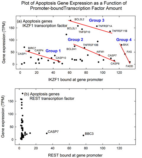
A plot of the apoptosis data yielded the expected result (Fig. 2a). The plot showed that the range examined was well-populated with points corresponding to individual apoptosis genes. For instance, of 56 apoptosis genes in the database, 43 are present in the plot. Red lines suggest the identify of genes that might be related to each other because their expression level is linearly related to IKZF1 level in the promoter. The genes between BCL2A1 and CASP8 are an example. Expression of these genes is inversely related to the level of IKZF1 bound to the promoter indicating that IKZF1 is acting repressively with the genes. For further analysis, a name was assigned to each of the four gene groups identified (Groups 1-4; see Fig. 2a). Only repressive relations are noted (red lines), but other groups including activating groups can be observed and are considered viable interpretations like the ones suggested.

Figure 2: Graph showing the level of apoptosis gene expression plotted against the level of promoter bound IKZF1 (a) and REST (b). Note that most genes are in the dynamic range of the IKZF1 plot indicating their expression is affected by the presence of IKZF1. In contrast, most genes in the REST plot are in the range expected if the transcription factor has little effect on gene expression.
A control experiment was performed in which ChIP-seq results from the transcription factor REST were substituted for IKZF1 (Fig. 2b). REST was considered to be an appropriate control transcription factor as it has not been implicated in regulation of apoptosis genes. Results demonstrated that few apoptosis genes are found in the same range of the plot observed for IKZF1 genes. Two are CASP7 and BBC3 (see Fig. 2b). The results are interpreted to indicate that other transcription factors do not have the same influence on apoptosis gene expression as IKZF1. Together the experimental and control studies suggest the identification of several apoptosis gene groups (Fig 2a) in which promoter bound IKZF1 is related to the level of gene expression
3.3 B cell development genes
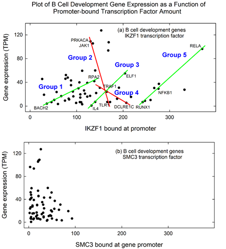
A similar analysis was carried out with genes in the B cell development database. Fig. 3a shows a plot of gene expression against IKZF1 promoter binding for 59 of the 64 B cell development genes. As in the case of apoptosis genes, groups are suggested in which gene expression is related to IKZF1 binding (Groups 1-5). Among the B cell development genes, two groups contain genes in which IKZF1 is interpreted to exert a repressive effect on transcription level (Groups 2 and 4) while in the other three groups IKZF1 is activating (Groups 1, 3 and 5). A control study was performed in which the transcription factor SMC3 was substituted for IKZF1, and the results were plotted against gene expression. The results demonstrated little evidence of genes with the same expression/transcription factor values observed with IKZF1 (Fig. 3b). As with the apoptosis genes, the results with B cell development genes are interpreted to suggest the existence of specific gene groups in which expression is related to IKZF1 binding in the promoter.

Figure 3: Graph showing the level of B cell development gene expression plotted against the level of promoter bound IKZF1 (a) and SMC3 (b). Note that most genes are in the dynamic range of the IKZF1 plot indicating their expression is affected by the presence of IKZF1. In contrast, most genes in the SMC3 plot are in the range expected if the transcription factor has little effect on gene expression.
Transcription factor binding sites in the promoters of expression group member genes Identification of apoptosis genes related by their response to IKZF1 suggested group members might be related in other ways that would be revealing about the control of their expression. Additional information about gene group members was therefore accumulated and compared. Features examined were gene chromosome, gene length, gene function, and the presence of non-IKZF1 binding sites in the promoter. The latter measure involved rank ordering TFs according to their binding site abundance in the promoter. TFs with higher rank were those that have greater abundance. Information accumulated about apoptosis group genes was also accumulated for B cell development genes.
3.4 Apoptosis gene groups
The results with apoptosis group genes show little similarity in chromosome or gene length (Table 1). For instance, no two genes are on the same chromosome in groups 2, 3 and 4. This result was expected and suggests much of the information relevant to control of gene expression is present in the local area of the gene. Some evidence of grouping by gene function was evident (Table 1). For instance, four of the six genes in group 1 encode caspases (i.e. CASP1, CASP4, CASP6 and CASP10). Three of the 5 genes in group 3 encode proteins involved in tumor necrosis factor function.
Table 1: Properties of apoptosis genes in proposed regulatory groups
|
Gene group |
Gene |
Gene |
|||||
|
Gene |
Chr |
Length (kb) |
TF rank 1 |
TF rank 2 |
TF rank 3 |
function |
|
|
1 |
CASP1 |
11 |
9.7 |
IKZF1 |
TAF1 |
ZBTB33 |
apoptosis effector |
|
BIRC7 |
20 |
4.6 |
RBM39 |
ASH2L |
SP1 |
inhibits apoptosis |
|
|
CASP4 |
11 |
25.7 |
ASH2L |
EP300 |
REST |
apoptosis effector |
|
|
CASP6 |
4 |
14.8 |
RBFOX2 |
AGO1 |
TAF1 |
apoptosis effector |
|
|
CASP10 |
2 |
38.5 |
CHD4 |
KDM1A |
HDAC1 |
apoptosis effector |
|
|
BCL2L10 |
15 |
3.5 |
YY1 |
HDAC6 |
ZFX |
regulates apoptosis |
|
|
2 |
BCL2A1 |
15 |
10.3 |
IKZF1 |
MTA3 |
MLLT1 |
regulates apoptosis |
|
TNFRSF10B |
8 |
48.9 |
IKZF1 |
RBFOX2 |
HDAC1 |
TNF receptor |
|
|
AIFM1 |
23 |
38.4 |
IKZF1 |
RBFOX2 |
RNF2 |
induces apoptosis |
|
|
CASP7 |
10 |
51.7 |
CTBP1 |
DPF2 |
IKZF1 |
apoptosis effector |
|
|
CASP8 |
2 |
53.1 |
IKZF1 |
ASH2L |
RBFOX2 |
apoptosis effector |
|
|
3 |
BCL2L2 |
14 |
9.4 |
IKZF1 |
PHF8 |
ASH2L |
regulates apoptosis |
|
BCL2L1 |
20 |
59.5 |
IKZF1 |
RBFOX2 |
ASH2L |
regulates apoptosis |
|
|
TNFRSF1A |
3 |
17.9 |
IKZF1 |
MAX |
REST |
TNF receptor |
|
|
TNFSF1A |
12 |
13.3 |
IKZF1 |
RBFOX2 |
HDAC1 |
TNF family cytokine |
|
|
TNFRSF11B |
8 |
28.3 |
IKZF1 |
KDM4A |
EZH2 |
TNF receptor |
|
|
4 |
BAX |
19 |
6.9 |
RBFOX2 |
HDAC1 |
IKZF1 |
regulates apoptosis |
|
FAS |
10 |
23.7 |
TAF1 |
IKZF1 |
EZH2 |
TNF receptor |
|
|
FADD |
11 |
4.1 |
RBFOX2 |
RNF2 |
MYC |
regulates apoptosis |
|
The role of TFs other than IKZF1 was probed by examining the abundance of their binding sites in apoptosis gene promoters. Binding site abundance was rank ordered beginning with data from ENCODE as described in Materials and Methods. The top three TFs are reported for each apoptosis gene (Table 1). As expected, IKZF1 had the highest abundance among rank 1 TFs with 10 of the 19 genes in the aggregate of the four apoptosis groups. A greater diversity was observed among rank 2 and 3 TFs. Among 15 rank 2 TFs, for instance, only two were represented more than once.
3.5 B cell development gene groups
As in the case of the apoptosis genes, genes in B cell development groups show little evidence of similarity in chromosome or gene length (Table 2). B cell development genes are enriched, however, in gene functions including transcription control and DNA repair. For example, 3 of 8 group 1 genes encode aspects of DNA repair (Table 2). Three of five group 5 genes are TFs.
Table 2: Properties of B cell development genes in proposed regulatory groups
|
Gene group |
Gene |
Chr |
Gene Length (kb) |
TF rank 1 |
TF rank 2 |
TF rank 3 |
Gene function |
|
|
1 |
BACH2 |
6 |
370.4 |
EZH2 |
RBFOX2 |
EP300 |
control transcription |
|
|
RUNX2 |
6 |
222.8 |
TAF1 |
FOS |
TAF7 |
transcription factor |
||
|
PTPRC |
1 |
118.4 |
MTA3 |
ATF7 |
MLLT1 |
protein phosphatase |
||
|
MRE11 |
11 |
78.6 |
ZFX |
SMAD5 |
HDAC1 |
DNA repair |
||
|
NHEJ1 |
2 |
91.5 |
KDM1A |
EP300 |
HDAC2 |
DNA repair |
||
|
GAB1 |
4 |
137.8 |
ASH2L |
RBFOX2 |
RBBP5 |
adaptor protein |
||
|
CREBBP |
16 |
155.7 |
RBFOX2 |
DPF2 |
TAF1 |
histone acetylation |
||
|
RPA2 |
1 |
23.1 |
RBFOX2 |
HNRNPLL |
L3MBTL2 |
DNA repair |
||
|
2 |
TLR1 |
4 |
8.5 |
IKZF1 |
MLLT1 |
MEF2B |
pathogen recognition |
|
|
POU2F2 |
19 |
46.5 |
EZH2 |
SMC3 |
PCBP1 |
transcription factor |
||
|
TRAF1 |
9 |
26.8 |
IKZF1 |
RBFOX2 |
ASH2L |
TNF receptor subunit |
||
|
JAK1 |
1 |
234.5 |
RBFOX2 |
HDAC2 |
RBBP5 |
protein tyr kinase |
||
|
PRKACA |
19 |
26.1 |
RBFOX2 |
ZBTB7A |
TAF1 |
protein kinase A |
||
|
3 |
IL4 |
5 |
8.7 |
IKZF1 |
GATAD2B |
DPF2 |
cytokine |
|
|
BATF |
14 |
24.5 |
IKZF1 |
RCOR1 |
ZNF217 |
transcription factor |
||
|
TRAF1 |
9 |
26.8 |
IKZF1 |
RBFOX2 |
ASH2L |
TNF receptor subunit |
||
|
ELF1 |
13 |
87.4 |
ASH2L |
HDAC1 |
CHD1 |
transcription factor |
||
|
4 |
DCLRE1C |
10 |
49.5 |
RBFOX2 |
DPF2 |
IKZF1 |
DNA repair |
|
|
CD86 |
3 |
65.8 |
IKZF1 |
DPF2 |
CBX5 |
T cell activation |
||
|
TRAF1 |
9 |
26.8 |
IKZF1 |
RBFOX2 |
ASH2L |
TNF receptor subunit |
||
|
TCF3 |
19 |
43.3 |
RBFOX2 |
KDM4A |
BRD4 |
transcription factor |
||
|
5 |
RUNX1 |
21 |
261.5 |
IKZF1 |
MTA3 |
ZBED1 |
transcription factor |
|
|
RUNX3 |
1 |
65.5 |
MLLT1 |
IKZF1 |
DPF2 |
transcription factor |
||
|
NFKB1 |
4 |
115.9 |
TAF1 |
RBFOX2 |
IKZF1 |
transcription factor |
||
|
NBN |
8 |
51.3 |
IKZF1 |
ATF2 |
EP300 |
DNA repair |
||
|
RELA |
11 |
9.3 |
IKZF1 |
ASH2L |
RBFOX2 |
NFKB subunit |
3.6 Comparison of TFs in apoptosis and B cell development gene promoters
The availability of rank ordered TF binding sites in apoptosis and B cell development promoters made it possible to compare binding sites in the two gene populations. While both populations were found to be enriched in IKZF1 binding sites in the promoter, one could now ask whether there was a similarity in less abundant TF binding sites as well. An analysis was performed beginning with all 57 binding sites recorded in grouped apoptosis genes (i.e., ranks 1-3), and the same binding sites (78) for B cell development genes. For each binding site present, the number was counted, expressed as a proportion of the total and the proportion plotted.
Results for the 8 most abundant TFs are shown in Fig. 4. They demonstrate a similarity between apoptosis and B cell development in 5 of the 8 TFs in the plot (i.e., IKZF1, RBFOX2, ASH2L TAF1 and EZH2). B cell development gene promoters are enriched in DPF2 and EP300 while apoptosis genes are enriched in HDAC1. The results are interpreted to emphasize the similarities in the apoptosis and B cell development gene promoters. The similarity in IKZF1 (Fig. 4) was expected as the gene populations were selected because of their response to IKZF1. The other four similarities are novel and suggest that high abundance of one of the five TFs in the promoter is correlated with high abundance of the others.
Figure 4: Plot comparing the transcription factor binding sites in apoptosis and B cell development genes. The plot includes all TF binding sites in the two gene populations, 57 for apoptosis genes and 78 for B cell development. Note the similarity observed in the proportion of IKZF1, RBFOX2, ASH2L, TAF1 and EZH2 in the two gene populations.
4. Discussion
A reliable strategy was employed here for exploring how the level of gene expression is controlled. Plot the level of gene expression against the level of promoter bound TF, and if a relationship exists, then the plot should reveal it. In the study described here, the chances of success were increased by the use of gene populations, apoptosis genes and B cell development genes, whose function was known to be influenced by IKZF1 [5-7]. The study also benefitted from the availability of information about the level of gene expression and about the extent of transcription factor binding at the promoter.
The results yielded a gratifying number of genes in which expression was related to IKZF1 promoter binding, 43 of 56 in the case of apoptosis genes and 59 of 64 for B cell development. This finding supports the view that IKZF1 has an important role in controlling the genes of the two systems examined. Also, the method proved very good in discriminating regulation due to IKZF1 from that of the control transcription factors, REST in the case of apoptosis genes and SMC3 for B cell development. A significantly greater number of responsive genes were identified with IKZF1 compared to the control TFs (see Figs. 2 and 3). The method therefore suggests itself for a role in future studies aiming to distinguish active from inactive TFs for a specific gene population.
It was expected that this study would reveal the observed abundance of IKZF1 binding sites in the promoters of apoptosis and B cell development genes. The two gene populations were chosen for study because of their dependence on Ikaros. Not expected, however, was the observed similarity in non-IKZF1 TF binding sites such as RBFOX, ASH2L and TAF1 (Fig. 4). The result suggests that similar gene regulatory elements may be found in genes in the same functional system [12]. Use of such similar control mechanisms may be an asset in integrating the elements of a functional pathway during evolutionary adaptation.
It was also unexpected to note the relative homogeneity in the most abundant TF in both the apoptosis and B cell development gene populations (10 of 19 genes in the case of apoptosis genes and 10 of 26 in B cell development). The result suggests there may be a special significance attached to the most abundant TF in the genes examined. Possibilities include specificity for co-factor binding or interaction with enhancers. The observation is an intriguing one that justifies further investigation.
Competing interests
The authors declare that there are no conflicts of interest.
References:
- Lambert SA, Jolma A, Campitelli LF, Das PK, Yin Y, Albu M, et al. The Human Transcription Factors. Cell 172 (2018): 650-65.
- Levine M, Cattoglio C, Tjian R. Looping back to leap forward: transcription enters a new era. Cell 157 (2014): 13-25.
- Morgan MAJ, Shilatifard A. Reevaluating the roles of histone-modifying enzymes and their associated chromatin modifications in transcriptional regulation. Nat Genet 52 (2020): 1271-81.
- Beagan JA, Phillips-Cremins JE. On the existence and functionality of topologically associating domains. Nat Genet 52 (2020): 8-16.
- Jourdan M, Reme T, Goldschmidt H, Fiol G, Pantesco V, De Vos J, et al. Gene expression of anti- and pro-apoptotic proteins in malignant and normal plasma cells. Br J Haematol 145 (2009): 45-58.
- Georgopoulos K, Bigby M, Wang JH, Molnar A, Wu P, Winandy S, et al. The Ikaros gene is required for the development of all lymphoid lineages. Cell 79 (1994): 143-56.
- Oliveira VC, Lacerda MP, Moraes BBM, Gomes CP, Maricato JT, Souza OF, et al. Deregulation of Ikaros expression in B-1 cells: New insights in the malignant transformation to chronic lymphocytic leukemia. J Leukoc Biol 106 (2019): 581-94.
- Sellars M, Kastner P, Chan S. Ikaros in B cell development and function. World J Biol Chem 2 (2011): 132-9.
- Calonga-Solis V, Amorim LM, Farias TDJ, Petzl-Erler ML, Malheiros D, Augusto DG. Variation in genes implicated in B-cell development and antibody production affects susceptibility to pemphigus. Immunology 162 (2021): 58-67.
- Dijon M, Bardin F, Murati A, Batoz M, Chabannon C, Tonnelle C. The role of Ikaros in human erythroid differentiation. Blood 111 (2008): 1138-46.
- Pulte D, Lopez RA, Baker ST, Ward M, Ritchie E, Richardson CA, et al. Ikaros increases normal apoptosis in adult erythroid cells. Am J Hematol 81 (2006): 12-8.
- Reiter F, Wienerroither S, Stark A. Combinatorial function of transcription factors and cofactors. Curr Opin Genet Dev 43 (2017): 73-81.
Supplementary Table 1: All apoptosis genes used in this study: 56 genes
|
Gene |
Gene expressiona |
IKZF1 ChIP-seq signalb |
REST ChIP-seq signalc |
|
AIFM1 |
38.2 |
96 |
4 |
|
APAF1 |
8.9 |
189 |
5 |
|
BAD |
66.7 |
427 |
3 |
|
BAK1 |
35.9 |
42 |
4 |
|
BAX |
80 |
123 |
3 |
|
BBC3 |
16.5 |
144 |
77 |
|
BCL2 |
23.8 |
173 |
7 |
|
BCL2A1 |
74.5 |
66 |
3 |
|
BCL2L1 |
180.4 |
61 |
4 |
|
BCL2L10 |
9.7 |
47 |
3 |
|
BCL2L11 |
17.9 |
66 |
7 |
|
BCL2L12 |
20.9 |
22 |
4 |
|
BCL2L13 |
19.7 |
73 |
3 |
|
BCL2L14 |
4.6 |
19 |
2 |
|
BCL2L2 |
158.1 |
70 |
2 |
|
BID |
37.7 |
82 |
4 |
|
BIK |
23.9 |
26 |
3 |
|
BIRC2 |
46.5 |
808 |
3 |
|
BIRC3 |
43.6 |
175 |
4 |
|
BIRC5 |
13.4 |
75 |
3 |
|
BIRC6 |
17.8 |
68 |
4 |
|
BIRC7 |
55.5 |
11 |
2 |
|
BMF |
14.3 |
172 |
4 |
|
BNIP3 |
73 |
429 |
4 |
|
BNIP3L |
122.8 |
56 |
3 |
|
BOK |
70.2 |
5 |
4 |
|
CASP1 |
53.6 |
10 |
3 |
|
CASP10 |
13.9 |
46 |
5 |
|
CASP2 |
3 |
325 |
3 |
|
CASP3 |
21.2 |
63 |
9 |
|
CASP4 |
45.6 |
23 |
4 |
|
CASP5 |
5.5 |
5 |
3 |
|
CASP6 |
25.8 |
38 |
3 |
|
CASP7 |
24.3 |
110 |
33 |
|
CASP8 |
14.9 |
114 |
3 |
|
CASP9 |
28.3 |
13 |
7 |
|
CFLAR |
68 |
277 |
4 |
|
CYCS |
93.9 |
94 |
4 |
|
DIABLO |
18.1 |
22 |
185 |
|
ENDOG |
15.1 |
75 |
5 |
|
FADD |
9.4 |
133 |
3 |
|
FAS |
42.3 |
127 |
4 |
|
FASLG |
5.8 |
41 |
2 |
|
HRK |
4.6 |
23 |
248 |
|
HTRA2 |
44 |
254 |
3 |
|
NAIP |
1 |
2 |
3 |
|
PMAIP1 |
25.3 |
184 |
13 |
|
TNF |
9 |
95 |
4 |
|
TNFRSF10A |
12.6 |
172 |
3 |
|
TNFRSF10B |
68 |
78 |
4 |
|
TNFRSF10C |
2.6 |
51 |
2 |
|
TNFRSF10D |
17.7 |
101 |
3 |
|
TNFRSF11B |
135.6 |
109 |
3 |
|
TNFRSF1A |
157.1 |
93 |
3 |
|
TNFSF10 |
144.6 |
75 |
3 |
|
XIAP |
15.7 |
34 |
5 |
a TPM; NIH Genotype-Tissue Expression Project, Version 8, August 2019.
b ChIP-sep signal p-value; GM12878 cells; downloaded from IGV, experiment
ENCSR874AFU, accession ENCFF678BHT.
c ChIP-seq signal p-value; GM12878 cells; downloaded from IGV, experiment
ENCSR000BGF, accession ENCFF898SKK.
Supplementary Table 2: All B cell development genes used in this study: 64 genes
|
Gene |
Gene expressiona |
IKZF1 ChIP-seq signalb |
SMC3 ChIP-seq signalc |
|
BACH2 |
3.7 |
36 |
40 |
|
BATF |
6.6 |
140 |
4 |
|
CD40 |
42.7 |
533 |
6 |
|
CD86 |
11.9 |
187 |
46 |
|
CHEK2 |
8.8 |
75 |
5 |
|
CREBBP |
38.7 |
123 |
11 |
|
CUX1 |
30.6 |
191 |
68 |
|
DCLRE1C |
5.1 |
207 |
10 |
|
E2F3 |
12.1 |
83 |
18 |
|
E2F6 |
9.8 |
259 |
32 |
|
ELF1 |
54.8 |
204 |
20 |
|
ERCC1 |
51.7 |
639 |
49 |
|
ETS1 |
93.5 |
168 |
11 |
|
GAB1 |
24.1 |
92 |
33 |
|
IKBKB |
42.9 |
508 |
46 |
|
IKZF2 |
10.7 |
84 |
43 |
|
IL15 |
4.9 |
99 |
25 |
|
IL4 |
0.5 |
132 |
5 |
|
INPP5D |
27.2 |
332 |
8 |
|
JAK1 |
107.3 |
169 |
21 |
|
JAK3 |
16 |
93 |
2 |
|
MAP3K14 |
19.3 |
108 |
85 |
|
MDC1 |
39.6 |
106 |
3 |
|
MRE11 |
16.4 |
67 |
31 |
|
NBN |
37.1 |
277 |
56 |
|
NFKB1 |
29 |
276 |
8 |
|
NHEJ1 |
18.7 |
30 |
50 |
|
PARP1 |
59.2 |
112 |
6 |
|
PIK3R1 |
59.7 |
140 |
41 |
|
PMS2 |
14 |
108 |
28 |
|
POU2F1 |
11 |
1095 |
48 |
|
POU2F2 |
6.4 |
172 |
93 |
|
PRKACA |
108.8 |
130 |
23 |
|
PRKDC |
25.9 |
116 |
4 |
|
PTPRC |
9.9 |
52 |
16 |
|
RAD50 |
16.8 |
129 |
17 |
|
RELA |
96.2 |
370 |
3 |
|
RELB |
29.6 |
104 |
18 |
|
RPA1 |
34.9 |
88 |
29 |
|
RPA2 |
41.7 |
138 |
5 |
|
RPA3 |
20.5 |
118 |
48 |
|
RUNX1 |
8.5 |
247 |
32 |
|
RUNX2 |
6.3 |
46 |
21 |
|
RUNX3 |
6.3 |
242 |
18 |
|
SMAD3 |
30.1 |
267 |
18 |
|
SMAD7 |
47.7 |
81 |
5 |
|
SP1 |
45.3 |
26 |
21 |
|
SPI1 |
23.3 |
57 |
55 |
|
SUPT5H |
105.4 |
135 |
20 |
|
SWAP70 |
42.8 |
88 |
33 |
|
TCF3 |
30.5 |
150 |
31 |
|
TGFB1 |
127.3 |
153 |
26 |
|
TGFBR1 |
21.6 |
142 |
65 |
|
TLR1 |
6.4 |
166 |
11 |
|
TLR5 |
6.3 |
107 |
50 |
|
TLR6 |
2.6 |
112 |
39 |
|
TLR9 |
3.1 |
60 |
71 |
|
TNFRSF8 |
3.9 |
526 |
60 |
|
TNFSF13 |
45.7 |
76 |
21 |
|
TNFSF13B |
6 |
110 |
5 |
|
TRAF1 |
23.8 |
165 |
3 |
|
TRAF2 |
15.5 |
123 |
11 |
|
TRAF3 |
15.1 |
217 |
8 |
|
XRCC4 |
52 |
77 |
4 |
a TPM; NIH Genotype-Tissue Expression Project, Version 8, August 2019.
b ChIP-seq signal p-value; GM12878 cells; downloaded from IGV, experiment
ENCSR874AFU, accession ENCFF678BHT.
c ChIP-seq signal p-value; GM12878 cells; downloaded from IGV, experiment
ENCSR000DZP, accession ENCFF179PKD.
