Colpocephaly in a 62-Year-Old Woman: A Case Report
Article Information
Emmanuel Kobina Mesi Edzie1,2*, Klenam Dzefi-Tettey3, Philip Narteh Gorleku1,2, Henry Kusodzi1, Abdul Raman Asemah1
1Department of Medical Imaging, School of Medical Sciences, College of Health and Allied Sciences, University of Cape Coast, PMB, Cape Coast, Ghana
2Department of Radiology, Cape Coast Teaching Hospital, Cape Coast, Ghana
3Department of Radiology, Korle-Bu Teaching Hospital, P.O.BOX KB 77, Accra, Ghana
*Corresponding Author: Dr. Emmanuel Kobina Mesi Edzie, Department of Medical Imaging, School of Medical Sciences, College of Health and Allied Sciences, University of Cape Coast, PMB, Cape Coast, Ghana
Received: 12 August 2020; Accepted: 15 September 2020; Published: 02 November 2020
Citation: Emmanuel Kobina Mesi Edzie, Klenam Dzefi-Tettey, Philip Narteh Gorleku, Henry Kusodzi, Abdul Raman Asemah. Colpocephaly in a 62-Year Old Woman: A Case Report. Archives of Clinical and Medical Case Reports 4 (2020): 1009-1013.
View / Download Pdf Share at FacebookAbstract
Colpocephaly is the disproportionate dilation of the occipital horns of the lateral ventricles and commonly diagnosed in infancy, but very rare in adults. We report a case of a 62-year old known hypertensive woman with a predominantly left sided colpocephaly which was incidentally diagnosed during a Computed Tomography (CT) Scan examination at the Cape Coast Teaching Hospital after admission with a history of palpitation, chest pains, and generalized bodily pains. She was found to be slightly confused with all other examination findings being normal except a blood pressure of 170/101 mmHg. A provisional diagnosis of acute confusional state secondary to uncontrolled hypertension was made, to rule out electrolyte imbalance and cerebrovascular accident, the investigation of which revealed this case of colpocephaly in an adult. Colpocephaly in adults is rare and often asymptomatic, therefore may be misdiagnosed as normal pressure hydrocephalus and an awareness of this will help prevent unnecessary diagnostic and therapeutic interventions.
Keywords
Colpocephaly; Adult; CT Scan; Lateral Ventricles
Colpocephaly articles, Adult articles, CT Scan articles, Lateral Ventricles articles
Colpocephaly articles Colpocephaly Research articles Colpocephaly review articles Colpocephaly PubMed articles Colpocephaly PubMed Central articles Colpocephaly 2023 articles Colpocephaly 2024 articles Colpocephaly Scopus articles Colpocephaly impact factor journals Colpocephaly Scopus journals Colpocephaly PubMed journals Colpocephaly medical journals Colpocephaly free journals Colpocephaly best journals Colpocephaly top journals Colpocephaly free medical journals Colpocephaly famous journals Colpocephaly Google Scholar indexed journals Adult articles Adult Research articles Adult review articles Adult PubMed articles Adult PubMed Central articles Adult 2023 articles Adult 2024 articles Adult Scopus articles Adult impact factor journals Adult Scopus journals Adult PubMed journals Adult medical journals Adult free journals Adult best journals Adult top journals Adult free medical journals Adult famous journals Adult Google Scholar indexed journals CT Scan articles CT Scan Research articles CT Scan review articles CT Scan PubMed articles CT Scan PubMed Central articles CT Scan 2023 articles CT Scan 2024 articles CT Scan Scopus articles CT Scan impact factor journals CT Scan Scopus journals CT Scan PubMed journals CT Scan medical journals CT Scan free journals CT Scan best journals CT Scan top journals CT Scan free medical journals CT Scan famous journals CT Scan Google Scholar indexed journals Lateral Ventricles articles Lateral Ventricles Research articles Lateral Ventricles review articles Lateral Ventricles PubMed articles Lateral Ventricles PubMed Central articles Lateral Ventricles 2023 articles Lateral Ventricles 2024 articles Lateral Ventricles Scopus articles Lateral Ventricles impact factor journals Lateral Ventricles Scopus journals Lateral Ventricles PubMed journals Lateral Ventricles medical journals Lateral Ventricles free journals Lateral Ventricles best journals Lateral Ventricles top journals Lateral Ventricles free medical journals Lateral Ventricles famous journals Lateral Ventricles Google Scholar indexed journals Ventricles articles Ventricles Research articles Ventricles review articles Ventricles PubMed articles Ventricles PubMed Central articles Ventricles 2023 articles Ventricles 2024 articles Ventricles Scopus articles Ventricles impact factor journals Ventricles Scopus journals Ventricles PubMed journals Ventricles medical journals Ventricles free journals Ventricles best journals Ventricles top journals Ventricles free medical journals Ventricles famous journals Ventricles Google Scholar indexed journals treatment articles treatment Research articles treatment review articles treatment PubMed articles treatment PubMed Central articles treatment 2023 articles treatment 2024 articles treatment Scopus articles treatment impact factor journals treatment Scopus journals treatment PubMed journals treatment medical journals treatment free journals treatment best journals treatment top journals treatment free medical journals treatment famous journals treatment Google Scholar indexed journals CT articles CT Research articles CT review articles CT PubMed articles CT PubMed Central articles CT 2023 articles CT 2024 articles CT Scopus articles CT impact factor journals CT Scopus journals CT PubMed journals CT medical journals CT free journals CT best journals CT top journals CT free medical journals CT famous journals CT Google Scholar indexed journals surgery articles surgery Research articles surgery review articles surgery PubMed articles surgery PubMed Central articles surgery 2023 articles surgery 2024 articles surgery Scopus articles surgery impact factor journals surgery Scopus journals surgery PubMed journals surgery medical journals surgery free journals surgery best journals surgery top journals surgery free medical journals surgery famous journals surgery Google Scholar indexed journals Pathogenesis articles Pathogenesis Research articles Pathogenesis review articles Pathogenesis PubMed articles Pathogenesis PubMed Central articles Pathogenesis 2023 articles Pathogenesis 2024 articles Pathogenesis Scopus articles Pathogenesis impact factor journals Pathogenesis Scopus journals Pathogenesis PubMed journals Pathogenesis medical journals Pathogenesis free journals Pathogenesis best journals Pathogenesis top journals Pathogenesis free medical journals Pathogenesis famous journals Pathogenesis Google Scholar indexed journals X-ray articles X-ray Research articles X-ray review articles X-ray PubMed articles X-ray PubMed Central articles X-ray 2023 articles X-ray 2024 articles X-ray Scopus articles X-ray impact factor journals X-ray Scopus journals X-ray PubMed journals X-ray medical journals X-ray free journals X-ray best journals X-ray top journals X-ray free medical journals X-ray famous journals X-ray Google Scholar indexed journals
Article Details
1. Introduction
Colpocephaly is a term used to describe the disproportionate dilation of the occipital horns associated with normal frontal horns [1]. This abnormality was first recognized by Benda in 1940 in a mentally retarded patient on neuropathological examination and described this abnormality as vesiculocephaly [2]. The term colpocephaly was suggested by Yakovlev and Wadsworth in 1946 after identifying this same patient as a case of fused lip schizencephaly [3]. There are various symptoms of colpocephaly and its effects on patients depend on the degree of severity [4]. Clinically, in the early phase of life, the patient may have delayed neuropsychomotor development, muscular spasms, seizures, visual and motor disorders [5]. Most cases of colpocephaly reported in scientific journals are typically cases discovered in infancy. Since the first report of colpocephaly in adults by Wunderlich et al in 1996, a total of ten cases have been reported so far in literature; the most recent ones reported by Srivastava and Parker et al in 2018 and 2019 respectively [6]. To add to scarcely available literature or reports on colpocephaly in adults, we report a peculiar case of a 62-year old woman with predominantly left sided colpocephaly.
2. Case Report
A 62-year-old female farmer who had been apparently well until she suddenly felt unwell and was brought to the emergency room of the Cape Coast Teaching Hospital (CCTH) because of complaints of chest pains, palpitations and generalized bodily pains of two days duration. She was a known hypertensive who had not been taking her medications for six weeks prior to her admission. Physical examinations revealed a normal respiratory system, gastro-intestinal system and musculoskeletal system. Her blood pressure was 170/101 mmHg and a pulse of 96 beats per minute was recorded. The heart sounds were normal. The Central Nervous System (CNS) revealed a conscious patient without any neurological deficits but was confused with a Glasgow Coma Scale (GCS) of 14/15 (Eye Opening Response (E) - 4/4, Motor Response (M) - 6/6 and a Verbal Response (V) – 4/5).
A provisional diagnosis of acute confusional state secondary to uncontrolled hypertension was made, to rule out electrolyte imbalance and cerebrovascular accident. The patient was admitted for a head CT Scan and other investigations including Chest X-ray, Electrocardiography (ECG), Liver Function Test, Kidney Function Test, Blood Sugar, Hepatitis B and C, Malaria and Full Blood Count to confirm the provisional diagnoses. She was put on bed rest and on her routine anti-hypertensive medication. Her GCS became normal (15/15) and a blood pressure of 130/70 mmHg was recorded just a day after the admission.
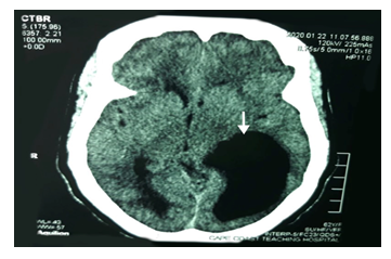
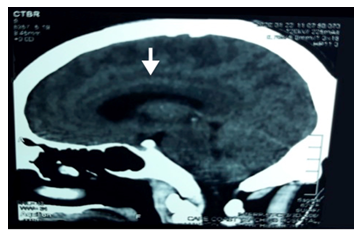
The CT scan of the head done on admission revealed this case of adult asymmetrical colpocephaly and essentially normal brain parenchyma as shown in [Figure 1] below and the presence of a normal corpus callosum shown with the white arrow on [Figure 2] below. All the other investigations were unremarkable. The patient was discharged three days after admission on her anti-hypertensive medication. A written informed consent was obtained from the patient after her discharge from the hospital, for her case to be written for publication with assurance of anonymity and confidentiality. The patient is currently well and going about her usual activities. She has been enrolled into the hypertensive clinic of the CCTH.

Figure 1 : 62-year-old woman who presented with palpitations, chest pains and generalized bodily pains, with a provisional diagnoses of acute confusional state secondary to uncontrolled hypertension. A non-contrast axial CT Scan of the head done, shows dilatation of the occipital horns of the lateral ventricles more prominent on the left side consistent with colpocephaly (white arrow). The brain parenchyma was unremarkable.

Figure 2: 62-year-old woman who presented with palpitations, chest pains and generalized bodily pains, with a provisional diagnoses of acute confusional state secondary to uncontrolled hypertension. A non-contrast sagittal CT Scan of the head done shows the presence of the corpus callosum (white arrow).
3. Discussion
Colpocephaly is commonly diagnosed in infants due to its association with neurological abnormalities [7]. Cases of colpocephaly diagnosed in asymptomatic adults are uncommon as such cases are misdiagnosed as normal pressure hydrocephalus (NPH) [8]. Colpocephaly may be as a result of a variety of congenital insults such as chromosomal abnormalities, exposure of mothers to some toxins, anoxic encephalopathy and infection in-utero [1, 5]. Radiologically, colpocephaly is characterized by an enlargement of the occipital horns and commonly associated with full or partial agenesis of the corpus callosum. Radiographically, an occipital-to-anterior horn ratio greater than 3 indicates the presence of colpocephaly, although it may have a relatively low sensitivity [6]. In our case, the occipital-to-anterior horn ratio was 3.5 indicating the presence of colpocephaly. Patients with corpus callosum agenesis may be asymptomatic and may be only discovered incidentally [6-9]. Agenesis of the corpus callosum has prevalence rates ranging from 2 to 7 of every 1,000 birth because the number of agenesis of the corpus callosum cases are symptom free and rarely come to the attention of diagnosticians and eventually making the wrong diagnosis [9]. For instance, in a case of an 88-year-old patient who was thought to be asymptomatic, his malformations was due to neuroplasticity, but when was later found to be symptomatic he was diagnosed as colpocephaly with agenesis of the corpus callosum [10].
In this case report, the patient was incidentally diagnosed as colpocephaly with an intact corpus callosum as shown in [Figure 2] above contrary to previous cases of normal adults colpocephaly reported in literature showing the presence of either a partial or complete agenesis of the corpus callosum. An asymptomatic adult with incidentally discovered colpocephaly requires no specific treatment.
4. Conclusion
A colpocephaly in a 62-year old woman is reported and the need for physicians to be aware of it because of its asymptomatic nature and not to be misdiagnosed as NPH. To the best of our knowledge, this is the very first case of this condition we are seeing in our region emphasizing the rarity of the condition in adults. Colpocephaly with or without agenesis of the corpus callosum is a rare disorder that is usually diagnosed within the initial years of life. This case report on colpocephaly in a female adult as well as the previous ones reported adds to the awareness of this type of disorder and a good understanding of these abnormalities will help prevent unnecessary diagnostic and therapeutic interventions.
Ethical Consideration
The authors certify that they have obtained all appropriate patient consent. Ethical clearance was not required for this report, as anonymity and confidentiality was assured hence no identifying detail was included.
Authors` Contribution
All authors made substantial contribution to conception, design, implementation, drafting, revising and approving the manuscript for publication.
Acknowledgement
The authors are grateful for the patient for consenting to make this report possible.
Financial Support and Sponsorship
No funding obtained for this report.
Conflicts of Interest
The authors have no conflicts of interest to declare.
References
- Cerullo A, Marini C, Cevoli S, et al. Colpocephaly in two siblings: further evidence of a genetic transmission. Developmental medicine and child neurology 42 (2000): 280-282.
- Esenwa CC, Leaf DE. Colpocephaly in adults. Case Reports (2013).
- Yakovlev PI, Wadsworth RC. Schizencephalies: A study of the congenital clefts in the cerebral mantle. I. Clefts with fused lips. Journal of Neuropathology and Experimental Neurology 5 (1946): 116-130.
- Saldanha RP, de Jesus JA, Silva BM, et al. Colpocephaly in newborn: case report and literature review. 7 (2017): 110-113.
- Puvabanditsin S, Garrow E, Ostrerov Y, et al. Colpocephaly: a case report. Am J Perinatol 23 (2006): 295-297.
- Parker C, Eilbert W, Meehan T, et al. Colpocephaly Diagnosed in a Neurologically Normal Adult in the Emergency Department. Clinical practice and cases in emergency medicine 3 (2019): 421.
- Herskowitz J, Rosman NP, Wheeler CB. Colpocephaly: clinical, radiologic, and pathogenetic aspects. Neurology 35 (1985): 1594-1598.
- Sambasivan M, Sanalkumar P, Basheer A. Colpocephaly. Kerala Med J 6 (2013): 49-50.
- Ciurea RB, Mihailescu G, Anton RM, et al. Corpus callosum dysgenesis and colpocephaly. Rom J Neurol 12 (2013): 160-163.
- Brescian NE, Curiel RE, Gass CS. Case study: a patient with agenesis of the corpus callosum with minimal associated neuropsychological impairment. Neurocase 20 (2014): 606-614.
