Colonoscopy View of Bulging Appendiceal Orifice
Article Information
Yusuf Nawras1*, Abdullah Kobeissy2
1Biology undergraduate student, University of Toledo, Toledo, Ohio, USA
2Department of Medicine, Division of Gastroenerology and Hepatology, University of Toledo Medical Center, Toledo, Ohio, USA
*Corresponding Author: Dr. Yusuf Nawras, Biology undergraduate student, University of Toledo, Toledo, Ohio, USA
Received: 25 March 2019; Accepted: 09 April 2019; Published: 06 May 2019
Citation: Yusuf Nawras, Abdullah Kobeissy. Colonoscopy View of Bulging Appendiceal Orifice. Archives of Clinical and Medical Case Reports 3 (2019): 77-79.
View / Download Pdf Share at FacebookKeywords
Colonoscopy, Abdominal pain, Inflammatory bowel disease
Colonoscopy articles Colonoscopy Research articles Colonoscopy review articles Colonoscopy PubMed articles Colonoscopy PubMed Central articles Colonoscopy 2023 articles Colonoscopy 2024 articles Colonoscopy Scopus articles Colonoscopy impact factor journals Colonoscopy Scopus journals Colonoscopy PubMed journals Colonoscopy medical journals Colonoscopy free journals Colonoscopy best journals Colonoscopy top journals Colonoscopy free medical journals Colonoscopy famous journals Colonoscopy Google Scholar indexed journals Abdominal pain articles Abdominal pain Research articles Abdominal pain review articles Abdominal pain PubMed articles Abdominal pain PubMed Central articles Abdominal pain 2023 articles Abdominal pain 2024 articles Abdominal pain Scopus articles Abdominal pain impact factor journals Abdominal pain Scopus journals Abdominal pain PubMed journals Abdominal pain medical journals Abdominal pain free journals Abdominal pain best journals Abdominal pain top journals Abdominal pain free medical journals Abdominal pain famous journals Abdominal pain Google Scholar indexed journals Inflammatory bowel disease articles Inflammatory bowel disease Research articles Inflammatory bowel disease review articles Inflammatory bowel disease PubMed articles Inflammatory bowel disease PubMed Central articles Inflammatory bowel disease 2023 articles Inflammatory bowel disease 2024 articles Inflammatory bowel disease Scopus articles Inflammatory bowel disease impact factor journals Inflammatory bowel disease Scopus journals Inflammatory bowel disease PubMed journals Inflammatory bowel disease medical journals Inflammatory bowel disease free journals Inflammatory bowel disease best journals Inflammatory bowel disease top journals Inflammatory bowel disease free medical journals Inflammatory bowel disease famous journals Inflammatory bowel disease Google Scholar indexed journals health articles health Research articles health review articles health PubMed articles health PubMed Central articles health 2023 articles health 2024 articles health Scopus articles health impact factor journals health Scopus journals health PubMed journals health medical journals health free journals health best journals health top journals health free medical journals health famous journals health Google Scholar indexed journals air insufflations articles air insufflations Research articles air insufflations review articles air insufflations PubMed articles air insufflations PubMed Central articles air insufflations 2023 articles air insufflations 2024 articles air insufflations Scopus articles air insufflations impact factor journals air insufflations Scopus journals air insufflations PubMed journals air insufflations medical journals air insufflations free journals air insufflations best journals air insufflations top journals air insufflations free medical journals air insufflations famous journals air insufflations Google Scholar indexed journals air insufflations articles air insufflations Research articles air insufflations review articles air insufflations PubMed articles air insufflations PubMed Central articles air insufflations 2023 articles air insufflations 2024 articles air insufflations Scopus articles air insufflations impact factor journals air insufflations Scopus journals air insufflations PubMed journals air insufflations medical journals air insufflations free journals air insufflations best journals air insufflations top journals air insufflations free medical journals air insufflations famous journals air insufflations Google Scholar indexed journals Diagnosis articles Diagnosis Research articles Diagnosis review articles Diagnosis PubMed articles Diagnosis PubMed Central articles Diagnosis 2023 articles Diagnosis 2024 articles Diagnosis Scopus articles Diagnosis impact factor journals Diagnosis Scopus journals Diagnosis PubMed journals Diagnosis medical journals Diagnosis free journals Diagnosis best journals Diagnosis top journals Diagnosis free medical journals Diagnosis famous journals Diagnosis Google Scholar indexed journals patient articles patient Research articles patient review articles patient PubMed articles patient PubMed Central articles patient 2023 articles patient 2024 articles patient Scopus articles patient impact factor journals patient Scopus journals patient PubMed journals patient medical journals patient free journals patient best journals patient top journals patient free medical journals patient famous journals patient Google Scholar indexed journals medicine articles medicine Research articles medicine review articles medicine PubMed articles medicine PubMed Central articles medicine 2023 articles medicine 2024 articles medicine Scopus articles medicine impact factor journals medicine Scopus journals medicine PubMed journals medicine medical journals medicine free journals medicine best journals medicine top journals medicine free medical journals medicine famous journals medicine Google Scholar indexed journals appendix articles appendix Research articles appendix review articles appendix PubMed articles appendix PubMed Central articles appendix 2023 articles appendix 2024 articles appendix Scopus articles appendix impact factor journals appendix Scopus journals appendix PubMed journals appendix medical journals appendix free journals appendix best journals appendix top journals appendix free medical journals appendix famous journals appendix Google Scholar indexed journals
Article Details
1. Case Description
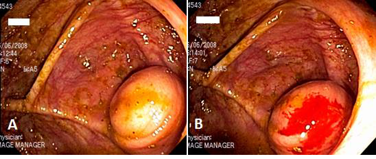
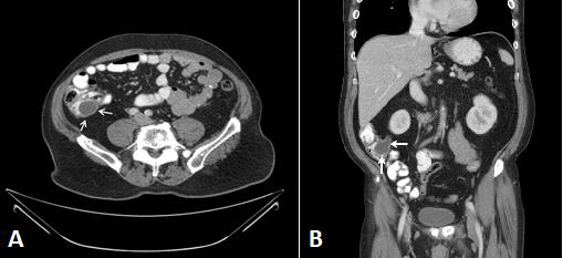
A 62-year-old male presented to the office with chronic right lower quadrant abdominal pain. He denied any diarrhea, constipation or family history of inflammatory bowel disease. Physical exam was unremarkable. A colonoscopy was normal except for a glossy, rounded protruding mass arising from the appendiceal orifice that didn’t flatten with air insufflations (Figure 1). Overlying mucosa was normal. Probing with the biopsy forceps reveals a hard lesion. The biopsy revealed normal mucosa. A CT scan was subsequently preformed and reported “a low attenuation, well-encapsulated round cystic mass in the right lower quadrant adjacent to the cecum with no other abnormalities” (Figure 2).
2. Questions Related To Our Case?
2.1 What is the most likely diagnosis?
- Carcinoid tumor of the appendix
- Lipoma
- Appendiceal mucocele
- Adenocarcinoma of the appendix
2.2 Which of the following is the next best step in her evaluation and treatment?
- Surgery evaluation for right hemicolectomy
- Surgery evaluation for appendectomy
- Close monitoring with repeat CT scan in 3-6 months
- Octreotide scan to determine presence of metastases

Figure 1 A and B: Colonoscopy view showing a glossy, rounded protruding mass arising from the appendiceal orifice. Before (A) and after biopsy (B).

Figure 2: A CT scan of the abdomen cross sectional (A) and coronal (B) showing a low attenuation, well-encapsulated round cystic mass (arrows) in the right lower quadrant adjacent to the cecum.
3. Answers and Discussion
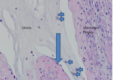
1- C and 2- B. Appendiceal mucoceles are a group of nonneoplastic or neoplastic lesions characterized by characterized by an enlarged, mucus-filled appendix. Retention cysts, mucosal hyperplasia, cystadenomas, and cystadenocarcinomas are the four histological subtypes. They are usually diagnosed in the 6th and 7th decades with a slight female predominance. Clinically, patients are often asymptomatic. Abdominal pain is the most common complaint in symptomatic patients. Incidental lesion found in endoscopic or imaging studies is the most common presentation. A Low attenuation, well-encapsulated round or tubular cystic mass in the right lower quadrant with normal cecum is the characteristic computed tomography (CT) finding [1]. On colonoscopy, appendiceal mucoceles have the appearance of a glossy, rounded protruding mass arising from the appendiceal orifice [2]. Histological exam of the appendix is the essential for diagnosis and excluding underlying malignancy (Figure 3). Differential diagnosis includes appendicitis, appendiceal tumors, and mesenteric or duplication cyst [2]. Standard appendectomy is the mainstay of treatment for all types except advanced cystadenocarcinomas with local invasion where right hemicolectomy should be considered [3]. Prognosis is favorable. Notably, some studies reported concurrent cancers like colorectal adenocarcinoma, ovary, and endometrium have been reported [4, 5].

Figure 3: Mucocele of the appendix. High power view microscopic tumor tissue section showing mucin deposition within the muscularis propria of the appendix. Note the muciphages (small arrows) and attenuated lining epithelium of the mucocele (large arrow). No neoplastic cells. Hematoxylin & Eosin stain.
References
- Madwed D, Mindelzun R, Jeffrey RB Jr. Mucocele of the appendix: imaging findings. AJR Am J Roentgenol 159 (1992): 69-72.
- Raijman I, Leong S, Hassaram S, Marcon NE. Appendiceal mucocele: endoscopic appearance. Endoscopy 26 (1994): 326-328.
- Stocchi L, Wolff BG, Larson DR, Harrington JR. Surgical treatment of appendiceal mucocele. Arch Surg 138 (2003): 585-589.
- Kalogiannidis I, Mavrona A, Grammenou S, Zacharioudakis G, et al. Endometrial adenocarcinoma and mucocele of the appendix: an unusual coexistence. Case Rep Obstet Gynecol (2013): 892378.
- Fujiwara T, Hizuta A, Iwagaki H, et al. Appendiceal mucocele with concomitant colonic cancer. Report of two cases. Dis Colon Rectum 39 (1996): 232-236.
