Case Report: An Extremely Rare Cause of Locally Recurrent Adenocarcinoma of the Spleen with Unknown Primary
Article Information
Dr Dhinagaren Narayanan1*, Dr Ahmed Hammouda1, Dr Junaid Syed Asghar2, Prof. Ruben Canelo3
Affiliation:
1North Cumbria Integrated Care NHS Foundation Trust
2The Newcastle University Hospitals NHS Foundation Trust
3University of Central Lancashire – UCLan
*Corresponding author: Dr Dhinagaren Narayanan, North Cumbria Integrated Care NHS Foundation Trust, United Kingdom.
Received: July 15, 2024 Accepted: July 22, 2024 Published: August 14, 2024
Citation: Dr Dhinagaren Narayanan, Dr Ahmed Hammouda, Dr Junaid Syed Asghar, Prof. Ruben Canelo. Case Report: An Extremely Rare Cause of Locally Recurrent Adenocarcinoma of the Spleen with Unknown Primary. Journal of Cancer Science and Clinical Therapeutics 8 (2024): 248-253.
View / Download Pdf Share at FacebookAbstract
The incidence of splenic tumour is relatively low compared to other organs. They are sometimes discovered incidentally on imaging. The majority of primary splenic tumours are diagnosed as benign. Primary malignant tumour of the spleen most commonly involves lymphosarcoma, reticulosarcoma, angiosarcoma and fibrosarcoma [1, 4]. The diagnostic algorithm to determine the nature of the splenic tumour should include series of laboratory tests, imaging studies, positron emission tomography (PET) scans as well as sometimes warranting the need for splenectomy followed by immunohistochemistry profiling. While the occurrence of primary splenic adenocarcinoma is rare, this case study reports recurrent primary splenic tumour with poorly differentiated adenocarcinoma in a patient who initially presented and investigated for anaemia of unknown cause. As to the best of our knowledge, this case has not been reported before and hence the present study provides insight into the response for standard treatment for locally recurrent adenocarcinoma of the spleen with unknown primary.
Keywords
Adenocarcinoma, Splenic tumour, Splenectomy, Immunohistochemistry.
Article Details
Introduction
The spleen is an important immune organ that has relatively low tumour incidence, accounting for only 0.03% of all types of tumours in humans due to the antitumor activity and an abundance of blood cells, a small amount of which are afferent lymphatic cells [8]. Splenic tumours can be divided into three types: Benign, malignant and metastatic, all of which are rare due to the aforementioned properties. As has been noted, primary tumours in the spleen are usually of lymphatic or vascular origin, whereas an epithelial tissue origin is rare as spleen lacks epithelial tissue. However, locally recurrent adenocarcinoma of the spleen with unknown primary has not been reported elsewhere to the best of our knowledge.
Case report
Case presentation
A 59 years old lady, with past medical history of depression and total abdominal hysterectomy with bilateral salpingo-oopherectomy for endometriosis, presented with pallor, weight loss and lethargy. She has had no history of trauma nor travel to any infectious endemic areas prior to presentation. Physical examination revealed a stable hemodynamic state, with no obvious abdominal tenderness, organomegaly or lymphadenopathy palpable elsewhere. Blood analysis showed mild normocytic anaemia with Hb 109g/l (115-165), MCV 80.6fL (80-100) and red cell width 15.5% (11.0-14.8) with normal LDH of 211 U/l (135-214). The liver function test was mildly cholestatic in picture with no elevation in bilirubin (T. Bill less than 5 umol/l, ALP 185U/l (30- 130), ALT 13 U/l (less than 40), GGT 61 U/l (less than 45)). She was referred to haematology team where she had further extensive workup for her anaemia, which revealed low iron and transferrin level; iron: 5 umol/l (6-3.5), transferrin: 1.5 g/l (2.0-3.6). Her ferritin and transferrin saturation were within normal range (Transferrin saturation: 15% (15-50), ferritin 262 ug/l (23-400)). Her FIT test was less than 5Hb/g faeces hence she was deemed not appropriate for colonoscopy. Her ESR was considerably high at 111mm/hr (0-20), as well as mild elevation in CA-125 level was seen (CA-125 - 73 kU/l (0- 34) with normal CEA, CA19-9 and AFP. Otherwise, the vasculitis, immunology and myeloma blood panel were all negative as follow; Light chain quantum kappa/lambda ratio 0.88 (0.26 - 1.65), Serum IgA 2.61g/l (0.7-4.9), IgG 13.27 (6.1-15.4) IgM 1.67 (0.3-1.8), ANA, AMA, ASMA ANTI LKM, anti LC, anti-gastric parietal cell, anti-cardiolipin- negative, Anti-MPO Abs and Anti-PR3 negative, rheumatoid factor less than 20 IU/ml. The patient then had computed tomography (CT) scan of her thorax, abdomen and pelvis to identify if there is any malignancy that could be responsible for her anaemia despite her initial blood workup unable to localise any causes. The CT scan revealed an enlarged spleen with multifocal low-density areas and prominent calcified ring in it, with no other suspicious lesions or nodal diseases seen elsewhere.
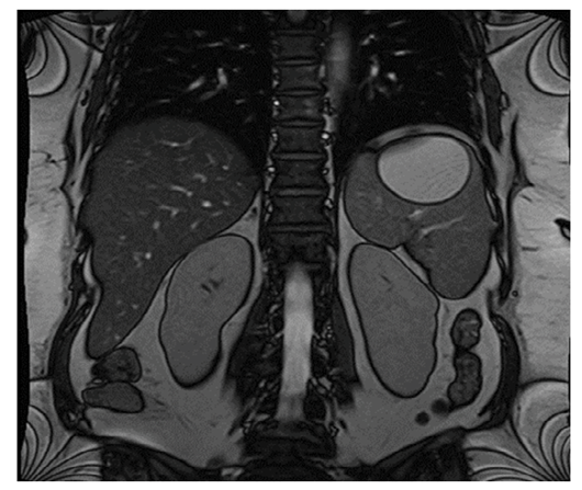
Patient eventually had MRI of the spleen to identify the nature of the lesion in the spleen, which identified the stable splenic cyst with another heterogenous lesion of indeterminate nature with high T2 signal seen anterior to the splenic cyst.
Patient was commenced on commenced on Albendazole and Praziquantel as treatment for hydatid splenic cyst based on radiological diagnosis. However, the hydatid serology came negative for ecchymosis and amoebiasis, hence the regime was stopped. The patient underwent nuclear medicine whole body positive emission tomography (NM whole body PET) scan which revealed abnormal increased Fludeoxyglucose F18 (FDG) uptake confined to spleen - intense FDG avid with internally necrotic splenic mass inseparable from posterior surface of stomach.
The case of this patient was then discussed in Cancer of Unknown Primary (CUP) multidisciplinary team meeting in Freeman Hospital, Newcastle where the team believes the radiological evidences are suggestive of solitary lesion of spleen with no evidence that it was a metastasis of unknown primary, and decided to proceed with splenectomy and sleeve gastrectomy.
Post-operative pathological analysis
The post-operative pathological analysis was done extensively to identify and formulate the nature and the primary cause of the lesion. Macroscopic examination of the specimen revealed an intact splenectomy weighing 640g and measuring 135mm superoinferiorly, 100mm mediolaterally and 85mm anteroposteriorly. The capsule shows an area of disruption on the inferoposterior aspect 20 x 10mm. Attached to the superior aspect is a stapled sleeve of stomach 50 x 30mm, which is slightly indrawn and adherent to a subcapsular bulging mass. The splenic capsule surrounding this has a firm, cream appearance. A small pedicle of anterior fat 85 x 35 x 8mm is present. Enlarged lymph nodes are noted at the stapled splenic hilum. Staple line of sleeve gastrectomy removed and sub-stapled margin inked blue. Staple line from splenic hilum removed and sub-stapled margin inked red. On slicing through the splenic capsule to allow fixation, the spleen drains light tan fluid. Slicing in the axial plane shows a large, firm, cream, well circumscribed nodular mass 83 x 75 x 72mm across the slices. This extends through the capsule into adherent fat superiorly and also suspicious of involving hilar fat. Embedded partly within the mass and partly extending into the surrounding splenic parenchyma is a large, intact calcified cyst 63 x 52 x 51 mm across the slices. This focally abuts the splenic capsule. Cruciate of the most superior slice with attached sleeve gastrectomy show tumour pushing against the muscularis with suspicion of involving muscularis focally. The carcinoma extends to within 1.4 mm of the stapled gastric resection margin (after removal of the staples). The splenic hilar margin appears to be tumour free, including four hilar lymph nodes.
Microscopic examination of the specimen revealed sections which show extensive cellular epithelioid tumour. The neoplastic cells seem to forms small solid packets and strands and have large round to oval vesicular nuclei with prominent nucleoli and moderate amounts of pale to clear cytoplasm. In places, the cells form anastomosing spaces. Elsewhere, they have a more spindled appearance, especially at the transition to areas of necrosis. There are more than 20 mitoses in 10 HPF. Variable numbers of chronic inflammatory cells are admixed. Areas of acute haemorrhage and multifocal haemosiderin deposition are also noticed. Extensive necrosis affects at least half of the entire mass. As described macroscopically, there is large cystic space closely associated with the tumour. The cystic space is lined by a band of hypocellular fibrous tissue with extensive calcification and ossification; no epithelial lining is identified.
As described macroscopically, the tumour widely involves the spleen and extends into the adherent gastric wall up to the muscularis mucosae, but the overlying mucosa is not involved. It extends into the peri-hilar fat with a pushing margin. Four hilar lymph nodes and the splenic hilar vessels are tumour-free. lmmunohistochemistry shows strong diffuse positivity for CKAE1/3 and PAX8, patchy staining for CK5D3, EMA and CA125 and very focal positivity for calretinin. The tumour is negative for the epithelial markers CK7, CK20, TTF1, Napsin, GATA3, oestrogen receptor, CDX2, HepPar1, Arginase and RCC. It is also negative for the mesothelial markers CK5/6, monoclonal WT1 and D2-40. There is no staining for the mesenchymal and melanocytic markers SMA, H-caldesmon, desmin, CD31, ERG, DOG1, ALK, vimentin, inhibin, MelanA, SOX10 or S100. INI is retained. There is no p16 overexpression in the tumour or the adjacent fat. Staining for p53 is wild-type. The Ki67 proliferation rate is very high.
Surveillance and treatment
Since all the visible disease has been resected completely, the hepatobiliary team (HPB) believed there was no role for adjuvant systemic therapy in either scenario. HPB team have agreed to keep the patient under imaging surveillance. Repeat CT abdomen and pelvis that was done 10 months after the initial splenectomy and sleeve gastrectomy revealed increased local soft tissue which could be regenerative splenic tissue or recurrent cancer, no disease elsewhere. The absence of any new primary or secondary disease elsewhere on the recent CT scan 10 months after surgery strongly suggests that the original splenic lesion was the primary rather than a metastasis of unknown primary. CUP MDT decided to proceed with PET scan to identify the nature of the recurrent lesion. The PET scan was performed and the study showed
Figure 6: NM Whole body PET FDG : There is marked FDG uptake at soft tissue in the left upper quadrant in contact with clips, the left adrenal, stomach and diaphragm (4.2 x 2.7 x 4.5 cm, SUVmax 9.8). Possible contact with the upper pole of the left kidney and distal pancreas. Central photopenia compatible with necrosis.
markedly FDG avid left upper quadrant mass in contact with the adjacent structures, suggestive of recurrent malignant process.
Patient was then seen in oncology clinic and findings of the PET scan was discussed. Patient agreed to chemotherapy with intended disease control after informed decision of the benefits and side effects of the regime. The regime that was initiated for the patient was cisplatin-gemcitabine (1+8 of 21-day cycle) for 6-8 cycles. Patient tolerated the treatment reasonably well and no major treatment related side effects was encountered except for mucositis and neuropathy grade 1 that resolves before each cycle. Patient had a repeat CT scan post cycle 4 to identify the response to the chemotherapy regime. The repeat scan showed partial response - the surgical bed recurrence has reduced from 39 to 31 mm. From patient’s perspective, the outcome of the chemotherapy regime was seen as very promising. Patient was planned to have continued chemotherapy with no modification to the regime and was planned for re-staging after the last cycle is completed.
Discussion
Primary neoplasms of the spleen include haemangioma, lymphangioma, hamartoma, haemangioendothelioma, hemangiopericytoma, angiosarcoma from vascular element, littoral cell angioma and lymphoma from lymphoid tissue [1]. The analysis of 194 splenic tumours by Zhan categorised 45 cases as metastatic tumours, 95 as primary malignant lymphoma and the remaining as mesenchymal malignant tumours such as liposarcoma, malignant fibrous sarcoma and angiosarcoma [2]. Primary adenocarcinoma of the spleen is extremely rare. In this present study, the histologic appearances are those of a poorly differentiated adenocarcinoma involving the spleen and the adherent gastric wall. The tumour does not appear to have arisen from the gastric mucosa. A diagnosis of primary peritoneal carcinoma was considered and cannot be entirely excluded, but the immunoprofile is not typical as they stained negative for oestrogen receptor (ER), WT1, CK7 and only had patchy staining for CA125. The staining pattern does not support a diagnosis of peritoneal mesothelioma as negative for the mesothelial markers CK5/6, monoclonal WT1 and D2-40. The tumour could represent a metastasis, but despite a comprehensive immunopanel, repeated radiological imaging and nuclear medicine as well as discussion with several consultant colleagues, we cannot determine the exact origin of this adenocarcinoma.
The spleen consists of a membrane, the trabeculae, white pulp, red pulp and the marginal zone, which are all mesenchymal [6]. Notably, there is no epithelial tissue in the spleen. Therefore, there are a few possibly hypothesis that can be postulated to explain the present study of adenocarcinoma in the spleen. The first hypothesis is similar to the seeding concept of endometriosis [3]. Fragments of epithelial tissue from other areas of the body could enter the spleen via the abundant blood supply of the spleen, eventually getting implanted in it. Non-specific inflammation and the action of certain hormones can induce the cells to undergo malignant transformation into adenocarcinoma or squamous cell carcinoma. The second hypothesis that would be relevant in this patient is the very unusual possibility that minute non- malignant tissue from an intra-abdominal organ such as ovary, stomach, colon, pancreas, or others might be dropped and accidentally implanted to the splenic surface during the surgery and transformed into cancer later [4, 5]. It has been proposed that an early blood-borne micro metastasis within the spleen may grow into an imaging-detectable metastasis after several years [7]. This idea could be a better explanation in this patient as this patient had previous total abdominal hysterectomy with bilateral salpingo-oopherectomy for her extensive endometriosis, though the timeline of 27 years apart from the current presentation might be against the justification. Therefore, the origin and the mechanism of the tumour in the present case study remain unknown despite detailed discussion and comparison with current available literature.
In conclusion, we have reported a case study of locally recurrent adenocarcinoma of the spleen, where no primary lesion was found despite extensive workup, which to the best of our knowledge, has not been reported previously. Furthermore, in this case, the chemotherapy cycles provide partial response in mid regime therefore reflects potentially useful information pertaining to the response for standard treatment after splenectomy for locally recurrent cases.
Abbreviations
- CEA = carcinoembryonic antigen
- AFP = alpha-fetoprotein
- Anti-LKM = anti- liver kidney microsomal
- Anti-LC = anti-liver cytosol
- Anti-MPO = anti-myeloperoxidase
- Anti-PR3 = anti-proteinase 3
- CKAE1/3 = cystokeratin AE1/AE3
- PAX8 = paired-box gene 8
- CK5D3 = cystokeratin 5D3
- EMA = epithelial membrane antigen
- CK7 = cystokeratin 7
- CK20 = cystokeratin 20
- TTF1 = thyroid transcription factor 1
- HepPar1 = hepatocyte paraffin 1
- RCC = renal cell carcinoma
- CK5/6 = cystokeratin 5/6
- WT1 = Wilm’s tumor 1
- D2-40 = podoplanin antibody
- SMA = smooth muscle actin
- CD31 = cluster of differentiation 31
- DOG1 = Discovered On GIST 1
- ALK = anaplastic lymphoma kinase
- SOX10 = SRY-Box Transcription Factor 10
Acknowledgements
Not applicable
Funding
No funding was received
Author’s contributions
Dr Syed Asghar was in charge of the patient from oncological point of view who liaised with his team to come up with the proposed investigation and decided the treatment for the patient. Prof Ruben Canelo came up with the idea of creating a case report and was involved in the management and coordination responsibility for the activity planning and execution. Dr Ahmed Hammouda was actively involved in data curation and collection. Dr Dhinagaren Narayanan assisted in data collection, as well as led the formal analysis of the data and writing the original draft, which was reviewed, revised and validated critically by all other authors for important intellectual content.
Consent
Written informed consent was obtained from the patient for publication of this case report and its accompanying images.
Competing interest
The authors declare that they have no competing interests.
References
- Kaza RK, Azar S, Al-Hawary MM, et al. Primary and secondary neoplasms of the spleen. Cancer Imaging 10 (2010): 173-182.
- Zhan Spleen cancer of 194 cases. Chin J Gen Surg 12 (1997): 183-184.
- Meola J, Rosa e Silva JC, Dentillo DB, et al. Differentially expressed genes in eutopic and ectopic endometrium of women with endometriosis. Fertil Steril 93 (2010): 1750-1773.
- Ohe C, Sakaida N, Yanagimoto Y, et A case of splenic low-grade mucinous cystadenocarcinoma resulting in pseudomyxoma peritonei. Med Mol Morphol 43 (2010): 235-240.
- Morinaga, Shojiroh, Ohyama, et Low-Grade Mucinous Cystadenocarcinoma in the Spleen. The American Journal of Surgical Pathology 16 (1992): 903-908.
- Lewis JT, Gaffney RL, Casey MB, et al. Inflammatory pseudotumor of the spleen associated with a clonal Epstein-Barr virus Case report and review of the literature. Am J Clin Pathol 120 (2003): 56-61.
- E Compérat, A Bardier-Dupas, P Camparo, et al. Splenic metastases: clinicopathologic presentation, differential diagnosis, and pathogenesis Arch Pathol Lab Med 131 (2007): 965-969.
- Coon Surgical aspects of splenic disease and lymphoma. Curr Probl Surg 35 (1998): 543-646.


