Cannonball Lung Metastases from A Retroperitoneal Sarcoma
Article Information
Paulo Luz*, Júlio Lemos Teixeira, Pedro Mendonça
Centro Hospitalar Universitário do Algarve, Serviço de oncologia médica, Rua Leão Penedo, 8000 – Faro, Portugal
*Corresponding Author: Dr. Paulo Luz, Centro Hospitalar Universitário do Algarve, Serviço de oncologia médica, Rua Leão Penedo, 8000 – Faro, Portugal
Received: 18 July 2019; Accepted: 02 August 2019; Published: 12 August 2019
Citation: Paulo Luz, Júlio Lemos Teixeira, Pedro Mendonça. Cannonball Lung Metastases from A Retroperitoneal Sarcoma. Journal of Cancer Science and Clinical Therapeutics 3 (2019): 102-104.
View / Download Pdf Share at FacebookAbstract
Sarcomas constitute a heterogeneous group of neoplasms, but with a very aggressive evolution in most cases. Presented, is the case of a male patient, 69 years old, with several emergency room visits in the past two months due to lumbar back pain complaints. Having presented a positive Murphy sign in his last visit to the Emergency Department, patient underwent renal ultrasound, showing the presence of a large hypoechogenic mass pushing the right kidney forwards. A thoracoabdominal-pelvic computed tomography scan was performed and showed a large mass in the retroperitoneum. A biopsy of the mass was performed revealing it to be a dedifferentiated liposarcoma. We presented the chest X-ray repeated after 18 days, showing an increase in pulmonary metastatic lesions. The patient died one month after the biopsy procedure. Since these cancers are rare, difficult to diagnose and treat, a collaborative effort from a team comprising of a surgical oncologist, a pathologist, a radiologist, a medical oncologist, a radiation oncologist and a palliative care specialist are mandatory for a positive outcome.
Keywords
Retroperitoneal sarcoma; Lung metastases
Article Details
1. Introduction
Sarcomas constitute a heterogeneous group of neoplasms (circa 80 different histological subtypes have been described), but with a very aggressive evolution in most cases. Therefore, a timely diagnosis is crucial. One of the most common sites where these neoplasms can be found is in the retroperitoneum, causing, albeit rarely, lower back pain [1, 2].
2. Case Report
Presented, is the case of a male patient, 69 years old, single and previously autonomous in daily life, with no relevant personal or family history, and with several emergency room visits in the past two months due to lumbar back pain complaints, radiating to the right anterolateral thigh. Patient denied having had a fever or any other complaints but reported a mild weight loss (<10% total body weight). Having presented a positive Murphy sign in his last visit to the Emergency Department (ED), patient underwent renal ultrasound, showing the presence of a large hypoechogenic mass pushing the right kidney forwards. This patient was hospitalised for further investigation of this mass and symptomatic control. During hospitalisation, a thoracoabdominal-pelvic computed tomography scan was performed. Results showed a mass in the retroperitoneum measuring 124 × 112 mm on the axial plane and 167mm on the longitudinal axis; an extensive thrombosis of the inferior vena cava, extending to the right common iliac vein; as well as multiple nodular pulmonary images with a mass of 45 × 34 mm in contact with the superior vena cava, altering its shape. A biopsy of the mass in the retroperitoneum was performed, with first analysis revealing it to be a fusocellular sarcoma whose immunocytochemical pattern was inconclusive in defining its histological subtype. The sample was therefore sent to a specialised centre for further analysis.
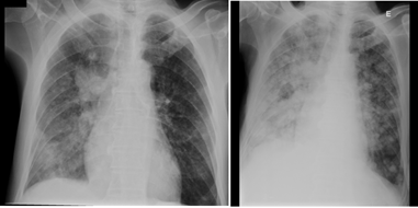
The patient was discharged from Internal Medicine after 17 days of hospitalisation and was referred for an urgent oncology appointment while awaiting the definitive result of the biopsy. Patient returned to the ED two days later reporting shortness of breath and lethargy. Chest X-ray was repeated, showing an increase in pulmonary metastatic lesions, as shown in Figure 1.
Patient was admitted to the Medical Oncology department for symptomatic control and ECOG (Eastern Cooperative Oncology Group) scale evaluation to decide whether he was a candidate to start treatment as soon as the definitive biopsy result was available. During hospitalisation, there was a progressive worsening of his general condition, showing respiratory difficulties and worsening edema of the lower limbs. Patient passed away on the 8th day of hospitalisation while still awaiting the anatomical pathology report, which ultimately proved to be a fusocellular sarcoma whose morphological pattern and immunocytochemical profile favoured a dedifferentiated liposarcoma.
This case aims to draw attention to two situations: first, the need for a detailed anamnesis on the approach to a patient with lower back pain with multiple visits to the ED. Although basic, we must not forget that it is essential to perform a thorough physical exam. Secondly, the difficulty that anatomical pathology services at secondary centres have in the diagnosis of these neoplasms (infrequent and quite heterogeneous), which often forces samples to be sent to a tertiary centre, further delaying the final diagnosis and consequent start of treatment for an already aggressive disease [2-4].
As previously mentioned, sarcomas constitute a heterogeneous group of neoplasms with a very aggressive evolution in most cases and therefore, a timely diagnosis is crucial. Since these cancers are rare, difficult to diagnose and treat, a collaborative effort from a team comprising of a surgical oncologist, a pathologist, a radiologist, a medical oncologist, a radiation oncologist and a palliative care specialist are mandatory for a positive outcome.
Competing Interests
The authors declare no competing interest.
Authors’ Contributions
The first three authors contributed equally to the conception and design, acquisition of data, analysis and interpretation of data.
References
- Trans-Atlantic RPS Working Group. Management of primary retroperitoneal sarcoma (RPS) in the adult: a consensus approach from the Trans-Atlantic RPS Working Group. Ann Surg Oncol 22 (2015): 256-263.
- Trojani M, Contesso G, Coindre JM, et al. Soft-tissue sarcomas of adults; study of pathological prognostic variables and definition of a histopatho- logical grading system. Int J Cancer 33 (1984): 37-42.
- Soft Tissue Sarcomas-The Pitfalls in Diagnosis and Management!!. Indian J Surg Oncol 2 (2011): 261-264.
- Ray-Coquard I, Montesco MC, Coindre JM, et al. Sarcoma: concordance between initial diagnosis and centralized expert review in a population- based study within three European regions. Ann Oncol 23 (2012): 2442-2449.

