Breast Lymphoma: A Report of 5 Cases and Literature Review
Article Information
Abidi Fethia1, Ons Chaibi1, Riahi Nihed2, Essghaier Sonia1, Ksentini Feriel3, Asma Zidi1
1Department of Radiology, Institut of Salah Azaiez, Tunis, Tunisia
2Department of Surgical Oncology, Institut of Salah Azaiez, Tunis, Tunisia
3Department of Medical Oncology, Institut of Salah Azaiez, Tunis, Tunisia
*Corresponding Author: Abidi Fethia, Chair, Department of radiology, Institut of Salah Azaiez, Tunis, Tunisia
Received: 14 April 2023; Accepted: 05 May 2023; Published: 19 May 2023
Citation: Abidi Fethia, Ons Chaibi, Riahi Nihed, Essghaier Sonia, Ksentini Feriel, Asma Zidi. Breast Lymphoma: A Report of 5 Cases and Literature Review. Journal of Cancer Science and Clinical Therapeutics. 7 (2023): 108-112.
View / Download Pdf Share at FacebookAbstract
Breast lymphoma may occur as either a primary or a secondary lesion and the secondary form is more common than the primitive form. We report five cases of breast lymphoma who underwent multimodal breast imaging studies in our institute between March 2018 and June 2020, one was primary and the others were secondary. The multimodal imaging consisted of mammography, ultrasonography (US) and dynamic computed tomography (CT). The objective of this paper was to illustrate the multimodal imaging findings in the primary and secondary forms of breast lymphoma and to describe the key clinical and radiological findings that allow it to be distinguished from other breast malignancies.
Keywords
Breast; Cancer; Lymphoma
Breast articles; cancer articles; lymphoma articles
Breast articles Breast Research articles Breast review articles Breast PubMed articles Breast PubMed Central articles Breast 2023 articles Breast 2024 articles Breast Scopus articles Breast impact factor journals Breast Scopus journals Breast PubMed journals Breast medical journals Breast free journals Breast best journals Breast top journals Breast free medical journals Breast famous journals Breast Google Scholar indexed journals Cancer articles Cancer Research articles Cancer review articles Cancer PubMed articles Cancer PubMed Central articles Cancer 2023 articles Cancer 2024 articles Cancer Scopus articles Cancer impact factor journals Cancer Scopus journals Cancer PubMed journals Cancer medical journals Cancer free journals Cancer best journals Cancer top journals Cancer free medical journals Cancer famous journals Cancer Google Scholar indexed journals Lymphoma articles Lymphoma Research articles Lymphoma review articles Lymphoma PubMed articles Lymphoma PubMed Central articles Lymphoma 2023 articles Lymphoma 2024 articles Lymphoma Scopus articles Lymphoma impact factor journals Lymphoma Scopus journals Lymphoma PubMed journals Lymphoma medical journals Lymphoma free journals Lymphoma best journals Lymphoma top journals Lymphoma free medical journals Lymphoma famous journals Lymphoma Google Scholar indexed journals mammography articles mammography Research articles mammography review articles mammography PubMed articles mammography PubMed Central articles mammography 2023 articles mammography 2024 articles mammography Scopus articles mammography impact factor journals mammography Scopus journals mammography PubMed journals mammography medical journals mammography free journals mammography best journals mammography top journals mammography free medical journals mammography famous journals mammography Google Scholar indexed journals ultrasonography articles ultrasonography Research articles ultrasonography review articles ultrasonography PubMed articles ultrasonography PubMed Central articles ultrasonography 2023 articles ultrasonography 2024 articles ultrasonography Scopus articles ultrasonography impact factor journals ultrasonography Scopus journals ultrasonography PubMed journals ultrasonography medical journals ultrasonography free journals ultrasonography best journals ultrasonography top journals ultrasonography free medical journals ultrasonography famous journals ultrasonography Google Scholar indexed journals breast imaging studies articles breast imaging studies Research articles breast imaging studies review articles breast imaging studies PubMed articles breast imaging studies PubMed Central articles breast imaging studies 2023 articles breast imaging studies 2024 articles breast imaging studies Scopus articles breast imaging studies impact factor journals breast imaging studies Scopus journals breast imaging studies PubMed journals breast imaging studies medical journals breast imaging studies free journals breast imaging studies best journals breast imaging studies top journals breast imaging studies free medical journals breast imaging studies famous journals breast imaging studies Google Scholar indexed journals lesion articles lesion Research articles lesion review articles lesion PubMed articles lesion PubMed Central articles lesion 2023 articles lesion 2024 articles lesion Scopus articles lesion impact factor journals lesion Scopus journals lesion PubMed journals lesion medical journals lesion free journals lesion best journals lesion top journals lesion free medical journals lesion famous journals lesion Google Scholar indexed journals computed tomography articles computed tomography Research articles computed tomography review articles computed tomography PubMed articles computed tomography PubMed Central articles computed tomography 2023 articles computed tomography 2024 articles computed tomography Scopus articles computed tomography impact factor journals computed tomography Scopus journals computed tomography PubMed journals computed tomography medical journals computed tomography free journals computed tomography best journals computed tomography top journals computed tomography free medical journals computed tomography famous journals computed tomography Google Scholar indexed journals malignancies articles malignancies Research articles malignancies review articles malignancies PubMed articles malignancies PubMed Central articles malignancies 2023 articles malignancies 2024 articles malignancies Scopus articles malignancies impact factor journals malignancies Scopus journals malignancies PubMed journals malignancies medical journals malignancies free journals malignancies best journals malignancies top journals malignancies free medical journals malignancies famous journals malignancies Google Scholar indexed journals lymphomatous articles lymphomatous Research articles lymphomatous review articles lymphomatous PubMed articles lymphomatous PubMed Central articles lymphomatous 2023 articles lymphomatous 2024 articles lymphomatous Scopus articles lymphomatous impact factor journals lymphomatous Scopus journals lymphomatous PubMed journals lymphomatous medical journals lymphomatous free journals lymphomatous best journals lymphomatous top journals lymphomatous free medical journals lymphomatous famous journals lymphomatous Google Scholar indexed journals
Article Details
1. Introduction
The breast is a rare site of lymphomatous involvement due to the low proportion of lymphoid tissue at this level [1,2]. Breast lymphoma accounts for only approximately 0.04% to 0.7% of all breast cancer cases [3-6]. A broad variety of histologic types have been reported. The majority are B-cell lymphomas, and the most common type is diffuse large B-cell lymphoma [6].
The imaging features of breast lymphoma are generally nonspecific. However, the treatment and prognosis differ from other breast malignancies; Surgery is not often used as a treatment of lymphoma because of the efficacy of chemotherapy, biological therapy and radiotherapy. To avoid unnecessary treatment, clinicians and radiologists should be familiarized with characteristic imaging features of breast lymphoma.
2. Case Reports
2.1 Case 1
A 42-year-old pregnant patient at 36 weeks consults for bilateral mastitis evolving for 2 months not improved by medical treatment. On examination, both breasts were increased in size, swollen, inflammatory and painful associated with bilateral skin thickening. Mammography objectived a totally opaque left breast, an increase in density of the right breast at the level of its anterior and middle two thirds and skin thickening. There were no detected microcalcifications (Figure 1). US revealed multiple confluent hypoechogenic masses in both breasts, irregular in shape, with indistinct margins, hypervascularized in the Doppler study without posterior attenuation. It also revealed skin thickening and multiple left axillary adenopathy (Figure 1). The patient underwent an ultrasound-guided microbiopsy of the masses and a fine-needle aspiration of the left adenopathy. Histopathological and immunohistochemical findings in the tumour and lymph nodes confirmed the diagnosis of a diffuse large B-cell primary breast lymphoma: CD20+, CD3-, cytokeratin negative.
The CT of the thorax and abdomen performed as a part of the extension workup did not reveal any other localization apart from bilateral mammary involvement and left axillary adenopathy (Figure 1). After four courses of chemotherapy, the CT control showed a partial response to treatment with partial regression of the breast infiltration.
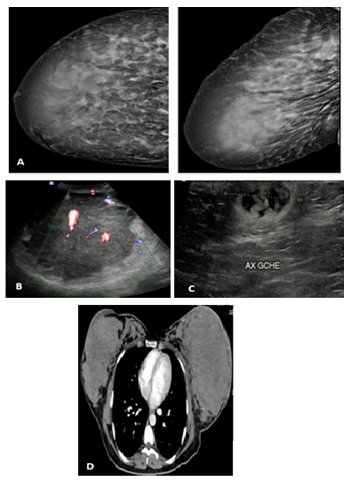
Figure 1: Patient with primary breast lymphoma.
A: Mammography: Increase in density of the right breast at the level of its anterior and middle two thirds; B: Ultrasound: hypoechogenic masse with increase of vascularization in the color Doppler study; C: Heteroechogenic left axillary adenopathy; D: CT: Significant increase in the volume of both breasts, diffuse infiltration without any other locations detected.
2.2 Case 2
A 17-year-old woman, without any pathological history, presented for left mastodynia evolving for 8 months associated with alteration of the general state. Physical examination revealed a firm immobile mass in the internal quadrants of the left breast associated with homolateral enlarged axillary lymph nodes. US showed a voluminous mass of the internal left breast quadrants, irregular in shape with microlobulated margins, mixed echogenicity and generalized increase in vascularisation in the color Doppler (Figure 2). Mediolateral oblique mammography views revealed generalized density increase of the left breast associated with skin thickening (Figure 2).
An abdominal US showed hepatic, pancreatic and ovarian masses with intraperitoneal effusion of medium abundance. Core-needle biopsy of the left breast mass revealed a secondary breast localization of a diffuse large B-cell lymphoma. The extension study with CT showed tumor involvement in multiple other locations.
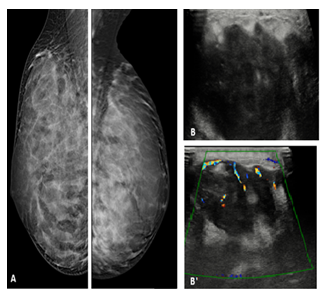
Figure 2: Secondary diffuse large B cell breast lymphoma.
A: Bilateral mediolateral oblique mammography views revealed generalized density increase of the left breast associated with skin thickening; B, B’: US: voluminous mass, irregular in shape with microlobulated margins, mixed echogenicity and generalized increase in vascularization in the color Doppler
2.3 Case 3
A 60-year-old woman, followed since 2018 for Burkitt lymphoma, presented on mammography bilateral breast masses in the right upper external quadrant and left lower internal quadrant with regular shape, circumscribed contours and medium density associated with adenopathies in the left axillary region without any clinical manifestation (Figure 3). US revealed oval-shaped masses with regular contours, heteroechogenic echostructure, increase Doppler vascularization and left axillary adenopathies of the same appearance (Figure 3).
CT showed bilateral nodular lesions with skin thickening of the left breast and homolateral axillary adenopathies (Figure 4). Breast biopsy of the masses was carried out, with anatomopathological diagnosis of secondary breast lymphoma localization.
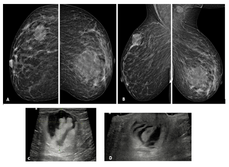
Figure 3: Adenopathies in the left axillary region without any clinical manifestation.
A, B: Mammography: Both craniocaudal and mediolateral oblique mammograms demonsrate bilateral masses with regular shape, circumscribed contours and medium density associated with adenopathies in the left axillary region; C: US: Breast oval-shaped masse with regular contours, heteroechogenic echostructure and increase Doppler vascularization; D: Axillary US: Left axillary adenopathies of the same appearance of the breast masses.

Figure 4: CT showed bilateral nodular lesions with skin thickening of the left breast and homolateral axillary adenopathies.
Staging CT: Bilateral breast nodular lesions (thick arrow) with skin thickening of the left breast and homolateral axillary adenopathies (thin arrow).
2.4 Case 4
A 33-year-old woman, followed for diffuse large B-cell lymphoma, presented right mastodynia with a swollen and inflammatory breast on examination. Mammography revealed diffuse density increase of the right breast, skin thickening and bilateral axillary adenopathies (Figure 5). US demonstrated multiple masses of the right breast of regular shape, well circumscribed contours, hypoechogenic without posterior attenuation, showing an increase of vascularization in the color Doppler study associated with a significant skin thickening and bilateral axillary nodes (Figure 5). The breast core needle biopsy with anatomopathological study concluded to a secondary lymphomatous breast localization.
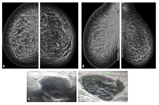
Figure 5: Mammography revealed diffuse density increase of the right breast, skin thickening and bilateral axillary adenopathies.
A, B: Cranio caudal and mediolateral oblique mammography showing generalized density increase of the right breast, bilateral skin thickening and axillary adenopathies; C, C’: Ultrasound: Masses with regular shape well circumscribed contours, hypoechogenic without posterior attenuation.
2.5 Case 5
A 62-year-old woman, followed for aggressive follicular lymphoma, presented on computed tomography that was done as part of the surveillance work up, multiples breast masses associated with axillary adenomegaly (Figure 6). Mammography revealed multiple masses in both breasts as well as bilateral axillary nodes. US showed bilateral heteroechogenic masses, mostly hypoechogenic, hypervascularized in the color Doppler (Figure 7).
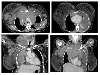
Figure 6: Computed tomography that was done as part of the surveillance work up, multiples breast masses associated with axillary adenomegaly.
Axial and coronal contrast enhanced CT showing a significant increase in the volume of the right breast, which shows multiple nodular lesions, and skin thickening. Conglomerate of mediastinal adenopathies extending into the supraclavicular and cervical space. Multiple bilateral axillary adenopathies.
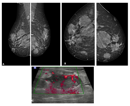
Figure 7: US showed bilateral heteroechogenic masses, mostly hypoechogenic, hypervascularized in the color Doppler.
A, B: Mammography: Multiple dense masses of regular shape and circumscribed contours in both breasts and bilateral axillary nodes; C: Ultrasound: Masse, heteroechogenic, mostly hypoechogenic, hypervascularized in the color Doppler associated with skin thickening and dilated dermal lymphatics.
3. Discussion
3.1 Epidemiology
Breast lymphoma occurs almost exclusively in women [1, 7]. It represents approximatly 2% of all extranodal malignant lymphomas [1, 8] and less than 0.7% of breast malignancy [9]. Primary breast lymphoma is very rare, accounting for only 0.85% to 2% of all extranodal non-Hodgkin's lymphoma. It has been defined as localized involvement of one or both breasts without metastatic disease except ipsilateral axillary nodal involvement [3, 10, 11]. Secondary breast lymphoma (SBL) is more common [3, 12] and falls within the scope of an extra mammary lymphomatous lesion already labelled [1].
The reported median age is in the 5th to 6th decades [3, 9, 13, 14], which is similar to the median age of breast carcinoma. Primary breast lymphoma (PBL) presents however some differences in the age distribution with a bimodal peak: Younger population showing bilateral involvement and older population showing unilateral involvement [15].
3.2 Pathophysiology
Breast lymphoma is subdivided in B-cell or T-cell form, based on the neoplastic cell of origin. The most common type is diffuse large B-cell lymphoma, mostly CD20+ [1, 7, 13] which accounts for more than 80% of the primitive form [7]. B-cell lymphoma may be subdivided into low-grade and high-grade tumors [9]. Less commonly, breast lymphoma can be present in other histopathologic types: follicular lymphoma (15%), MALT lymphoma (12.2%) and Burkitt's lymphoma (10.3%) [7, 16].
Primary breast lymphoma behave similarly to lymphoma of similar histologic types and stages in other sites [6] however PBL in pregnant or recently pregnant woman presents generally high-grade of malignancy, rapidly dissemination and poor prognosis [17].
Breast T-cell lymphoma is rare but his incidence may soon increase: Implant-associated anaplastic large cell lymphoma (ALCL) is emerging as a new clinical entity and both saline and silicone implants have been implicated [9]. They have been shown to induce inflammatory T-cell reaction and lymphoma may occur in the fibrous capsule of the breast implant [3, 9, 18-20]. Some morphologic similarities between lymphoma cells, invasive lobular carcinoma and medullary carcinoma have been reported. Histological misinterpretation may lead to an erroneous diagnosis and incorrect treatment [9].
References
- Jroundi L, Fikri M, Barkouchi F, et al. Imagerie du lymphome mammaire. Imagerie de la Femme. déc 14 (2004): 307-312.
- Sabaté JM, Gómez A, Torrubia S, et al. Lymphoma of the breast: clinical and radiologic features with pathologic correlation in 28 patients. Breast J 8 (2002): 294-304.
- Shim E, Song SE, Seo BK, et al. Lymphoma Affecting the Breast: A Pictorial Review of Multimodal Imaging Findings. J Breast Cancer 16 (2013): 254.
- Brustein S, Filippa DA, Kimmel M, et al. Malignant lymphoma of the breast. A study of 53 patients. Ann Surg. 205 (1987): 144-150.
- Giardini R, Piccolo C, Rilke F. Primary non-Hodgkin’s lymphomas of the female breast. Cancer. 1 févr 69 (1992): 725-735.
- Brogi E, Harris NL. Lymphomas of the breast: pathology and clinical behavior. Semin Oncol. juin 26 (1992): 357-364.
- Joks M, Mysliwiec K, Lewandowski K. Primary breast lymphoma - a review of the literature and report of three cases. Aoms 1 (2011): 27-33.
- Topalovski M, Crisan D, Mattson JC. Lymphoma of the breast. A clinicopathologic study of primary and secondary cases. Arch Pathol Lab Med. Déc 12 (1999): 1208-1218.
- Nicholson BT, Bhatti RM, Glassman L. Extranodal Lymphoma of the Breast. Radiologic Clinics of North America. juill 54 (2016): 711-726.
- Gupta V, Bhutani N, Singh S, et al. Primary non-Hodgkin’s lymphoma of breast - A rare cause of breast lump. Human Pathology: Case Reports. mars 7 (2017): 47-50.
- Jinming X, Qi Z, Xiaoming Z, et al. Primary non-Hodgkin’s lymphoma of the breast: mammography, ultrasound, MRI and pathologic findings. Future Oncology. janv 8 (2012): 105-109.
- Surov A, Holzhausen H-J, Wienke A, et al. Primary and secondary breast lymphoma: prevalence, clinical signs and radiological features. Br J Radiol. juin 85 (2012): e195-205.
- Yang WT, Lane DL, Le-Petross HT, et al. Breast Lymphoma: Imaging Findings of 32 Tumors in 27 Patients. Radiology. déc 245 (2007): 692-702.
- Avilés A, Delgado S, Nambo MJ, et al. Primary breast lymphoma: results of a controlled clinical trial. Oncology 69 (2005): 256-260.
- Moujahid M, Ziadi T, Ouzzad O, et al. Lymphome malin non hodgkinien primitif du sein. Imagerie de la Femme. mars 21 (2011): 31-34.
- [En ligne]. Primary breast lymphoma: the role of mastectomy and the importance of lymph node status - PubMed 12 (2021).
- Carmona M. Lymphoma of the breast. Our experience. European Congress of Radiology 10 (2019): 1210.
- Aguilera NS, Tavassoli FA, Chu WS, et al. T-cell lymphoma presenting in the breast: a histologic, immunophenotypic and molecular genetic study of four cases. Mod Pathol. juin 13 (2000): 599-605.
- Jewell M, Spear SL, Largent J, et al. Anaplastic large T-cell lymphoma and breast implants: A review of the literature. Plastic and reconstructive surgery. Lippincott Williams and Wilkins; sept 128 (2011): 651-661.
- Aladily TN, Medeiros LJ, Amin MB, et al. Anaplastic large cell lymphoma associated with breast implants: a report of 13 cases. Am J Surg Pathol. juill 36(7) (2012): 1000-1008.
- Talwalkar SS, Miranda RN, Valbuena JR, et al. Lymphomas involving the breast: a study of 106 cases comparing localized and disseminated neoplasms. Am J Surg Pathol. sept 32 (2008): 1299-1309.
- Ganjoo K, Advani R, Mariappan MR, et al. Non-Hodgkin lymphoma of the breast. Cancer. 1 juill 110 (2007): 25-30.
- Liberman L, Giess CS, Dershaw DD, et al. Non-Hodgkin lymphoma of the breast: imaging characteristics and correlation with histopathologic findings. Radiology. juill 192 (1994): 157-160.
- Jackson FI, Lalani ZH. Breast lymphoma: radiologic imaging and clinical appearances. Can Assoc Radiol J. févr 42 (1991): 48-54.
- Lyou C, Yang S, Choe D, et al. Mammographic and sonographic findings of primary breast lymphoma. Clinical imaging. 1 juill 31 (2007): 234-238.
- Demirkazik FB. MR imaging features of breast lymphoma. Eur J Radiol. avr 42 (2002): 62-64.
- Fatnassi R, Bellara I. Les lymphomes malins non-hodgkiniens primitifs du sein: À propos de deux cas. Journal de Gynécologie Obstétrique et Biologie de la Reproduction. 1 nov 34 (2005): 721-724.
- Ribrag V, Bibeau F, El Weshi A, et al. Primary breast lymphoma: a report of 20 cases: Primary Breast Lymphoma. British Journal of Haematology. nov 115 (2001): 253-256.
- Ve D, Vv R, Nc J, et al. Non-Hodgkin’s lymphoma of the breast: a review of 18 primary and secondary cases. Ann Diagn Pathol. 1 juin 10 (2006): 144-148.
- Wong WW, Schild SE, Halyard MY, et al. Primary non-Hodgkin lymphoma of the breast: The Mayo Clinic Experience. J Surg Oncol. mai 80 (2002): 19-25.
- Miller TP, Dahlberg S, Cassady JR, et al. Chemotherapy alone compared with chemotherapy plus radiotherapy for localized intermediate- and high-grade non-Hodgkin’s lymphoma. N Engl J Med. 2 juill 339 (1998): 21-26.
- Caon J, Wai ES, Hart J, et al. Treatment and outcomes of primary breast lymphoma. Clin Breast Cancer. déc 12 (2012): 412-419.
- Jeanneret-Sozzi W, Taghian A, Epelbaum R, et al. Primary breast lymphoma: patient profile, outcome and prognostic factors. A multicentre Rare Cancer Network study. BMC Cancer 1 (2008): 86.
