Assessment of Anterior Abdominal Wall Layers Thickness under the influence of Age and Sex, Using Computed Tomography Imaging in Ibn-Sina Hospital, Khartoum, Sudan 2021
Article Information
Ayman E Abbas1, Mohammad Hatem Alrawi2, Esra Ali Mahjoub Saeed3*, Mohammed Abdelmtalab4
1Faculty of medicine and health sciences, Omdurman Islamic university, Omdurman, Sudan
2Faculty of medicine and health sciences, International University of Africa, Sudan
3Faculty of medicine, University of Khartoum, Khartoum, Sudan
4Assistant Professor, Anatomy Department, Faculty of Medicine, International University of Africa, Sudan
*Corresponding author: Esra Ali Mahjoub Saeed, Faculty of medicine, University of Khartoum, Khartoum, Sudan.
Received: 03 November 2022; Accepted: 15 November 2022; Published: 08 December 2022
Citation: Ayman E Abbas, Mohammad Hatem Alrawi, Esra Ali Mahjoub Saeed. Assessment of Anterior Abdominal Wall Layers Thickness under the influence of Age and Sex, using Computed Tomography Imaging in Ibn-Sina Hospital, Khartoum, Sudan 2021. Archives of Clinical and Biomedical Research 6 (2022): 982-992.
View / Download Pdf Share at FacebookAbstract
Background: The anterior abdominal wall is made up of skin, superficial fascia, deep fascia, muscles, extraperitoneal fascia, and parietal peritoneum. The radiological assessment of muscle and fat properties is fundamental in muscle diseases and obesity. A few studies in particular measured the thickness of anterior abdominal wall muscles and fatty layer.
Purpose: To study the measurements of anterior abdominal wall layers thickness using CT KUB scan.
Material and Methods: This is a retrospective descriptive radiology centerbased- study of patients come for CT-KUB in Ibn Sina Specialized Hospital during the period July 2021 to December 2021. It includes 232 male and female patients of different age groups. Thickness measurements were taken from a single slice of CT-KUB scan at the level of L3 –L4. The influence of age and gender was investigated, as well as muscular asymmetry.
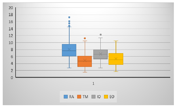
Results: The thickness of abdominal subcutaneous fatty layer in male is 17.5 mm and 24.8 in females. The mean thickness of subcutaneous fatty layer in mm is 13.28, 23.56, 25.95 and 22.57 in all four groups respectively. The mean thickness of abdominal wall muscles in all patients in RA = 8.08 with a p value = .003, TA = 4.88 with a p value = .011, IO = 6.7 with a p value = .000 and EO = 5.39 with a p value = .000. The RT side show more thickness that is more obvious in RA (10.1 mm for right and 6 mm for left) and IO (8.7 mm for right and 4.7 mm for left). Pearson correlation test was used to test the correlation between the thickness of subcutaneous fatty layer and the muscles. The results show r=-.157 for TM, -.233 for IO, and -.210 for EO. The results were insignificant for RT & LT RA and LT TA muscles.
Conclusion: The infl
Keywords
Anterior Abdominal Wall; Abdominal Surgery; Anatomy; CTKUB; Radiology
Anterior Abdominal Wall articles; Abdominal Surgery articles; Anatomy articles; CT-KUB articles; Radiology articles
Article Details
Abbreviations:
CT KUB- Computed Tomography of Kidneys, Ureters and Bladder; RA- Rectus Abdominus muscle; EO- External Oblique muscle; IO- Internal Oblique muscle; TA- Transversus Abdominus muscle; RT & LT- Right and Left; L- Lumbar vertebrae; BMI- Body Mass Index
Introduction
The anterior abdominal wall develops from both somatic and lateral plate mesoderm. It is made up of skin, superficial fascia, deep fascia, muscles, extraperitoneal fascia, and parietal peritoneum.The superficial fascia is divided into a superficial fatty layer (fascia of Camper) and a deep membranous layer (Scarpa’s fascia). The fatty layer is continuous with the superficial fat over the rest of the body and may be extremely thick (3 in. [8 cm] or more in obese patients) [1]. The superficial fascia of Camper contains a varying quantity of adipose tissue. Subcutaneous adipose tissue is composed of two different subcompartments: a "superficial" Subcutaneous adipose tissue and "deep" Subcutaneous adipose tissue, separated by a connective plane namedfascia superficialis,orScarpa's fascia [2]. The deep fascia in the anterior abdominal wall is merely a thin layer of connective tissue covering the muscles; it lies immediately deep to the membranous layer of superficial fascia [1]. The muscles of the anterior abdominal wall consist of three broad thin sheets that are aponeurotic in front; from exterior to interior they are the external oblique, internal oblique, and transversus. On either side of the midline anteriorly is, in addition, a wide vertical muscle, the rectus abdominis. As the aponeuroses of the three sheets pass forward, they enclose the rectus abdominis to form the rectus sheath. The lower part of the rectus sheath might contain a small muscle called the pyramidalis [1].
The radiological assessment of muscle and fat properties is fundamental in muscle diseases and obesity. Measurements of muscles and fat thickness were assessed in different populations and for different purposes. Detailed Knowledge of the layered anatomy of the abdomen in patient is essential for the successful performance of surgery and pleasing outcome, and better understanding of medical conditions that affect the anterior abdominal wall e.g. obesity, hernia.
A morphometric analysis of the abdominal wall can be performed precisely using computed tomography (CT) scans. As they allow for an accurate separation of the various tissue types based on their attenuation characteristics [3] and provide good assessment of muscle properties (size, mass, density, composition, andadipose tissueinfiltration) [4]. Computed tomography (CT) generates very delicate cross sections which is very helpful in the research studies. CT abdomen images are permitting the measurement of the abdominal fatty layer and differentiate it from the intra-abdominal fat [5]. On the other hand, many articles used ultra-sonographic anthropometric analysis to study the effect of age, sex and obesity on male anterior abdominal wall muscles.
Literature review presents a complete morphometric analysis of the healthy abdominal wall, confirms the great variability in abdominal wall anatomy, and the influence of several factors like age, sex and BMI. For example, Jourdan et al. conducted retrospective study in 2019 that included 120 patients over 18 years old and CT scans indicated for renal colic on an outpatient basis (1). Engelke et al. suggest that recent systematic reviews and met-analyses has demonstrated that muscle imaging by quantitative CT plays an important role in a large variety of diseases (2). For example, in 2018, Han, J.Y. and his team established clinical correlation of respiratory muscle with severity of emphysema visualized on CT scan.
Numerous studies that was concerned about quantification of subcutaneous fat and muscle tissue at abdominal computed tomography suggests a precise anatomical landmark in the L3–L4 disc region to predict more accurate measurements (5,7,8). According to the literature review this type of studies were not conducted in Sudanese population. This study is focusing on normal population measurements; therefore, further studies that study diseases that affect muscles and fat can make use of it.
Objectives
Broad Objectives: To assess the measurements of anterior abdominal wall layers thickness under the influence of age and sex, using CT KUB scan.
Specific Objectives:
- To define reference values for the thickness of anterior abdominal wall layers, in age-matched individuals.
- To compare the difference values for the thickness of anterior abdominal wall layers in different sex.
- To determine the asymmetry between right and left anterior abdominal wall muscles ( EO,IO,TA,RA)
- To determine the relationship between subcutaneous fatty layer and abdominal wall muscles.
- To compare the anterior abdominal wall layers measurements in Sudanese population to other population.
Materials and Methods:
Methods:
Study Design:
This is a retrospective descriptive hospital-based-study to assess the anterior abdominal wall of patients come for computed topography of kidney, ureter and bladder (CT-KUB).
Study Area:
The study was conducted in Ibn-Sina Specialized Hospital radiology center which is a tertiary public hospital located in the center of Khartoum. It has a dedicated imaging center and receives imaging requests from all over Sudan.
Study Duration:
The study was conducted during the period from July to December 2021.
Study Population:
This study was carried on Sudanese patients who presented to Ibn-Sina Specialized Hospital Radiology Center for CT-KUB imaging and aged between 19 – 71 years.
Sample Size:
The study sample size was calculated according to this formula:
Sample Size = z2 * p(1-P)/e2 / 1 + [ z2 * p(1-P)/e2 * N]
- z-score = 1.96
- e = margin of error = 5%
- p = population percentage = 50%
- N = population size = 500
The sample size =232
Materials:
Ethical Consideration:
The study commenced after approval from the ethical committee of Omdurman Islamic University and the need for an informed consent was waived. Confidentiality has been preserved.
Selection Criteria:
Inclusion Criteria:
Male or female patient aged more than 18 years old, who marked for CT-KUB scan and has no abnormalities in the anterior abdominal wall.
Exclusion Criteria:
The study excluded patients who did not meet the inclusion criteria e.g. Pediatrics and any patient with abdominal pathology that affect the anterior abdominal wall e.g. ascites or abdominal tumor.
Data Categorization and Management:
The patient’s abdominal CT scan was visualized and data regarding anterior abdominal wall thickness was collected with radiology technician assistance.
Data Collection Tools:
The study data was collected through computerized search of the radiology database, each 4 CT images were copied to a DVD.
Data Collection Technique:
The study data was collected from radiology data base according and saved to external DVD. Each DVD contained 4 images of CT scans. The CT scan images were a single slice of cross sectional view of abdomen at the level of L3 and L4 (where all muscles are observed clearly). DICOM viewer program was used to view the CT scan and take the required measurements.
Statistical Analysis:
The study data were analyzed by statistic expert using SPSS to calculate important values for muscles and subcutaneous tissue e.g. mean and SD. ANOVA test and Pearson correlation test were used where appropriate. P<0.05 was considered statistically significant.
Results
Demographic data
The study results showed that the average age was 43.08 ± 14.408 years old. The total number of patients is 232 male and female aged between 19 to 71. Four age groups were identified (19 – 32) (32 - 45) (45 – 58) (58 – 71) and presented in the table (1).
Table (1) Frequency distribution of age
|
Age group |
Frequency |
Percentage |
|
19-32years |
62 |
26.7% |
|
32-45years |
72 |
31.0% |
|
45-58years |
55 |
23.7% |
|
58-71years |
43 |
18.5% |
|
Total |
232 |
100 % |
The total number of patients contains equal number of male and female = 116 as shown in table (2)
Table (2): Frequency distribution of gender
|
Gender |
Frequency |
Percentage |
|
Male |
116 |
50% |
|
Female |
116 |
50% |
|
Total |
232 |
100% |
Measurements and variability:
Table (3) shows the minimum and maximum age of patients involved in the study, the minimum and maximum measurements of all anterior abdominal wall muscles including the right and the left side and the mean and standard deviation of each.
|
Descriptive Statistics |
|||||
|
N |
Minimum |
Maximum |
Mean |
Std. Deviation |
|
|
Age |
232 |
19 |
70 |
43.08 |
14.408 |
|
RT RA |
232 |
1.02 |
31.10 |
10.1049 |
3.18711 |
|
LT RA |
232 |
1.26 |
16.20 |
6.0610 |
3.64083 |
|
RA |
232 |
2.71 |
17.25 |
8.0829 |
2.70406 |
|
RT TM |
232 |
1.52 |
11.40 |
5.1377 |
2.36142 |
|
LT TM |
232 |
1.57 |
12.30 |
4.6247 |
2.13074 |
|
TM |
232 |
1.55 |
11.20 |
4.8812 |
2.10147 |
|
RT IM |
232 |
1.00 |
20.90 |
8.7459 |
3.15873 |
|
LT IM |
232 |
1.55 |
11.40 |
4.7450 |
2.09721 |
|
IM |
232 |
2.75 |
12.25 |
6.7455 |
1.93382 |
|
RT EO |
232 |
1.89 |
10.90 |
5.5919 |
2.11594 |
|
LT EO |
232 |
1.58 |
11.50 |
5.1924 |
2.02724 |
|
EO |
232 |
1.74 |
10.55 |
5.3922 |
1.94195 |
|
Subcutaneous fatty layer |
232 |
2.66 |
47.30 |
21.2023 |
10.64099 |
|
Valid N (list wise) |
232 |
||||
Influence of age:
Table (3): show the mean of the measured thickness of subcutaneous fatty layer and muscles for each age group. The mean thickness of subcutaneous fatty layer in mm is 13.28, 23.56, 25.95 and 22.57 in all four groups respectively. The mean thickness of all age groups is 21.2.
Table (3): ANOVA Test to compare the mean of the measured thickness in (mm) of (Subcutaneous fatty layer) between age groups.
|
Report |
|||
|
Subcutaneous fatty layer |
|||
|
Age group |
Mean |
N |
Std. Deviation |
|
19-32years |
13.2887 |
62 |
8.82723 |
|
32-45years |
23.5647 |
72 |
10.17956 |
|
45-58years |
25.9571 |
55 |
8.57830 |
|
58-71years |
22.5751 |
43 |
10.30850 |
|
Total |
21.2023 |
232 |
10.64099 |
|
p-Value .000 |
|||
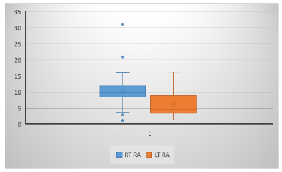
As demonstrated in table (4) the mean thickness of each muscle that was measured in right and left side in all four age groups. The total mean thickness of RA in the four age groups is 9, 8.1, 7.5 and 7.33 respectively and the mean thickness in all patients is 8.08 with a p value .003. The RT RA show a mean of 11.46, 10.16, 9.5 and 8.77 respectively with a p value .000. The LT RA show a mean of 6.66, 6, 5.5 and 5.89 respectively with a p value of .406. The mean thickness for RT and LT RA in all patients is 10.1 and 6 respectively.
Table (4): ANOVA Test to compare the mean of the measured thickness in (mm) of (Rt.RA <.RA& RA) between age groups
|
Report |
||||
|
Age group |
RT RA |
LT RA |
TOTAL RA |
|
|
19-32years |
Mean |
11.4611 |
6.6621 |
9.0616 |
|
N |
62 |
62 |
62 |
|
|
Std. Deviation |
3.16855 |
4.05128 |
2.71541 |
|
|
32-45years |
Mean |
10.1674 |
6.0432 |
8.1053 |
|
N |
72 |
72 |
72 |
|
|
Std. Deviation |
2.64043 |
3.49295 |
2.43498 |
|
|
45-58years |
Mean |
9.5355 |
5.5369 |
7.5362 |
|
N |
55 |
55 |
55 |
|
|
Std. Deviation |
3.87439 |
3.33436 |
2.77347 |
|
|
58-71years |
Mean |
8.7730 |
5.8944 |
7.3337 |
|
N |
43 |
43 |
43 |
|
|
Std. Deviation |
2.28853 |
3.63685 |
2.68567 |
|
|
Total |
Mean |
10.1049 |
6.0610 |
8.0829 |
|
N |
232 |
232 |
232 |
|
|
Std. Deviation |
3.18711 |
3.64083 |
2.70406 |
|
|
P value |
.000 |
.406 |
.003 |
|
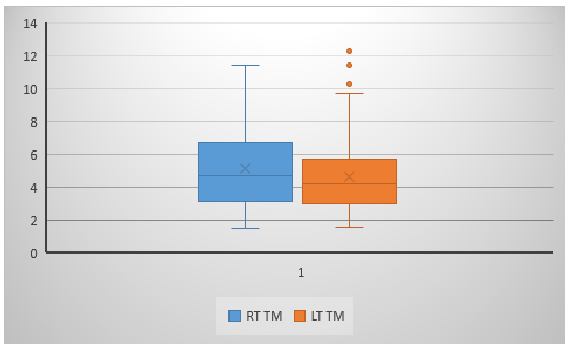
Table (5) shows the total mean thickness of TA in the four age groups is 5.56, 4.88, 4.5 and 4.35 respectively and the mean thickness in all patients is 4.88 with a p value .011. The mean thickness of TA muscle was measured in the right and left side in all four age groups. The RT TA show a mean of 5.89, 5.16, 4.8 and 4.4 respectively with a p value .009. The LT TA show a mean of 5.2, 4.6, 4.2 and 4.3 respectively with a p value of .041. The mean thickness for RT and LT TA in all patients is 5.1 and 4.6 respectively.
Table (5): ANOVA Test to compare the mean of the measured thickness in (mm) of (Rt.TA <.TA& Total TA) between age groups
|
Report |
||||
|
Age group |
RT TA |
LT TA |
TOTAL TA |
|
|
19-32 years |
Mean |
5.8905 |
5.2356 |
5.5631 |
|
N |
62 |
62 |
62 |
|
|
Std. Deviation |
2.59040 |
2.23215 |
2.20999 |
|
|
32-45years |
Mean |
5.1693 |
4.6090 |
4.8892 |
|
N |
72 |
72 |
72 |
|
|
Std. Deviation |
2.37698 |
2.23395 |
2.15451 |
|
|
45-58years |
Mean |
4.8189 |
4.2051 |
4.5120 |
|
N |
55 |
55 |
55 |
|
|
Std. Deviation |
2.29289 |
1.89180 |
2.02223 |
|
|
58-71years |
Mean |
4.4072 |
4.3070 |
4.3571 |
|
N |
43 |
43 |
43 |
|
|
Std. Deviation |
1.75943 |
1.95207 |
1.71595 |
|
|
Total |
Mean |
5.1377 |
4.6247 |
4.8812 |
|
N |
232 |
232 |
232 |
|
|
Std. Deviation |
2.36142 |
2.13074 |
2.10147 |
|
|
P value |
.009 |
.041 |
.011 |
|
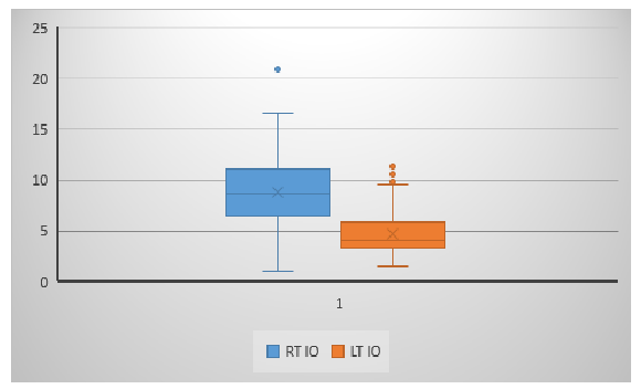
As demonstrated in table (6) the total mean thickness of IO in the four age groups 7.8, 6.8, 6.2 and 5.68 respectively and the mean thickness in all patients is 6.7 with a p value .000. The mean thickness of IO muscle was measured in the right and left side in all four age groups. The RT IO show a mean of 10.27, 8.89, 8.1 and 7.05 respectively with a p value .000. The LT IO show a mean of 5.35, 4.78, 4.33 and 4.32 respectively with a p value of .028. The mean thickness for RT and LT IO in all patients is 8.7 and 4.7 respectively.
Table (6): ANOVA Test to compare the mean of the measured thickness in (mm) of (Rt.IO <.IO & Total IO) between age groups
|
Report |
||||
|
Age group |
RT IO |
LT IO |
TOTAL IO |
|
|
19-32 years |
Mean |
10.2718 |
5.3508 |
7.8113 |
|
N |
62 |
62 |
62 |
|
|
Std. Deviation |
3.34706 |
2.40568 |
1.90328 |
|
|
32-45 years |
Mean |
8.8975 |
4.7860 |
6.8417 |
|
N |
72 |
72 |
72 |
|
|
Std. Deviation |
3.23516 |
2.15600 |
2.01891 |
|
|
45-58 years |
Mean |
8.1498 |
4.3395 |
6.2446 |
|
N |
55 |
55 |
55 |
|
|
Std. Deviation |
2.73779 |
1.82035 |
1.60337 |
|
|
58-71 years |
Mean |
7.0547 |
4.3214 |
5.6880 |
|
N |
43 |
43 |
43 |
|
|
Std. Deviation |
2.09896 |
1.65019 |
1.39468 |
|
|
Total |
Mean |
8.7459 |
4.7450 |
6.7455 |
|
N |
232 |
232 |
232 |
|
|
Std. Deviation |
3.15873 |
2.09721 |
1.93382 |
|
|
P value |
.000 |
.028 |
.000 |
|
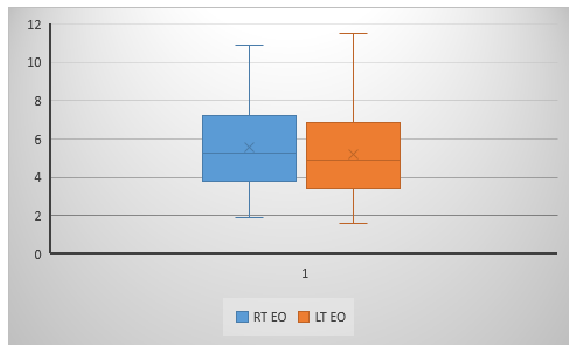
Table (7) shows the total mean thickness of EO in the four age groups is 6.17, 5.48, 4.8 and 4.79 respectively and the mean thickness in all patients is 5.39 with a p value .000. The mean thickness of EO muscle was measured in the right and left side in all four age groups. The RT EO show a mean of 6.3, 5.7, 5.09 and 4.87 respectively with a p value .001. The LT EO show a mean of 6.02, 5.2, 4.59 and 4.7 respectively with a p value of .000. The mean thickness for RT and LT EO in all patients is 5.59 and 5.19 respectively.
Table (7): ANOVA Test to compare the mean of the measured thickness in (mm) of (Rt. EO <.EO ) between age groups
|
Report |
||||
|
Age group |
RT EO |
LT EO |
TOTAL EO |
|
|
19-32years |
Mean |
6.3356 |
6.0223 |
6.1790 |
|
N |
62 |
62 |
62 |
|
|
Std. Deviation |
2.17086 |
1.97912 |
1.93558 |
|
|
32-45years |
Mean |
5.7589 |
5.2108 |
5.4849 |
|
N |
72 |
72 |
72 |
|
|
Std. Deviation |
2.24903 |
2.14758 |
2.07375 |
|
|
45-58years |
Mean |
5.0976 |
4.5998 |
4.8487 |
|
N |
55 |
55 |
55 |
|
|
Std. Deviation |
2.02199 |
1.71551 |
1.76252 |
|
|
58-71years |
Mean |
4.8723 |
4.7228 |
4.7976 |
|
N |
43 |
43 |
43 |
|
|
Std. Deviation |
1.50282 |
1.90557 |
1.53590 |
|
|
Total |
Mean |
5.5919 |
5.1924 |
5.3922 |
|
N |
232 |
232 |
232 |
|
|
Std. Deviation |
2.11594 |
2.02724 |
1.94195 |
|
|
P value |
.001 |
.000 |
.000 |
|
Influence of sex:
As shown in table (8) the total mean thickness of RA in males and females is 9.1 and 7.03 respectively, with a p value .000. The mean thickness of RA muscle was measured in the right and left side in both sex. The RT RA show a mean of 11.3 in males and 8.89 in females, with a p value .000. The LT RA show a mean of 6.95 in males and 5.1 in females with a p value of .000.
Table (8): T- Test to compare the mean of the measured thickness in (mm) of (Rt.RA <.RA& RA) between male and female
|
Group Statistics |
P value |
|||||
|
Gender |
N |
Mean |
Std. Deviation |
Std. Error Mean |
||
|
RT RA |
Male |
116 |
11.3173 |
3.47962 |
.32307 |
.000 |
|
Female |
116 |
8.8924 |
2.30873 |
.21436 |
||
|
LT RA |
Male |
116 |
6.9544 |
3.93153 |
.36503 |
.000 |
|
Female |
116 |
5.1676 |
3.09182 |
.28707 |
||
|
RA |
Male |
116 |
9.1359 |
2.78555 |
.25863 |
.000 |
|
Female |
116 |
7.0300 |
2.16601 |
.20111 |
||
Table (9) demonstrate the total mean thickness of TA in males and females is 5.55 and 4.21 respectively, with a p value .000. The mean thickness of TA muscle was measured in the right and left side in both sex. The RT TA show a mean of 5.8 in males and 4.44 in females, with a p value .000. The LT TA show a mean of 5.27 in males and 3.97 in females with a p value of .000.
Table (9): T- Test to compare the mean of the measured thickness in (mm) of (RT TA < TA & TA) between male and female
|
Group Statistics |
P value |
|||||
|
Gender |
N |
Mean |
Std. Deviation |
Std. Error Mean |
||
|
RT TA |
Male |
116 |
5.8314 |
2.63215 |
.24439 |
.000 |
|
Female |
116 |
4.4441 |
1.81718 |
.16872 |
||
|
LT TA |
Mae |
116 |
5.2731 |
2.38148 |
.22111 |
.000 |
|
Female |
116 |
3.9764 |
1.61247 |
.14971 |
||
|
TA |
Mae |
116 |
5.5522 |
2.36076 |
.21919 |
.000 |
|
Female |
116 |
4.2102 |
1.54571 |
.14352 |
||
Table (10): show the total mean thickness of IO in males and females is 7.4 and 6.07 respectively, with a p value .000. The mean thickness of IO muscle was measured in the right and left side in both sex. The RT IO show a mean of 9.47 in males and 8.01 in females, with a p value .000. The LT IO show a mean of 5.3 in males and 4.1 in females with a p value of .000.
Table (10): T- Test to compare the mean of the measured thickness in (mm) of (RT IO < IO & IO) between male and female
|
Group Statistics |
P value |
|||||
|
Gender |
N |
Mean |
Std. Deviation |
Std. Error Mean |
||
|
RT IO |
Mae |
116 |
9.4738 |
3.46729 |
.32193 |
.000 |
|
Female |
116 |
8.0181 |
2.63648 |
.24479 |
||
|
LT IO |
Male |
116 |
5.3609 |
2.23235 |
.20727 |
.000 |
|
Female |
116 |
4.1290 |
1.75669 |
.16310 |
||
|
IO |
Male |
116 |
7.4174 |
2.01516 |
.18710 |
.000 |
|
Female |
116 |
6.0735 |
1.59378 |
.14798 |
||
Table (11): demonstrate the total mean thickness of EO in males and females is 6.16 and 4.61 respectively, with a p value .000. The mean thickness of EO muscle was measured in the right and left side in both sex. The RT EO show a mean of 6.37 in males and 4.8 in females, with a p value .000. The LT EO show a mean of 5.95 in males and 4.42 in females with a p value of .000.
Table (11): T-Test to compare the mean of the measured thickness in (mm) of (RT EO < EO & EO) between male and female
|
Group Statistics |
P value |
|||||
|
Gender |
N |
Mean |
Std. Deviation |
Std. Error Mean |
||
|
RT EO |
Mae |
116 |
6.3736 |
2.28404 |
.21207 |
.000 |
|
Female |
116 |
4.8103 |
1.59494 |
.14809 |
||
|
LT EO |
Male |
116 |
5.9590 |
2.09291 |
.19432 |
.000 |
|
Female |
116 |
4.4258 |
1.63991 |
.15226 |
||
|
EO |
Male |
116 |
6.1663 |
2.03786 |
.18921 |
.000 |
|
Female |
116 |
4.6180 |
1.48769 |
.13813 |
||
Table (12): show the mean thickness of subcutaneous fatty layer in males and females is 17.59 and 24.8 respectively, with a p value .000.
Table (12): T-Test to compare the mean of the measured thickness in (mm) of Subcutaneous fatty layer between male and female
|
Group Statistics |
P value |
|||||
|
Gender |
N |
Mean |
Std. Deviation |
Std. Error Mean |
||
|
Subcutaneous fatty layer |
Male |
116 |
17.5959 |
10.37666 |
.96345 |
.000 |
|
Female |
116 |
24.8087 |
9.67119 |
.89795 |
||
Correlation:
Table (13): show the Pearson correlation test result that was used to test the correlation between the thickness of subcutaneous fatty layer and the muscles. The results show r=-.157 for TM, -.233 for IO , and -.210 for EO.
Table (13): Correlation between Subcutaneous fatty layer with abdominal muscle wall thickness in mm
Table (14): demonstrate the Pearson correlation test result that was used to test the correlation between the thickness of fatty layer with abdominal muscle wall (RA&TM& &IO & EO) thickness in mm. The results were insignificant for RT & LT RA and LT TA muscles.
Table (14): Correlation between Subcutaneous fatty layer with abdominal muscle wall (RA&TM& &IO & EO) thickness in mm
|
Correlations |
||||||
|
Subcutaneous fatty layer |
RA |
TM |
IO |
EO |
||
|
Subcutaneous fatty layer |
Pearson Correlation |
1 |
-.080 |
-.157* |
-.233** |
-.210** |
|
Sig. (2-tailed) |
.225 |
.017 |
.000 |
.001 |
||
|
N |
232 |
232 |
232 |
232 |
232 |
|
|
*. Correlation is significant at the 0.05 level (2-tailed). |
||||||
|
**. Correlation is significant at the 0.01 level (2-tailed). |
||||||
Muscle Asymmetry:
The Figure shows the difference between the mean thickness of right and left RA muscles and the rate of asymmetry.
Discussion
After careful literature review, this study is the first of its kind to be done in Sudan, emphasizing the value of the use of CT in measurement of anterior abdominal wall thickness, the uniqueness of this study emerges from its representative study sample and the accuracy of the measurements, in contrast to previous studies were ultrasonography was used for the measurement of thickness which is exposed to more subjectivity and the need for exclusion of intra-abdominal pathologies and gynecological conditions [13,14]. Clearly knowing the normal anterior abdominal wall measurements will have a big role in helping in preparation of abdominal surgeries especially in patients with abnormal abdominal wall. According to what is mentioned before, this data represents a reference value for Sudanese population anterior abdominal wall measurements because it includes four age groups between the age 19 and 71, and the gender is represented equally. Because of the wide range of measurements performed in this work, it is now possible to explain this variability through the influence of age and sex, in contrast to Khan et al were subjects were grouped according to age and obesity ,obesity was measured using the help of BMI which later showed little correlation with abdominal muscles thickness [8]. The influence of age on abdominal subcutaneous fatty layer is noted and found to be increased with age, this is consistent with the Jourdan et al, and Khan et al. Jourdan et al show nearly similar results in all four age groups of 21.5, 22.1, 25.1, 21.3 mm respectively. In contrast to muscle, which show a decrease in thickness with age (RA, TA, IO, EO), which is consistent with previous studies [8,10]. This was explained by Khan et al, the slight decrease in abdominal muscle thickness with advancing age is observed by simultaneous increase in intramuscular fat content. Comparing the mean thickness of all muscles of all ages to Jourdan et al, the mean thickness was higher than the mean we found in EO and IO, 8.5 and 8 respectively, and lower than TA and RA, 6.1 and 3.7 respectively. In comparing the right and left abdominal musculatures, we used the thickness in mm unlike Jourdan et al in which the asymmetry was evaluated with the muscle area. In contrast to Mannion et al., which suggests no significant differences between left and right sides, the RT side show more thickness that is more obvious in RA ( 10.1 mm for right and 6 mm for left )and IO ( 8.7 mm for right and 4.7 mm for left ). Although this could be explained by hand dominance, the lack of asymmetry based on hand dominance was not supported in previous studies [14]. The influence of sex on abdominal subcutaneous fatty layer is showing female predilection and this is in accordance with previous work. The thickness of subcutaneous fatty layer in female in our study is 24.8 mm which is consistent with Jourdan et al which show 24.2 mm and Kim et al study which show subcutaneous fat thicknesses of 24.31 mm (right) and 23.39 mm (left). However, a slight difference in male was found, 17.6 mm and 20.8 mm respectively. In contrast, the muscles in males have more thickness compared to females. Using ultrasound image Springer et. al. illustrated the thickness of TA in male and female and found out similar values 5+/-0.9 and 4.2 +/- 0.7 respectively. Tahan et al. also used ultrasound imaging to evaluate EO, IO, TA and RA thickness in healthy adult male and female subjects and found similar values to ours (Rt EO 5.7 mm, Lt EO 5.4 Rt IO 8.9 mm, Lt IO 8.5, Rt TA 4.5 mm, Lt TA 3.8 mm, Rt RA 10.3, Lt RA 10.4 for males and 4.8, 4.8, 6.1, 5.8, 3.5, 3.3, 8.7, 8.3 for females respectively). A Pearson correlation show weak negative correlation between the thickness of subcutaneous fatty layer and the muscles r=-.157 for TM, -.233 for IO, and -.210 for EO. The correlation is insignificant between RM and subcutaneous fatty layer, in contrast to Kim et al study which demonstrate positive correlation, clear association was found between age and wall thickness, and the influence of sex was also significant, in relevance to Mannion et al which showed that the main effect of gender was significant for all the OI parameters, OE change in thickness from rest to hollowing , and the sum of all lateral parameters during hollowing , but in contrast the differences were mainly significant to left side [13]. This study is not devoid of certain limitations. All measurements were taken on single slice which could limit the accuracy of the results. Additionally, human error could happen during measurements.
Conclusion
The study was performed among 232 patients who underwent CT KUB scan in Ibn Sina Specialized Hospital in the second half of 2021 with equal number of male and female. It demonstrates the mean thickness of subcutaneous fatty layer and anterior abdominal wall muscles in both right and left group. The muscle thickness is decreasing with age, in contrast to subcutaneous fatty layer thickness which increases with age. Males have thicker muscles than female however, females have thicker subcutaneous fat. The asymmetry is present between all muscles with right group dominance. The RT RA has the highest mean thickness followed by IO then EO and then TA. The correlation between abdominal wall muscles and fatty layer is weakly negative.
Recommendation
The study gives reference measurements which can be useful to surgeons in different conditions like when they are dealing with ventral hernia, abdominal wall flaps. Because it can improve the outcome, increasing the awareness of this muscles measurements and variations is one of our recommendations. Additionally, we recommend further researches to assess the abdominal wall muscles in relation to BMI. Furthermore, more focus on subcutaneous fatty layer to study its accuracy in diagnosing obesity instead of BMI. Additionally further studies regarding the correlation between hand dominance and muscle wall thickness could be of a value, finally we recommend studying the effects of various conditions on abdominal wall muscle thickness.
Statements and Declarations
Authors declare that they have no competing interests.
Funding
no fund.
References
- RS S. Clinical anatomy by regions. Philadelphia, Lippincott Williams & Wilkins (2012).
- Cancello R, Zulian A, Gentilini D, et al. Molecular and morphologic characterization of superficial- and deep-subcutaneous adipose tissue subdivisions in human obesity. Obesity (Silver Spring) 21 (2013): 2562-2570.
- Mazonakis M, Damilakis J. Computed tomography: What and how does it measure?.European journal of radiology85 (2016): 1499-1504.
- Engelke K, Museyko O, Wang L, et al. Quantitative analysis of skeletal muscle by computed tomography imaging—State of the art.Journal of orthopaedic translation 15 (2018): 91-103.
- Borkan GA, Gerzof SG, Robbins AH, et al. Assessment of abdominal fat content by computed tomography.The American journal of clinical nutrition36 (1982): 172-177.
- Park J, Gil JR, Shin Y, et al. Reliable and robust method for abdominal muscle mass quantification using CT/MRI: An explorative study in healthy subjects.PloS one 14 (2019): e0222042.
- Faron A, Luetkens JA, Schmeel FC, et al. Quantification of fat and skeletal muscle tissue at abdominal computed tomography: associations between single-slice measurements and total compartment volumes. Abdominal Radiology 44 (2019): 1907-1916.
- Khan MA, Qureshi K, Rehman Z. Effect of Age and Obesity on Human Male Anterior Abdominal Wall Muscles: an Ultrasonographic Anthropometric Analysis. Pakistan Journal of Medical and Health Sciences 6 (2012): 406-412.
- Sun J, Lv H, Zhang M, et al. The Appropriateness Criteria of Abdominal Fat Measurement at the Level of the L1-L2 Intervertebral Disc in Patients With Obesity.Frontiers in endocrinology12 (2021).
- Jourdan A, Soucasse A, Scemama U. et al. Abdominal wall morphometric variability based on computed tomography: Influence of age, gender, and body mass index. Clin Anat 33 (2020): 1110-1119.
- Engelke K, Museyko O, Wang L, et al. 2018. Quantitative analysis of skeletal muscle by computed tomography imaging—State of the art.Journal of orthopaedic translation 15 (2018): 91-103.
- Han JY, Lee KN, Kang EJ, et al. Quantitative Computed Tomography Assessment of Respiratory Muscles in Male Patients Diagnosed with Emphysema.Journal of the Korean Society of Radiology78 (2018): 371-379.
- Mannion AF, Pulkovski N, Toma V. et al. Abdominal muscle size and symmetry at rest and during abdominal hollowing exercises in healthy control subjects.Journal of anatomy213 (2008): 173-182.
- Springer BA, Mielcarek BJ, Nesfield TK, et al. Relationships among lateral abdominal muscles, gender, body mass index, and hand dominance.Journal of Orthopaedic & Sports Physical Therapy36 (2006): 289-297.
- Tahan N, Khademi-Kalantari K, Mohseni-Bandpei MA, et al. Measurement of superficial and deep abdominal muscle thickness: an ultrasonography study.Journal of physiological anthropology35 (2016): 1-5.
- Frank K, Hamade H, Casabona G, et al. Influences of age, gender, and body mass index on the thickness of the abdominal fatty layers and its relevance for abdominal liposuction and abdominoplasty.Aesthetic Surgery Journal 39 (2019): 1085-1093.
- Kim J, Lim H, Lee SI, et al. Thickness of rectus abdominis muscle and abdominal subcutaneous fat tissue in adult women: correlation with age, pregnancy, laparotomy, and body mass index.Archives of Plastic Surgery39 (2012): 528.
- Chang YWW, Murphy K, Yackzan D, et al. Abdominal wall thickness is a predictor for surgical site infections in patients undergoing colorectal operations.The American Surgeon87 (2021): 1155-1162.
- Gibson D, Burden S, Strauss B,et al.The role of computed tomography in evaluating body composition and the influence of reduced muscle mass on clinical outcome in abdominal malignancy: a systematic review.Eur J Clin Nutr69 (2015): 1079-1086.
- Kim DW, Ha J, Ko Y, et al. Reliability of skeletal muscle area measurement on CT with different parameters: a phantom study.Korean Journal of Radiology22 (2021): 624.
- Mekonen, H.K., Hikspoors, J.P., Mommen, G., Köhler, S.E. and Lamers, W.H., 2015. Development of the ventral body wall in the human embryo.Journal of anatomy,227(5), pp.673-685.

