Arthroscopic Assisted Mini Open Rotator Cuff Repair using Acromial Split: An Ethical Hack to Flatten the Shoulder Surgery Learning Curve
Article Information
Zafar Israil1*, Pradyumna Sharma2, Rohit Vikas3, Narender Reddy4
1DNB Ortho, Military Hospital Bareilly, Natraj Cinema Hall, Civil Lines, Bareilly, Uttar Pradesh, India
2MS Ortho, Military Hospital Jalandhar, Nalwa Street, New S A Colony, Jalandhar Cantt, Jalandhar, Punjab, India
3MS Ortho, Army Hospital Research & Referral, Military Hospital Road, Subroto Park, Dhaula Kuan, New Delhi, India
4BPT, Military Hospital, Jalandhar, Nalwa Street, New S A Colony, Jalandhar Cantt, Jalandhar, Punjab, India
*Corresponding Author: Zafar Israil, DNB Ortho, Military Hospital Bareilly, Natraj Cinema Hall, Civil Lines, Bareilly, Uttar Pradesh, India.
Received: 20 January 2025; Accepted: 29 January 2025; Published: 07 February 2025
Citation: Zafar Israil, Pradyumna Sharma, Rohit Vikas, Narender Reddy. Arthroscopic Assisted Mini Open Rotator Cuff Repair using Acromial Split: An Ethical Hack to Flatten the Shoulder Surgery Learning Curve. Journal of Orthopedics and Sports Medicine. 7 (2025): 39-47.
View / Download Pdf Share at FacebookAbstract
Mini Open Rotator cuff repair is a dynamic term that encompasses Diagnostic arthroscopy of shoulder, sub acromial decompression, identification and tagging of torn rotator cuff tendons followed by flexible conversion to open repair by extending the lateral portal. Acromial split of anterior edge of acromion gives full exposure of entire footprint of rotator cuff tendons aided by rotation of the arm in lateral decubitus position.
Frequently, rotator cuff tendon tears have complex patterns and size which makes all arthroscopic procedure complex and tedious. Adding acromial split to 5 cms of anterolateral deltoid raphe split and 1cm lateral end clavicle excision gives about 10 cms exposure to do complex repairs. The sliver of acromion is fixed with #2 ethibond sutures at the time of closure and heals onto the acromion surface. This mini open approach enables to flatten the shoulder surgery learning curve because of combining the benefits of arthroscopic visualization to tag the tear and completing the repair with open techniques. Complex repairs like biceps SCR, Fascia lata SCR, subscapularis and infraspinatus double row repair and lower trapezius tendon transfer have been performed by this study group with excellent results.
42 patients, 26 males and 16 females with MRI confirmed various rotator cuff tear patterns were operated with this technique in Military Hospitals Jalandhar and Bareilly in India from 2020 to 2024. The average age of patients was 52.5 years and follow up was variable from 6-24 months.
38% of patients had complete supraspinatus tear, 23.8% has partial suprasinatus tear, 16.6 % had supraspinatus with infraspinatus tear, 9.5% has supra+infra+partial subscapularis tears and 12% had supraspinatus with partial subscapularis tears.
40 % of patients with 6 months follow up had average Constant score improved from 26 to 71.5 after the surgery. Another set of 40 % of patients with 12 months follow up had average Constant score improved from 22 to 86.4 after the surgery. 15% of patients with upto 2 years follow up had their average Constant scores improved from 18.7 to 89.7. The results are consistent with similar mini open rotator cuff surgeries in literature.
This study proves the hypothesis that Arthroscopic assisted Mini open Rotator cuff surgeries performed with Acromial split gives enough exposure to do a myriad pattern of Repairs with excellent outcomes and is an ethical hack to do complex repairs.
Keywords
Mini open; Rotator cuff repair; Acromial split; Constant score
Article Details
1. Introduction
Rotator cuff tear is a fairly common diagnosis in Orthopaedics out patient department. It is known to cause varying levels of shoulder dysfunction accompanied with pain, loss of sleep and negative effect on activities of daily living. The goal of rotator cuff repair is elimination of pain along with gain of shoulder strength and function. Optimal repair of tendon to bone requires robust surgical repair, achievement of high fixation strength and maintenance of the repair under cyclical loading. All arthroscopic repair is frequently marred by intermittent visualisation due to thick and hyperaemic bursa. Often it is a slow learning curve and surgeons frequently resort to arthroscopic assisted mini open rotator cuff repair to avoid various perils. Arthroscopic assisted Mini open rotator cuff repair is dynamic and can be accomplished by extending the lateral portal. Deltiod split in between anterior and lateral raphe is easy and safe upto 5cms. Adding a mumford excision of 1cm of lateral end of clavicle in cases of AC joint arthritis and splitting of Acromion with a bony sliver is done. Since the surgery is done in lateral decubitus position, arthroscopic gleno humeral work is done followed by sub acromial decompression. The rotator cuff preparations, biceps tenotomy, tagging of torn ends, releases and mobilization are done arthroscopic. Followed by converting it into a mini open repair which allows deltoid to fall anteriorly and rotator cuff repair to be undertaken in a easy and robust way. In our case, we were able to do 42 mini open rotator cuff repairs from 2020 to 2024 with this technique which works on the premise that bone to bone healing of acromial sliver precedes the rotator cuff tendon healing. This study introduces the hypothesis that Arthroscopic assisted Mini open Rotator cuff surgeries performed with Acromial split gives enough exposure to do a myriad pattern of Repairs with excellent outcomes and is an ethical hack to do complex repairs.
2. Materials and Methods
This was a retrospective study done from 2020 to 2024 in Military Hospitals Jalandhar and Bareilly. MRI confirmed rotator cuff tear patients were recruited for this study and Pre and Post op Constant scores were recorded. The follow up was variable from 6 -24 months post-surgery.
Institutional medical ethics guidelines were followed and written informed consent was obtained from all participating patients.
Inclusion criteria:
- a) All patients with MRI diagnosed Rotator cuff pathologies
- b) Age between 35 to 70 years
- c) Consent for the study with regular follow up of atleast 6 months post-surgery
Exclusion criteria:
- a) patients with traumatic glenohumeral instability
- b) Age <35 and >70
- c) Humeral head collapse and rotator cuff arthropathy
3. Surgical Technique
Diagnostic Arthroscopy, Biceps tenotomy and subacromial decompression is carried out initially in this technique in lateral decubitus position. The difficulties encountered in visualisation of subacromial areas frequently require hypotensive anaesthesia to keep the bleeding minimal. Also, if extensive and thick bursa is present, major operative time is lost in visualisation of the anatomy. A good arthroscopic shaver system is essential to resect the inflammatory tissue and supplemented by extensive use of radio frequency ablation. Sub acromial decompression using the burr also is subjective and many times inadequate. Tackling acromioclavicular joint degenerative arthritis frequently requires an excision of 1cm of lateral end clavicle which is much easier to be done open and is the first step of the mini - open part of the procedure.
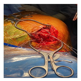
The incision starts from lateral end clavicle going over to AC joint, anterior edge of the Acromion and 4 cms beyond along anterolateral edge of the acromion. The deltoid origin is preserved by making a 4cm split in the raphe between anterior and lateral deltoid. However this access is deep, small and requires a self retaining mastoid retractor to keep the bulky musculature apart.
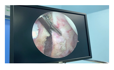
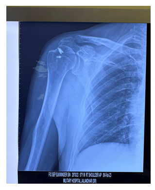
A takedown of a sliver of acromion with the anterior deltoid allows further visualisation of the muscular portion of supraspinatus belly. Further extension of this approach by adding excision of 1cm of lateral end clavicle in concomitant arthritic AC joint adds up to visualisation of even the superior glenoid rim. External Rotation of the arm brings bicipital groove and superior portion of lesser tuberosity in view. Arthroscopic subscapularis tagged sutures can be repaired with knotless anchors. Internal rotation of the arm brings footprint of infraspinatus over greater tuberosity in view and double row repair can be done. Closure involves suturing the acromial sliver to its anatomical position using #2 ethibond sutures followed by subcutaneous sutures and skin staples (Figure 1-18).
4. Results
A total of 63 cases were undertaken for Arthroscopic assisted Mini Open Repair using Acromial split. 42 cases were followed up for an average of 12 months (range 6 – 24 months) and unfortunately rest were lost to follow up. 26 males (62%) and 16 females (38%) were operated for various rotator cuff pathologies which were confirmed by MRI and X ray and were followed up with Constant scores being recorded. The advantage of this technique is that it is versatile and dynamic and all consecutive patients with rotator cuff tears were included in this study design (Table 1). The average age of patients was 52 in this study.
|
Sl. No. |
Rotator cuff pathology |
Type of surgery |
Percentage of patients |
|
1 |
Complete supraspinatus tear |
Arthroscopic assisted mini open supraspinatus repair (double row repair) + biceps tenodesis +mumford |
38% |
|
2 |
Small tear supraspinatus /PASTA/Partial bursal surface tear/sub acromial impingement |
Arthroscopic/ mini open Sub acromial decompression with single row/in situ repair |
23.8% |
|
3 |
Supraspinatus and infraspinatus tear |
Arthroscopic assisted mini open double row supraspinatus and infraspinatus repair + biceps tenodesis+ mumford procedure |
16.6 % |
|
4 |
Supraspinatus and Infraspinatus tear with partial subscapularis tear |
Arthroscopic assisted mini open subscapularis repair with double row supraspinatus and infraspinatus repair + biceps tenodesis + mumford procedure. |
9.5 % |
|
5 |
Partial subscapularis tear with supraspinatus tear |
Athroscopic assisted mini open subscapularis double row repair with supraspinatus double row repair + biceps tenodesis + mumford |
12% |
Table 1: Breakup of types of rotator cuff tear pathologies.
Acromio clavicular joint arthritis was a frequent entity in shoulders with middle age and 85.7% of patients underwent mumford procedure of excision of 1cm of lateral end clavicle. This not only aided the process of sub acromial decompression but increased the proximal exposure especially in medially retracted tendons upto glenoid rim (Patte 3).
Mini open supra pectoral Biceps tenodesis was performed in 85% cases with evidence of biceps tenosynovitis. In few cases, unicortical fixation with endobutton was done but in majority, soft tissue tenodesis aided with anchorage to a knotless anchor near bicipital groove was used.
Acromion sliver of 2 to 3mm was split in 92 % of cases to expose proximal portion of medially retracted supraspinatus and infraspinatus tendons. At the time of closure, # 2 ethibond was used to fix it back using towel clip and aided with k wire to hold it. The hypothesis that bone to bone healing of acromion sliver will occur in 3 months and as the rotator cuff rehabilitation is a slow process, with arm sling for 6 weeks, it is enough time for Acromion to heal up.
We encountered only 1 case (2.3%) with anterior deltoid wasting at 4 months post op. Also 1 case had delayed superficial infection pertaining to ethibond closure of acromial sliver which was debrided. Post op X rays in axillary lateral view and scapular Y view showed bony/fibrous healing of acromial sliver. 3d CT scan was done in few doubtful cases of fibrous union but their recovery as measured by Constant scores were not affected by fibrous/bony union.
Complex repairs like Biceps SCR(2 cases), Fascia lata SCR (2 cases) and Lower Trapezius tendon transfer (1 case) were undertaken which highlight the versatile nature of exposure allowed by this approach.
38% of patients had complete supraspinatus tear, 23.8% has partial suprasinatus tear, 16.6 % had supraspinatus with infraspinatus tear, 9.5% has supra+infra+partial subscapularis tears and 12% had supraspinatus with partial subscapularis tears.
40 % of patients with 6 months follow up had average Constant score improved from 26 to 71.5 after the surgery. Another set of 40 % of patients with 12 months follow up had average Constant score improved from 22 to 86.4 after the surgery. 15% of patients with upto 2 years follow up had their average Constant scores improved from 18.7 to 89.7. The results are consistent with similar mini open rotator cuff surgeries in literature.
On Grading the Constant Shoulder score, (the difference between normal and Abnormal side of shoulder ) at the end of 6 -24 months followup had 68% Excellent and Good results, 21% Fair and 11% poor results. There were 3 patients with poor scores, out of which 2 had massive irreparable cuff tears and one in this cohort had a repeat arthroscopic surgery at a different center.
5. Discussion
A study of Mini Open Repair with a lateral deltoid –splitting approach by Ricardo et al. [1] has shown good long-term results but attempts to repair a large or massive tear can still lead to significant deltoid and axillary nerve injury from excessive traction. Assessment of medially retracted tendons or subscapularis tears is also difficult. Assessment of posterior cuff tears by anterior approach is difficult as mentioned in this study.
In our study we were able to repair medially retracted supraspinatus tendon by using arthroscopic or open release. The exposure overlying glenoid rim was made possible by doing a mumford procedure of excision of 1cm of lateral end clavicle using a microsaw. Splitting the Acromion by taking a 2 to 3 mm sliver of anterolateral edge of Acromion allows gentle exposure of muscular portion of supraspinatus. In cases of retraction of supraspinatus upto humeral head (Patte 2) and upto glenoid rim (Patte 3), open releases can be done using bankart elevators on both articular and bursal sides.
16 cases (38 %) with complete supraspinatus tear were easily repaired with this technique as it provides excellent visualization of the footprint and double row repair can be done by beginner shoulder surgeons also.
Adding a mumford procedure of excision of 1cm lateral end clavicle for acromio clavicular joint arthritis which is a frequent condition in elderly patients, decompresses the sub acromial space. If arthroscopic sub acromial decompression is marred by poor visualization, an attempt to supplement it by removing any bony spur on the anterior undersurface of acromion can be undertaken using a rongeur /microsaw/burr/ file to allow adequate passage of the surgeon’s finger in the posterior sub acromial space. Care should be taken to avoid exuberant bone removal which can predispose to have an intra op acromial fracture.
In this study, 7 cases (16.6%) were a combination of supraspinatus and infraspinatus tears with variable retractions leaving the stump proximally. Internal rotation of the arm held in traction brings the greater tuberosity footprint of infraspinatus in view. Double row repair of infraspinatus and supraspinatus was undertaken with ease and needs to be highlighted in comparison to the tedious suture madness of all-arthroscopic technique.
As the spectrum of Rotator cuff pathologies unfolds, there were massive and irreparable cuff tear situations. This approach is a masterstroke for a young surgeon with limited arthroscopic experience as it allows complex repairs like Superior capsular reconstruction and tendon transfers. In this study we had 4 cases (9.5%) with irreparable supraspinatus, infraspinatus tears along with upper 1/3 subscapularis tears.
We strongly advocate the use of combination of mumford, acromial sliver and upto 5cms split of anterior and lateral deltoid giving a total of 10 cms exposure. We performed 2 cases of SCR using fascia lata autograft and 1 case of lateral trapezius tendon transfer using peroneus longus tendon autograft. Thus, this approach flattens the learning curve of shoulder surgeons and has results consistent with similar mini open studies as the bony sliver of acromion gets healed in 3 months time leaving no residual anterior deltoid wasting.
Frequently we did arthroscopic Biceps tenotomy (85%) for tenosynovitis or routinely in subscapularis tears to improve visualization of Comma tissue. The biceps is delivered into the wound and open suprapectoral tenodesis was done in all such cases, thus obliterating the occurrence of Popeye sign in the arm.
Also, bicipital groove is a marker for attachment of subscapularis to lesser tuberosity which is brought in view by simply external rotation of the arm hung in traction. Exposure of subscapularis tear anteriorly and posteriorly and decompression of the subcoracoid space ‘inside the box’ and releases ‘out of the box’ can be done arthroscopically. As the surgeon evolves his/her arthroscopic skills, this procedure is dynamic and can be shifted to arthroscopic assisted mini open after tagging the comma tissue [2-6]. The procedure of mumford followed by acromial split and extension of deltoid split in between anterior and lateral raphe to upto 5 cms gives enough exposure of lesser tuberosity. Single or double row subscapularis repair can be done for any kind of Lafosse tears. In our study we did 9 subscapularis repairs (21%) which had excellent and good outcomes in 1 to 2 years follow up as measured by Constant scores.
Nageswararao et al. [2] have published significant improvement in active ROM at 6 months follow up. Ghorpade et al. [7] have also used arthroscopic assisted mini open rotator cuff repair albeit in a beach chair position and have shown statistically significant improvement in Constant scores with an average of 82.4 at 6 months.
Similar results are present in our study with average post op Constant score 71.5 at 6 months (40% patient cohort). At 12 months average post op Constant score was 86.4 which was a cohort of 40% of patients. 15% of patients with upto 2 year follow up had average post op Constant score of 89.7
D. Peruguia et al. [4] in a study of 112 patients with massive cuff tears and open approach to rotator cuff repair, reported 8% of patients with deltoid detachment. The approach was lateral para-acromion and deltoid was always detached from bony insertion using electrocautery. The deltoid detachment happened at an average of 3 months (range 1-5 months) during the active functional rehabilitation phase. The average post op Constant score was 67.1 in these patients with deltoid detachment and they had lesser flexion and abduction. We also report of 1 case of anterior deltoid wasting at 6 months post op with decreased flexion and abduction with a post op Constant score of 70 and a Fair result. The premise that bone to bone healing of Acromial sliver needs to be further studied with a post op CT evaluation at 6 months which is a limitation of this study as only post op X rays were evaluated.
Chul-Hyun et al. [5] in their study mention that mini open repair may lead to early post operative shoulder stiffness [5] and the incidence is between 11 % to 20 %. Youm et al. [6] compared all arthroscopic repair versus mini-open repair and found no evidence of shoulder stiffness with mini open surgery.
Rokito et al. [8] have studied the postoperative protocol after large and massive rotator cuff surgeries. They have proposed passive ROM exercises and the use of an arm sling pouch for an initial 6 weeks. In our case, we also propose to delay active ROM exercises for first 4 to 6 weeks and use of arm sling pouch for 6 weeks to 3 months. After majority of passive ROM is achieved, then active ROM exercises is started along with strengthening exercises over the next 3 to 6 months.
The downside of this technique is that accelerated rehabilitation protocol can not be adopted as deltoid repair by Acromial bony sliver healing will take initial 4 to 6 weeks.[9].
Severud et al [10] also have 93 % good to excellent results at 44 month followup. They have tailored their protocol for mini open cuff repair with (1) arm sling immobilization and passive ROM exercises for 6 weeks (2) active assisted ROM exercises and progression to active motion in next 6 to 12 weeks (3) resistive exercises starting at 12 weeks (4) return to full activity at 6 months. Our rehabilitation protocol was quite similar to the one described by Severud et al [10].
6. Conclusion
Arthroscopic assisted mini open rotator cuff repair is an ethical hack to do complex rotator cuff repairs and has functional outcomes with excellent and good results.
Acknowledgements
We would like to acknowledge our patients who consented for this study. There was no funding or grant taken from any commercial interest.
Conflict of Interest
The authors have no conflict of interest.
References
- Ricardo M. Anterolateral Mini-open Approach to Repair Rotator Cuff tears: Mini Review. Ortho and Rheum Open Access J 17 (2020): 555956.
- Nageswararao Y, Shinde GM, et al. A Study on Outcomes of Mini Open Procedures on Rotator Cuff Injuries. J Med Res Surg 4 (2023): 43-45.
- Constant CR, Murley AH. A clinical method of functional assessment of the shoulder. Clin Orthop Relat Res 214 (1987): 160-4.
- Gumina S, Di Giorgio G, Perugia D, et al. Deltoid detachment consequent to open surgical repair of massive rotator cuff tears. International Orthopaedics (SICOT) 32 (2008): 81-84.
- Chul-Hyun C, Kwang-Soon S, Byung-Woo M, et al. Anterolateral approach for mini-open rotator cuff repair. International Orthopaedics (SICOT) 36 (2012): 95-100.
- Youm T, Murray DH, Kubiak EN, et al. Arthroscopic versus mini-open rotator cuff repair: a comparison of clinical outcomes and patient satisfaction. J Shoulder Elbow Surg 14 (2005): 455-459.
- Ghorpade K, Patil J, Patil T, et al. Functional Outcome Of Arthroscopic Assisted Mini Open Rotator Cuff Tear Repair. Asian Journal of Pharmaceutical And Clinical Research 16 (2023): 58-62.
- Rokito AS, Cuomo F, Gallagher MA, et al. Long-term functional outcome of repair of large and massive chronic tears of the rotator cuf. J Bone Joint Surg Am 81 (1999): 991-997
- Provencher MT, Ghodara N, Romeo AA. Open, Mini-open, and All-Arthroscopic Rotator Cuff Repair Surgery: Indications and Implications for Rehabilitation. Journal of Orthopaedic and Sports Physical Therapy 39 (2009): 81-9.
- Severud EL, Ruotolo C, Abbott DD, et al. All-arthroscopic versus mini-open rotator cuf repair: a long-term retrospective outcome comparison. Arthroscopy 19 (2003): 234-238.


