Abnormal Cortical Thickness in Epilepsy Compared to Simple Febrile Seizures in Children: Voxel-Based Morphometric Study
Article Information
Mustafa Salimeen Abdelkareem Salimeen1,2, Congcong Liu1, Miaomiao Wang1, Lu Gao1, Xianjun Li1*, and Jian Yang1*
1Department of Radiology, The First Affiliated Hospital of Xi'an Jiaotong University, Xi'an, China
2Department of Radiology, Dongola Teaching Hospital, University of Dongola, Dongola, Republic of Sudan, Sudan
*Corresponding Author: Xianjun Li, Department of Radiology, The First Affiliated Hospital of Xi'an Jiaotong University, Xi'an, PR China, Xi'an, 710049, China
Jian Yang, Department of Radiology, The First Affiliated Hospital of Xi'an Jiaotong University, Xi'an 710061, China
Received: 07 April 2021; Accepted: 20 April 2021; Published: 28 April 2021
Citation: Mustafa Salimeen Abdelkareem Salimeen, Congcong Liu, Miaomiao Wang, Lu Gao, Xianjun Li, Jian Yang. Abnormal Cortical Thickness in Epilepsy Compared to Simple Febrile Seizures in Children: Voxel-Based Morphometric Study. Journal of Radiology and Clinical Imaging 4 (2021): 057-066.
View / Download Pdf Share at FacebookAbstract
Abstract Background and Aim: Epilepsy is currently considered a common neurological disease associated with excessive neuronal damage. Recent functional MRI studies indicate that frontal cortex activity occurs before thalamic involvement in epileptic discharges, implying that the frontal cortex may play a role in childhood seizures. Therefore, this study aimed to investigate differences in gray matter (GM) structural alteration between epilepsy and simple febrile seizures (simple FS).
Material and Methods: In this retrospectively, we includeed 34 children with epilepsy, 27 children with simple FS, and 29 controls aged 6–60 months with magnetic resonance imaging (MRI). Voxel-Based morphometry (VBM) was used to compare GM voxel among the above groups. T13D-weighted images were used to segment GM through a custom-designed automated method.
Results: GM cortical reduction was significantly detected in the right precentral gyrus, right middle frontal gyrus, right frontal gyrus (opercular), right frontal gyrus (medial), bilateral orbitofrontal cortex (medial), and bilateral anterior cingulate gyrus in the epilepsy group (P < 0.05). There were no deep nuclei volume changes in the epilepsy group than in control (P > 0.05). Compared to controls, there was no significant change in GM deep nuclei or cortical thickness of children with simple FS (P > 0.05).
Conclusions: VBM is an effective method to differentiate epilepsy from simple FS. In the epilepsy group, the diseases' initiation started from cortical neurons; deep nuclei were not involved. Simple FS cannot cause deep GM volume reduction or cortical thickness changes and is expected to have a good outcome.
Keywords
Epilepsy; Simple Febrile Seizure; Gray Matter; Voxel-Based Morphometry
Epilepsy articles; Simple Febrile Seizure articles; Gray Matter articles; Voxel-Based Morphometry articles
Epilepsy articles Epilepsy Research articles Epilepsy review articles Epilepsy PubMed articles Epilepsy PubMed Central articles Epilepsy 2023 articles Epilepsy 2024 articles Epilepsy Scopus articles Epilepsy impact factor journals Epilepsy Scopus journals Epilepsy PubMed journals Epilepsy medical journals Epilepsy free journals Epilepsy best journals Epilepsy top journals Epilepsy free medical journals Epilepsy famous journals Epilepsy Google Scholar indexed journals Simple Febrile Seizure articles Simple Febrile Seizure Research articles Simple Febrile Seizure review articles Simple Febrile Seizure PubMed articles Simple Febrile Seizure PubMed Central articles Simple Febrile Seizure 2023 articles Simple Febrile Seizure 2024 articles Simple Febrile Seizure Scopus articles Simple Febrile Seizure impact factor journals Simple Febrile Seizure Scopus journals Simple Febrile Seizure PubMed journals Simple Febrile Seizure medical journals Simple Febrile Seizure free journals Simple Febrile Seizure best journals Simple Febrile Seizure top journals Simple Febrile Seizure free medical journals Simple Febrile Seizure famous journals Simple Febrile Seizure Google Scholar indexed journals Gray Matter articles Gray Matter Research articles Gray Matter review articles Gray Matter PubMed articles Gray Matter PubMed Central articles Gray Matter 2023 articles Gray Matter 2024 articles Gray Matter Scopus articles Gray Matter impact factor journals Gray Matter Scopus journals Gray Matter PubMed journals Gray Matter medical journals Gray Matter free journals Gray Matter best journals Gray Matter top journals Gray Matter free medical journals Gray Matter famous journals Gray Matter Google Scholar indexed journals Voxel-Based Morphometry articles Voxel-Based Morphometry Research articles Voxel-Based Morphometry review articles Voxel-Based Morphometry PubMed articles Voxel-Based Morphometry PubMed Central articles Voxel-Based Morphometry 2023 articles Voxel-Based Morphometry 2024 articles Voxel-Based Morphometry Scopus articles Voxel-Based Morphometry impact factor journals Voxel-Based Morphometry Scopus journals Voxel-Based Morphometry PubMed journals Voxel-Based Morphometry medical journals Voxel-Based Morphometry free journals Voxel-Based Morphometry best journals Voxel-Based Morphometry top journals Voxel-Based Morphometry free medical journals Voxel-Based Morphometry famous journals Voxel-Based Morphometry Google Scholar indexed journals febrile seizures articles febrile seizures Research articles febrile seizures review articles febrile seizures PubMed articles febrile seizures PubMed Central articles febrile seizures 2023 articles febrile seizures 2024 articles febrile seizures Scopus articles febrile seizures impact factor journals febrile seizures Scopus journals febrile seizures PubMed journals febrile seizures medical journals febrile seizures free journals febrile seizures best journals febrile seizures top journals febrile seizures free medical journals febrile seizures famous journals febrile seizures Google Scholar indexed journals magnetic resonance imaging articles magnetic resonance imaging Research articles magnetic resonance imaging review articles magnetic resonance imaging PubMed articles magnetic resonance imaging PubMed Central articles magnetic resonance imaging 2023 articles magnetic resonance imaging 2024 articles magnetic resonance imaging Scopus articles magnetic resonance imaging impact factor journals magnetic resonance imaging Scopus journals magnetic resonance imaging PubMed journals magnetic resonance imaging medical journals magnetic resonance imaging free journals magnetic resonance imaging best journals magnetic resonance imaging top journals magnetic resonance imaging free medical journals magnetic resonance imaging famous journals magnetic resonance imaging Google Scholar indexed journals precentral gyrus articles precentral gyrus Research articles precentral gyrus review articles precentral gyrus PubMed articles precentral gyrus PubMed Central articles precentral gyrus 2023 articles precentral gyrus 2024 articles precentral gyrus Scopus articles precentral gyrus impact factor journals precentral gyrus Scopus journals precentral gyrus PubMed journals precentral gyrus medical journals precentral gyrus free journals precentral gyrus best journals precentral gyrus top journals precentral gyrus free medical journals precentral gyrus famous journals precentral gyrus Google Scholar indexed journals Apgar scores articles Apgar scores Research articles Apgar scores review articles Apgar scores PubMed articles Apgar scores PubMed Central articles Apgar scores 2023 articles Apgar scores 2024 articles Apgar scores Scopus articles Apgar scores impact factor journals Apgar scores Scopus journals Apgar scores PubMed journals Apgar scores medical journals Apgar scores free journals Apgar scores best journals Apgar scores top journals Apgar scores free medical journals Apgar scores famous journals Apgar scores Google Scholar indexed journals fluid-attenuated inversion recovery articles fluid-attenuated inversion recovery Research articles fluid-attenuated inversion recovery review articles fluid-attenuated inversion recovery PubMed articles fluid-attenuated inversion recovery PubMed Central articles fluid-attenuated inversion recovery 2023 articles fluid-attenuated inversion recovery 2024 articles fluid-attenuated inversion recovery Scopus articles fluid-attenuated inversion recovery impact factor journals fluid-attenuated inversion recovery Scopus journals fluid-attenuated inversion recovery PubMed journals fluid-attenuated inversion recovery medical journals fluid-attenuated inversion recovery free journals fluid-attenuated inversion recovery best journals fluid-attenuated inversion recovery top journals fluid-attenuated inversion recovery free medical journals fluid-attenuated inversion recovery famous journals fluid-attenuated inversion recovery Google Scholar indexed journals idiopathic generalized epilepsy articles idiopathic generalized epilepsy Research articles idiopathic generalized epilepsy review articles idiopathic generalized epilepsy PubMed articles idiopathic generalized epilepsy PubMed Central articles idiopathic generalized epilepsy 2023 articles idiopathic generalized epilepsy 2024 articles idiopathic generalized epilepsy Scopus articles idiopathic generalized epilepsy impact factor journals idiopathic generalized epilepsy Scopus journals idiopathic generalized epilepsy PubMed journals idiopathic generalized epilepsy medical journals idiopathic generalized epilepsy free journals idiopathic generalized epilepsy best journals idiopathic generalized epilepsy top journals idiopathic generalized epilepsy free medical journals idiopathic generalized epilepsy famous journals idiopathic generalized epilepsy Google Scholar indexed journals
Article Details
1. Introduction
Epilepsy is one of the most common neurological diseases nowadays, affecting more than 70 million people of all ages around the world. It's mainly endemic in low and middle-income countries, with 2,4 million almost being newly diagnosed each year [1, 2]. Childhood epilepsy is associated with children who are vulnerable to seizures during their rapidly developing brain period. Epilepsy usually has different clinical presentations, according to a distinct pathophysiological mechanism [3, 4]. Childhood epileptic seizures accounted for 50% of all diagnoses, with the majority of cases marked by generalized tonic-clonic seizures and loss of consciousness similar to simple febrile seizures (simple FS) [5, 6]. This similar clinical presentation makes it difficult to differentiate between epilepsy and simple FS, particularly in early life seizures [7].
Simple FS are usually benign and self-limited, and they don't appear to cause long-term neurodevelopmental delayed [8]. On the other hand, simple FS children have a slightly higher risk of developing epilepsy than normal children (1% vs. 0.5% ) [8]. Furthermore, simple FS represents 70% of all febrile seizures (FS) seen in the pediatrics emergency clinic [9]. The current brain damage, family history of seizures, genetic susceptibility, or neonatal complications such as low Apgar scores may develop epilepsy after FS [3]. Moreover, seizure episode also causes distress to parents and family members [8]. However, it is unknown if simple FS will increase the risk of seizure-induced brain damage or changes in gray matter (GM) thickness. GM maturation in early life is essential for the developing brain, and it is susceptible to a variety of etiologic influences [10, 11]. FS can cause a fragile brain state during the maturation process [8]. Previous research has shown that abnormal cortical GM changes in the brain are related to the etiological factors of childhood epilepsy [10, 11]. Although most epileptic patients have an FS history, it is unknown if this increases their brain damage susceptibility [12, 13]. While visual inspection of structural magnetic resonance imaging (MRI) in epileptic patients typically reveals a normal appearance, a more advanced analysis technique based on quantitative MRI evaluations can improve sensitivity and enable a more detailed examination of brain structural abnormalities [14-17]. Many studies using voxel-based morphometry (VBM) analysis have discovered structural abnormalities in the thalamus and frontal lobe in epileptic patients [18, 19]. However, pathological studies showed no association between structural brain damage in epilepsy and clinical presentation [19]. There has been no previous study relating cortical GM alterations in epileptic patients compared to simple FS children and controls. We claim that structural abnormalities in epilepsy begin in the developing brain's cortical GM area without involving deep nuclei. Therefore, this study aimed to investigate differences in GM structural alteration between epilepsy and simple FS using quantitative VBM analysis.
2. Material and Methods
This retrospective study was approved by, local institutional review board in the Clinical Research Ethics Committee of the First Affiliated Hospital of Xi'an Jiaotong University. All possible risks of an MRI scan, such as excessive noise, were explained to the children's parents. The parents signed the written informed consent.
2.1 Clinical and demographic data of the participants
Children aged 6–60 months who underwent MRI examinations as part of the screening for brain disease at the Department of Radiology in Xi'an Jiaotong University First Affiliated Hospital were sequentially enrolled between June 2014 and December 2020. In the current study, participants including 34 newly diagnosed epileptic children without a history of antiepileptic drugs, 27 simple FS, and 29 controls were enrolled based on inclusions and exclusion criteria. The epileptic children who were admitted had to meet the following criteria: (i) primary epilepsy diagnosed according to the International League Against Epilepsy diagnostic criteria [5, 20]; (ii) newly diagnosed epilepsy without a history of antiepileptic medications; (iii) gestational age ≥ 37 weeks; (iv) no other neurological disorders, such as autism or attention deficit hyperactivity disorder; and (v) a two weeks delay between the MRI scan and the last seizure before the MRI scan. The following were used as exclusion criteria: (i) insufficient clinical details on the course following seizure onset and seizure duration; (ii) a history of intracranial infection or head trauma; and (iii) MRI anomalies, such as T2 FLAIR hyperintensity. Simple FS children were identified as those who met the following criteria: (i) were diagnosed with simple FS using the criteria of the American Academy of Pediatrics [9, 21]; (ii) had a gestational age ≥ 37 weeks; (iii) had no history of brain injury, head trauma, or central nervous system infections; and (iv) had a period less than two weeks between seizure onset and MRI scan. Exclusion criteria were as follows: (i) insufficient clinical information on the course of the seizure after onset; (ii) insufficient clinical information on the duration of the seizure; and (iii) MRI anomalies, such as hyperintensity on T2 fluid-attenuated inversion recovery (FLAIR) [22]. The children in the control group met the following criteria: (i) gestational age of ≥ 37 weeks; (ii) no history of epilepsy or other forms of seizures; and (iii) no MRI anomalies. Children whose images showed artifacts were not allowed to participate. Additionally, children with developmental disorders (such as facial palsy and tic disorders) and intracranial infection are also at risk. The course duration after seizures onset is the time interval is referred to time between seizure onset and MRI scan, to make sure all diseases children were scan in the same circumstances, the previous study in idiopathic generalized epilepsy (IGE) found it can affect brain fluid circulation and Glymphatic system [23].
|
Epilepsy (n=34) |
Simple FS (n=27) |
Control (n=29) |
P value |
|||
|
Epilepsy vs. Simple FS |
Epilepsy vs. Control |
Simple FS vs. Control |
||||
|
Age (months) |
29.98 ± 12.14 |
27.92 ± 12.39 |
26.75 ± 12.23 |
0.87 |
0.94 |
0.98 |
|
Gender(male) |
22(63.63%) |
17(61.54%) |
18(60.71%) |
0.80 |
0.86 |
0.91 |
|
GA (weeks) |
38.21 ± 1.08 |
38.35 ± 1.23 |
38.32 ± 1.36 |
0.487 |
0.515 |
0.93 |
|
Seizure duration (minutes) |
7.88 ± 7.62 |
4.54 ± 2.45 |
NA |
<0.001** |
NA |
NA |
|
Course duration after seizures onset (days) |
5.42 ± 1.43 |
4.69 ± 1.38 |
NA |
0.427 |
NA |
NA |
Note: mean ± standard deviation; NA, Not applicable
Table 1: Demographics and clinical data of the participants.

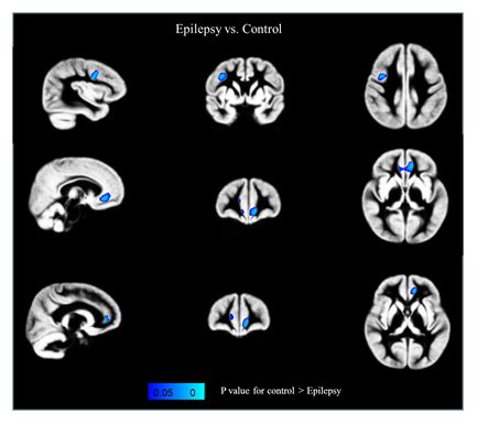
Figure 1: Statistical VBM reveals cortical GM reductions in the brains of epilepsy compared with control.

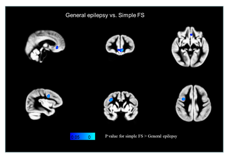
Figure 2: Statistical VBM demonstrates cortical GM atrophy in the brains of epilepsy compared with simple FS.
|
Epilepsy vs. control |
Epilepsy vs. Simple FS |
|||||
|
Cluster number |
Voxels number |
P-value (minimum) |
Cluster number |
Voxels number |
P-value (minimum) |
|
|
Precentral gyrus right |
1 |
318 |
0.015* |
1 |
342 |
0.017* |
|
Middle frontal gyrus right |
1 |
70 |
0.025* |
1 |
31 |
0.032* |
|
Inferior frontal gyrus (opercular) right |
1 |
508 |
0.015* |
1 |
519 |
0.017* |
|
Superior frontal gyrus (medial) right |
1 |
39 |
0.045* |
- |
- |
- |
|
Orbitofrontal cortex (medial) left |
1 |
456 |
0.015* |
1 |
360 |
0.032* |
|
Orbitofrontal cortex (medial) right |
2 |
374 |
0.030* |
1 |
49 |
0.038* |
|
Anterior cingulate gyrus left |
2 |
1008 |
0.015* |
1 |
463 |
0.032* |
|
Anterior cingulate gyrus right |
2 |
499 |
0.040* |
- |
- |
- |
P < 0.05.
Table 2: VBM revealed significant cortical GM atrophy in epilepsy compared to control and simple FS.
At the First Affiliated Hospital of Xi'an Jiaotong University, all participants underwent MRI exams using the same 3.0-T scanner (Signa HDxt, GE Healthcare, Milwaukee, WI) with an 8-channel head coil. The single-shot echo-planar are three-dimensional fast spoiled gradient-recalled echo T1-weighted imaging (T1WI) was performed by using the following parameters: Repetition time = 10.468 ms; Echo time = 4.764 ms; Inversion time: 400 ms; Field of view = 240 × 240 mm2; Acquisition matrix = 240 × 240; Slice thickness = 1 mm.
2.3 Image processing and statistical analysis
2.3.1 Voxel-based morphometry (VBM): Firstly, 3D T1WI images are segmented into GM, white matter (WM), and cerebrospinal fluid (CSF) regions using the Statistical Parametric Mapping software (SPM, version 12, https:/www.fil.ion.ucl.ac.uk / spm). Secondly, a study-specific GM template is generated using a group-wise approach based on the control group's images [24]. Thirdly, by using the registration, all the individual GM images are normalized to template space. Fourthly, all reported GM images are multiplied by the warp Jacobian and smoothed with a Gussian kernel (sigma = 3mm). The statistical analysis of VBM was performed by using the general linear model. P values > 0.05 were considered statistical significance after correction of the family-wise error (FWE) rate and the threshold-free cluster enhancement (TFCE).
2.3.2 Statistical analysis: The intergroup was analyzed using a separate t-test, using substantially different demographic and clinical data between epilepsy, simple FS, and control groups. For the comparison of categorical variables, the chi-square (x2) method was used. The Non-parametric Mann-Whitney U test was used to evaluate the continuous variables with non-normal group distribution, identifying the statistical significance (P < 0.05). For statistical analysis, a commercial software package (SPSS version 21.0 IBM, Armonk, NY, USA) was used.
3. Results
There were 34 epileptic children and 27 simple FS children, and 29 control children in this section met selection requirements and consented to participate in the study. After monitoring seizures among these groups, there were no substantial differences in age, sex, gestational age, and scan time after seizures onset (Table 1).
3.1 Epilepsy with control
Relative to the control group, epileptic children displayed decreased cortical GM thickness on the right precentral gyrus, right middle frontal gyrus, right frontal gyrus (opercular), right frontal gyrus (medial), bilateral orbitofrontal cortex (medial), and bilateral anterior cingulate gyrus compared to control group (P < 0.05) (Figure 1, Table 2). However, no significant difference was observed in the deep GM nucleus volume, such as bilateral thalami, basal ganglions, hippocampus, or cerebellum.
3.2 Epilepsy with simple FS
Comparing epileptic children with a simple FS group, the findings showed a marked reduction in cortical GM thickness on the right precentral gyrus, right middle frontal gyrus, right inferior frontal gyrus (opercular), bilateral orbitofrontal cortex (medial), and left anterior cingulate gyrus relative to a simple group of FS (P < 0.05) (Figure 2, Table 2). There were significant diffrences in seizure durations between epilepsy and simple FS (P < 0.05). No significant changes in volumes of bilateral thalami, basal ganglions, hippocampus, or cerebellum were seen.
3.3 Simple FS with control
No significant differences in cortical GM thickness or subcortical nuclei volumes were observed in children with simple FS than the control group (P > 0.05).
4. Discussion
This study attempted to explore the cortical GM's structural changes in children who had recently been diagnosed with epilepsy using the VBM approach that considers volumetric changes. The study's main findings are cortical GM atrophy in bilateral orbitofrontal cortex (medial), and bilateral anterior cingulate gyrus, while in the right precentral gyrus, right middle frontal gyrus, right frontal gyrus (opercular), right frontal gyrus (medial), focally damaged in epileptic children compared to control. However, in comparing to simple FS, no significant differences were found in the superior frontal gyrus (medial) right and anterior cingulate gyrus right in the epileptic group. Besides, there were no substantial changes in the volume of the thalamus, basal ganglia, and hippocampus in epilepsy or simple FS in all comparisons. Because our epilepsy group was heterogynous, our results showed some regions are bilaterally damage, and some are focal. These results may help in understanding the neuroanatomical modifications that contribute to epilepsy during brain development. These results indicated that newly diagnosed epileptic children have frontal lobe cortical GM atrophy, suggesting that epilepsy affects this brain region directly.
Furthermore, our children's initial lesion is cortical, with no
thalamic or deep nuclei volume changes. Again, previous research using quantitative MRI failed to identify structural defects in the thalamus of patients with IGE, similar to our findings [14, 25]. The volumes of the thalamus in our study did not change substantially between the patient's and control groups, in contrast to a large number of animal and, to a lesser extent, clinical studies suggesting that the thalamus plays a significant role in this disease. These findings are consistent with the report of a previous volumetric study of IGE patients [25]. Consequently, anomalies in the thalamocortical network, which have been observed in animals [26] and humans[27], do not seem to be related to thalamic structural changes. Maybe, VBM cannot detect mild changes in thalamic volume; at the same time, MR spectroscopy (MRS) of the thalamus has shown defects in patients with IGE [27], but VBM did not [25]. As a consequence, the absence of structural changes does not rule out the possibility of functional abnormalities. Furthermore, our results indicate that structural reduction of the cortical cortex occurs outside of the thalamus. Moreover, the thalamus is not only essential but also the most impaired organ in people with epilepsy. According to animal and clinical studies, the thalamocortical circuitry is involved in seizure generalization and EEG discharge maintenance [28, 29]. Our study, standard VBM examination, revealed no significant GM volume alterations in the bilateral thalami, basal ganglions, hippocampus, or cerebellum.
The frontal cortical GM thickness was reduced in the epilepsy group compared to the simple FS and control groups. The current findings were consistent with the previous cortical reduction in generalized tonic-clonic (GTC) and juvenile myoclonic epilepsy (JMC), [18, 30, 31] suggesting that epileptic children may have brain changes in the frontal lobe. Multisensory inputs converge in the cortical, which connects to subcortical regions. Furthermore, VBM studies in patients with juvenile myoclonic epilepsy (JME) have revealed increased GM in the superior frontal gyrus [32] and orbitofrontal regions [33]. In children with JME, fMRI showed frontal lobe hyperactivity in the prefrontal cortex (PFC) and superior frontal gyrus [34]. These differences may suggest that epilepsy children have minor anatomical defects in their developing brains. The second possibility may be due to the subjects' age group and the fact that their epilepsy was newly diagnosed and had never taken antiepileptic drugs before. In comparing epilepsy to simple FS, the study finds inconsistencies in the superior frontal gyrus (medial) and the right and anterior cingulate gyrus that we can't even clarify. It's possible that the simple FS maybe is not benign. Simple FS children may develop neurological disorders such as cognitive decline, emotional memory loss, [35] or behavioral disturbances, [36] based on nuclei functions, and a recommended cohort follow-up study. Our study has a range of limitations. First, it was a retrospective cross-sectional analysis with a heterogynous epilepsy group. Second, we did not follow up with children who had simple FS or examine the risk factors for simple FS developing into epilepsy. Third, clinical evidence such as cytokine levels was not assessed in patients with simple FS and epilepsy. However, our results showed that simple FS does not cause GM abnormalities after onset, which may relieve families' concerns about their children with simple FS.
5. Conclusion
This VBM approach has a significant advantage in terms of distinguishing these two seizures. The frontal lobe cortex of children with epilepsy was substantially reduced without involving deep nuclei. There were no cortical thickness changes in simple FS, suggesting that the disease would have a positive outcome. Our results contribute to a better understanding of neuroimaging processes in the field, leading to new therapeutic approaches that consider seizure mechanisms.
Conflicts of Interest
The authors declared there are no financial conflicts of interest.
Acknowledgment
The first author would like to express his gratitude to all Medical Imaging and Nuclear Medicine department members at Xi'an Jiaotong University's first affiliated hospital.
References
- Covanis A, Guekht A, Li S, et al. From global campaign to global commitment: The World Health Assembly’s Resolution on epilepsy. Epilepsia 56 (2015): 1651-1657.
- Koh S. Role of Neuroinflammation in Evolution of Childhood Epilepsy. clin transl neurol 33 (2018): 64-72.
- Seinfeld SA, Pellock JM, Kjeldsen MJ, et al. Epilepsy after Febrile Seizures: Twins Suggest Genetic Influence. Pediatr Neurol 55 (2016): 14-16.
- Grill MF, Ng YT. Simple febrile seizures plus (SFS+): More than one febrile seizure within 24hours is usually okay. Epilepsy Behav 27 (2013): 472-476.
- Falco-Walter JJ, Scheffer IE, Fisher RS. The new definition and classification of seizures and epilepsy. Epilepsy Res 139 (2018): 73-79.
- Hampers LC, Spina LA. Evaluation and Management of Pediatric Febrile Seizures in the Emergency Department. Emerg Med Clin North Am 29 (2011): 83-93.
- Pavone P, Corsello G, Ruggieri M, et al. Benign and severe early-life seizures: A round in the first year of life. Ital J Pediatr 44 (2018): 1-11.
- Fetveit A. Assessment of febrile seizures in children. Eur J Pediatr 167 (2008): 17-27.
- Ma L, McCauley SO. Management of Pediatric Febrile Seizures. J Nurse Pract 14 (2018): 74-80.
- Curwood EK, Pedersen M, Carney PW, et al Abnormal cortical thickness connectivity persists in childhood absence epilepsy. Ann Clin Transl Neurol 10 (2015): 178-188
- Karalok ZS, Öztürk Z, Gunes A. Cortical thinning in benign epilepsy with centrotemporal spikes (BECTS) with or without attention-deficit/hyperactivity (ADHD). J Clin Neurosci 68 (2019): 123-127.
- Auer T, Barsi P, Bone B, et al. History of simple febrile seizures is associated with hippocampal abnormalities in adults. Epilepsia 49 (2008): 1562-1569.
- Dubé C, Vezzani A, Behrens M, et al. Interleukin-1β contributes to the generation of experimental febrile seizures. Ann Neurol 57 (2005): 152-155.
- Seeck M, Dreifuss S, Lantz G, et al. Subcortical nuclei volumetry in idiopathic generalized epilepsy. Epilepsia 46 (2005): 1642-1645.
- Kim JH, Lee JK, Koh SB, et al. Regional grey matter abnormalities in juvenile myoclonic epilepsy: a voxel-based morphometry study. Neuroimage 37 (2007): 1132-1137.
- Pardoe H, Pell GS, Abbott DF, et al. Multi-site voxel-based morphometry: Methods and a feasibility demonstration with childhood absence epilepsy. Neuroimage 42 (2008): 611-616.
- Bonilha L, Montenegro MA, Rorden C, et al. Voxel-based morphometry reveals excess gray matter concentration in patients with focal cortical dysplasia. Epilepsia 47 (2006): 908-915.
- Huang W, Lu G, Zhang Z, et al. Gray-matter volume reduction in the thalamus and frontal lobe in epileptic patients with generalized tonic-clonic seizures Réduction de volume de la substance grise dans le thalamus et le lobe. J Neuroradiol 38 (2011): 298-303.
- Ciumas C, Savic I. Structural changes in patients with primary generalized tonic and clonic seizures. Neurology 67 (2006): 683-686.
- Fisher RS, Acevedo C, Arzimanoglou A, et al. ILAE Official Report: A practical clinical definition of epilepsy. Epilepsia 55 (2014): 475-482.
- Dougherty D, Duffner PK, Baumann RJ, et al. Febrile seizures: Clinical practice guideline for the long-term management of the child with simple febrile seizures. Pediatrics 121 (2008): 1281-1286.
- Liauw L, Grond J Van Der, Slooff V, et al. Differentiation between peritrigonal terminal zones and hypoxic-ischemic white matter injury on MRI. Eur J Radiol 65 (2008): 395-401.
- Liu C, Habib T, Salimeen M, et al. Quantification of visible Virchow–Robin spaces for detecting the functional status of the glymphatic system in children with newly diagnosed idiopathic generalized epilepsy. Seizure 78 (2020): 12-17.
- Li X, Gao J, Wang M, et al. Rapid and reliable tract-based spatial statistics pipeline for diffusion tensor imaging in the neonatal brain: Applications to the white matter development and lesions. Magn Reson Imaging 34 (2016): 1314-1321.
- Natsume J, Bernasconi N, Andermann F, et al. MRI volumetry of the thalamus in temporal, extratemporal, and idiopathic generalized epilepsy. Neurology 60 (2003): 1296-1300.
- Afifi AK Topical. Review: Basal Ganglia: Functional Anatomy and Physiology. Part 1. J Child Neurol 9 (1994): 249-260.
- Bernasconi A, Bernasconi N, Natsume J, et al. Magnetic resonance spectroscopy and imaging of the thalamus in idiopathic generalized epilepsy. Brain 126 (2003): 2447-2454.
- Gotman J, Grova C, Bagshaw A, et al. Generalized epileptic discharges show thalamocortical activation and suspension of the default state of the brain. PNAS (2005): 10-14.
- Review N Evolving Concepts on the Pathophysiology of Absence Seizures. JAMA Neurol 62 (2005): 267-278.
- Sun J, Gao Y, Miao A, et al. Multifrequency Dynamics of Cortical Neuromagnetic Activity Underlying Seizure Termination in Absence Epilepsy. Front Hum Neurosci 14 (2020): 1-9.
- Tae WS, Kim SH, Joo EY, et al. Cortical thickness abnormality in juvenile myoclonic epilepsy. J Neurol 255 (2008): 561-566.
- Woermann FG, Free SL, Koepp MJ, et al. Abnormal cerebral structure in juvenile myoclonic epilepsy demonstrated with voxel-based analysis of MRI. Brain 122 (1999): 2101-2107.
- Lin K, Jackowski AP, Carrete H, et al. Voxel-based morphometry evaluation of patients with photosensitive juvenile myoclonic epilepsy. Epilepsy Res 86 (2009): 138-145.
- Wandschneider B, Centeno M, Vollmar C, et al. Risk-taking behavior in juvenile myoclonic epilepsy. Epilepsia (2013): 1-8.
- Rolls ET. The cingulate cortex and limbic systems for action, emotion, and memory, 1st ed. Elsevier B.V. (2019).
- Li C, Wang XQ, Wen CH, et al. Association of degree of loss aversion and grey matter volume in superior frontal gyrus by voxel-based morphometry. Brain Imaging Behav 14 (2020): 89-99.
