A Sleeping Giant - A Case Report of Echinococcosis
Article Information
Madalena Sousa Silva*, António Carneiro, Isa Silva, Luís Miguel Pereira, Natália Fernandes
Hospital de Cascais, Alcabideche, Portugal
*Corresponding Author: Dr. Madalena Sousa Silva, Hospital de Cascais, Alcabideche, Portugal
Received: 24 November 2019; Accepted: 04 December 2019; Published: 13 January 2020
Citation: Madalena Sousa Silva, António Carneiro, Isa Silva, Luís Miguel Pereira, Natália Fernandes. A Sleeping Giant - A Case Report of Echinococcosis. Archives of Clinical and Medical Case Reports 4 (2020): 075-079.
View / Download Pdf Share at FacebookAbstract
Echinococcosis is a parasitary disease produced by Echinococcus spp. It is endemic in some European regions such as Portugal. We present the case of a patient who was admitted for de-novo ascites. In the course of the investigation, a bulky hepatic hydatid cyst was diagnosed. The cyst was in the inactive phase of the disease, calcified and it bore no symptoms. The ascites was due to heart failure and resolved with appropriate treatment. The relevance of the case relates to the exuberance of the cyst and the interesting images obtained.
Keywords
Echinococcosis; Hepatic cyst hydatidosis; Ascites; Eosinophilia
Echinococcosis articles, Hepatic cyst hydatidosis articles, Ascites articles, Eosinophilia articles
Echinococcosis articles Echinococcosis Research articles Echinococcosis review articles Echinococcosis PubMed articles Echinococcosis PubMed Central articles Echinococcosis 2023 articles Echinococcosis 2024 articles Echinococcosis Scopus articles Echinococcosis impact factor journals Echinococcosis Scopus journals Echinococcosis PubMed journals Echinococcosis medical journals Echinococcosis free journals Echinococcosis best journals Echinococcosis top journals Echinococcosis free medical journals Echinococcosis famous journals Echinococcosis Google Scholar indexed journals Hepatic cyst hydatidosis articles Hepatic cyst hydatidosis Research articles Hepatic cyst hydatidosis review articles Hepatic cyst hydatidosis PubMed articles Hepatic cyst hydatidosis PubMed Central articles Hepatic cyst hydatidosis 2023 articles Hepatic cyst hydatidosis 2024 articles Hepatic cyst hydatidosis Scopus articles Hepatic cyst hydatidosis impact factor journals Hepatic cyst hydatidosis Scopus journals Hepatic cyst hydatidosis PubMed journals Hepatic cyst hydatidosis medical journals Hepatic cyst hydatidosis free journals Hepatic cyst hydatidosis best journals Hepatic cyst hydatidosis top journals Hepatic cyst hydatidosis free medical journals Hepatic cyst hydatidosis famous journals Hepatic cyst hydatidosis Google Scholar indexed journals cyst articles cyst Research articles cyst review articles cyst PubMed articles cyst PubMed Central articles cyst 2023 articles cyst 2024 articles cyst Scopus articles cyst impact factor journals cyst Scopus journals cyst PubMed journals cyst medical journals cyst free journals cyst best journals cyst top journals cyst free medical journals cyst famous journals cyst Google Scholar indexed journals Hepatic articles Hepatic Research articles Hepatic review articles Hepatic PubMed articles Hepatic PubMed Central articles Hepatic 2023 articles Hepatic 2024 articles Hepatic Scopus articles Hepatic impact factor journals Hepatic Scopus journals Hepatic PubMed journals Hepatic medical journals Hepatic free journals Hepatic best journals Hepatic top journals Hepatic free medical journals Hepatic famous journals Hepatic Google Scholar indexed journals hydatidosis articles hydatidosis Research articles hydatidosis review articles hydatidosis PubMed articles hydatidosis PubMed Central articles hydatidosis 2023 articles hydatidosis 2024 articles hydatidosis Scopus articles hydatidosis impact factor journals hydatidosis Scopus journals hydatidosis PubMed journals hydatidosis medical journals hydatidosis free journals hydatidosis best journals hydatidosis top journals hydatidosis free medical journals hydatidosis famous journals hydatidosis Google Scholar indexed journals treatment articles treatment Research articles treatment review articles treatment PubMed articles treatment PubMed Central articles treatment 2023 articles treatment 2024 articles treatment Scopus articles treatment impact factor journals treatment Scopus journals treatment PubMed journals treatment medical journals treatment free journals treatment best journals treatment top journals treatment free medical journals treatment famous journals treatment Google Scholar indexed journals Ascites articles Ascites Research articles Ascites review articles Ascites PubMed articles Ascites PubMed Central articles Ascites 2023 articles Ascites 2024 articles Ascites Scopus articles Ascites impact factor journals Ascites Scopus journals Ascites PubMed journals Ascites medical journals Ascites free journals Ascites best journals Ascites top journals Ascites free medical journals Ascites famous journals Ascites Google Scholar indexed journals patient articles patient Research articles patient review articles patient PubMed articles patient PubMed Central articles patient 2023 articles patient 2024 articles patient Scopus articles patient impact factor journals patient Scopus journals patient PubMed journals patient medical journals patient free journals patient best journals patient top journals patient free medical journals patient famous journals patient Google Scholar indexed journals Eosinophilia articles Eosinophilia Research articles Eosinophilia review articles Eosinophilia PubMed articles Eosinophilia PubMed Central articles Eosinophilia 2023 articles Eosinophilia 2024 articles Eosinophilia Scopus articles Eosinophilia impact factor journals Eosinophilia Scopus journals Eosinophilia PubMed journals Eosinophilia medical journals Eosinophilia free journals Eosinophilia best journals Eosinophilia top journals Eosinophilia free medical journals Eosinophilia famous journals Eosinophilia Google Scholar indexed journals tumor articles tumor Research articles tumor review articles tumor PubMed articles tumor PubMed Central articles tumor 2023 articles tumor 2024 articles tumor Scopus articles tumor impact factor journals tumor Scopus journals tumor PubMed journals tumor medical journals tumor free journals tumor best journals tumor top journals tumor free medical journals tumor famous journals tumor Google Scholar indexed journals
Article Details
1. Case Report
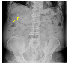
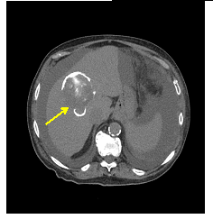
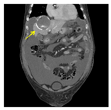
The authors present the clinical case of an independent 90 years-old man with medical history of hypertensive and ischemic cardiopathy with heart failure; colon cancer in 2008 with complete remission and MGUS IgG/k. The patient was admitted in the medical infirmary with de novo ascites. During the course of clinical investigation some results were prominent – mild eosinophilia, hyperbilirubinemia with conjugated bilirubin elevation. The abdomen radiography showed a nodular image on the right hypochondrium (Figure 1), which was, then reviwed by computed tomography (CT scan). The CT scan revealed a 9.5 cm multiseptated cyst in the right lobe with calcified walls suggestive of hidatid cyst (Figure 2). Echinococcus granulosus serology was negative. The case was discussed in a multidisciplinary reunion and a conservative approach was adopted as the cyst was calcified. The patient was medicated for 21 days with albendazole wih resolution of eosinophilia. The etiology of ascites was associated with heart failure and resolved with diuretics.

Figure 1: Abdomen radiography, yellow arrow shows a calcified cyst on right hypochondrium.

Figure 2: Abdomen CT scan, horizontal cut, yellow arrow shows hydatid cyst on the right lobe of the liver – the contrast enhances the round lesion compatible with a ringlike pattern of calcification.

Figure 3: Abdomen CT scan, sagital cut, yellow arrow shows hydatid cyst.
2. Discussion
Hepatic hydatid cyst is a small spectrum of the disease produced by Echinococcus parasites. There are four species of Echinococcus that can produce disease in humans - E. granulosus, E. multilocularis, the most common; E. vogeli and E. oligarthrus, extremely rare. E. granulosus is responsible for the hepatic disease, whereas E. multilocularis is accountable for the pulmonary hydatid cyst. A definite host (usually the dog) transmits the parasite to the human (fecal-oral transmission), and, once in the blood stream it can reach and accommodate in several different organs where a cyst will be produced.
The clinical course will depend on the affected tissue due to mass effect (e.g., cough, dispnea, hemoptysis if it affects the lung; abdominal pain, nausea and vomiting in the case of hepatic cyst). The host immune response creates a fibrous capsule that evolves the cyst, and, in time becomes calcified. The major associated complication is cyst rupture that can lead to sepsis or anaphylactic shock by spreading of the cyst continent. The right lobe is the most frequently involved portion of the liver. The natural evolution of the disease is the calcification of the entire cyst, with death of the parasite. A classification system has been developed to adjust the best therapeutic option. The classification is based on the cyst ultrasound characteristics.
The diagnosis is made by image technique – ultrasound, CT or magnetic resonance – and the lesions can vary from pure cyst to completely calcified lesions. Calcification is seen in X-rays in 20-30% of hydatid cysts, shown as a ring-like hyoptransparent lesion. The serology for the Echinococcus is useful for diagnosis and to survey treatment response.
Surgical removal and anti-parasite medication is the definite treatment. The drug of choice is albendazole. The second options are mebendazole and praziquantel. Albendazol is given in a 400 mg dose two times per day. If the cyst is completely solidified it is thought to be inactive and surveillance is the best option in absence of symptoms.
|
WHO- Stage |
Description |
Stage |
Size |
Preferred treatment |
|
CE 1 |
Unilocular unechoic cystic lesion with double line sign |
Active |
< 5 cm |
Albendazole |
|
> 5 cm |
Albendazole + PAIR¥ |
|||
|
CE 2 |
Multiseptated, “rosette-like” “honeycomb cyst” |
Active |
Any |
Albendazole + modified catheterization or surgery |
|
CE 3a |
Cyst with detached membranes (water-lily sign) |
Transitional |
< 5 cm |
Albendazole + PAIR¥ |
|
> 5 cm |
||||
|
CE 3b |
Cyst with daughter cysts in solid matrix |
Transitional |
Any |
Albendazole + modified catheterization or surgery |
|
C4 |
Cyst with heterogenous hypoechoic/hyperechoic contents; no daughter cysts |
Inactive |
Any |
Observation |
|
C5 |
Solid plus calcified wall |
Inactive |
Any |
Observation |
¥PAIR – puncture, aspiration, injection, reaspiration; endoschopic technique
Table 1: World Health Organization classification of cystic echinococcosis and treatment stratified by cyst stage.
3. Conclusion
Hydatid cyst is an increasingly less frequent form of disease due to the awareness of animal vaccination in urban as well as rural environments. Nevertheless, it is important to remember the existence of the disease so that we can adequately treat our patients. In this clinical case, the diagnosis of hepatic cyst hydatidosis was incidental for it bared no associated symptoms nor was implied in the process of ascites. Even so, we wanted to share the interesting images as well as to highlight some aspects of the disease.
References
- Ramos RM, Videira C, Traila J, et al. Liver Hydatid Cyst – Revisited, Acta Radiológica Portuguesa 22 (2010): 47-51.
- Botezatu C, Mastalier B, Patrascu T. Hepatic hydatis cyst diagnose and treatment algorithm. Journal of Medicine and Life 11 (2018): 203-209.
- Pakala T, Molina M, Wu GY. Hepatic Echinococcal Cysts: A Review. J Clin Transl Hepatol 4 (2016): 39-46.
- Nunnari G, Pinzone MR, Gruttadauria S, et al. Hepatic echinococcosis: Clinical and therapeutic aspects. World J Gastroenterol 18 (2012): 1448-1458.
- Savioli L, Daumerie D, WHO Department of Control of Neglected Tropical Diseases. Sustaining the drive to overcome the global impact of neglected tropical diseases: second WHO report on neglected tropical diseases. World Health Organization (2013).
- Srinivas MR, Deepashri B, Lakshmeesha MT. Imaging Spectrum of Hydatid Disease: Usual and Unusual Locations. Pol J Radiol 81 (2016): 190-205.
- Scherer V, Weinzierl M, Sturm R, et al. Computed tomography in hydatid disease of the liver: a report on 13 cases. J Comput Assist Tomogr 2 (1978): 612-617.
- de Diego J, Lecumberri FJ, Franquet T, et al. Computed tomography in hepatic echinococcosis. AJR Am J Roentgenol 139 (1982): 699-702.
- Pedrosa I, Saíz A, Arrazola J, et al. Hydatid Disease: Radiologic and Pathologic Features and Complications RadioGraphics 20 (2000): 795-817.
- Brunetti E, Kern P, Vuitton DA. Writing Panel for the WHO-IWGE. Expert consensus for the diagnosis and treatment of cystic and alveolar echinococcosis in humans. Acta Trop 114 (2010): 1.
