A Rare Tumour of Ileo-transverse Colon Presenting As an Incarcerated Incisional Hernia in Right Iliac Fossa: A Case Report
Article Information
Rukmini Waghmare, Kavita Jadhav*, Rajalakshmi Venkateswaran, Amit Thombare, Lalramchhana, Megha Kinake
Department of General Surgery, Grant Government Medical College & Sir J.J. Group of Hospitals, Mumbai, India 400008
*Corresponding Author: Kavita Jadhav, Department of General Surgery, Grant Government Medical College & Sir J.J. Group of Hospitals, Mumbai, India 400008
Received: 06 August 2024; Accepted: 20 August 2024; Published: 28 August 2024
Citation: Rukmini Waghmare, Kavita Jadhav, Rajalakshmi Venkateswaran, Amit Thombare, Lalramchhana, Megha Kinake. A Rare Tumour of Ileo-transverse Colon Presenting As an Incarcerated Incisional Hernia in Right Iliac Fossa: A Case Report. Journal of Surgery and Research 7 (2024): 359-362.
View / Download Pdf Share at FacebookAbstract
Incisional hernias are delayed complications of abdominal surgery and occur in 0.5-13.9% of patients. Neoplasm of intestine presenting as a content of hernia is rare. Few cases have been reported in literature. Sigmoid colon and caecal malignancies presenting as inguinal hernia have been reported. In present case, a 60 years old female patient presented with a strangulated incisional hernia in the right iliac fossa with gangrenous changes of the overlying skin. Patient underwent emergency surgery wherein the content was found to be a tumour of the small bowel firmly adherent to the proximal transverse colon. Extended right hemicolectomy was done. Histopathology revealed inflammatory myo-fibroblastic tumour of the ileum involving transverse colon which is rare. A brief case report with review of literature is presented here.
Keywords
Incarcerated incisional hernia, Inflammatory myo-fibroblastic tumor of ileo-transeverse colon
Incarcerated incisional hernia articles; Inflammatory myo-fibroblastic tumor of ileo-transeverse colon articles
Article Details
Introduction
Malignant tumors as content of hernia sac are rare. The incidence of 0.07% to 0.7% reported in literature [1]. Rarely, the hernia can be the first presentation of an unknown malignancy.
Inflammatory myo-fibroblastic tumors (IMTs) are very rare lesions affecting most commonly children and young adults. IMTs have various names, including inflammatory pseudo-tumor, plasma cell granuloma, fibrous histiocytoma, solitary mast cell tumor, and fibro-xanthoma. The clinical behavior of IMTs is similar to that of tumors of uncertain malignant potential, so IMTs are considered tumors of borderline malignancy. The lung is the most commonly affected location, but lesions can be seen intraabdominal.
Small bowel tumors are particularly rare, and even more rarely they are located in the anti-mesenteric edge of the small bowel [2].
Case Presentation
A 60 years old female patient presented to the casualty with a lump in right iliac fossa since 1 month. Patient developed localised pain and fever since 5 days. Patient had history of open appendicectomy 6 years back.
On clinical examination, patient was febrile, tachypnoeic, with a pulse rate was 120 beats/minute, blood pressure was 90/60 mm of Hg.
Local examination revealed a hard lump at the scar site in the right iliac fossa with tenderness over the lump and surrounding induration. Overlying skin was gangrenous and not pinch-able.
Clinically, she was diagnosed to have an incarcerated incisional hernia with omentum as content. Urine output was less than 30ml/hour and arterial blood gas analysis showed metabolic acidosis.
Biochemical investigations showed leucocytosis (18,700 cells/mm3), rest blood investigations were within normal limits. X-ray abdomen and chest was reported to be normal. An ultrasound abdomen revealed an abdominal wall abscess of 40-50cc with a incarcerated incisional hernia, omentum being the content. Hence, patient was taken for emergency local exploration. An elliptical incision around scar was taken. Intraoperatively there was thickness of subcutaneous tissue forming a capsule of abscess (Fig 1).
Pus was drained and it was noted that there was a hard mass arising from a segment of bowel. On tracing the bowel, the distal ileum nearly 20cm from ileo-caecal junction had a perforating mass which was communicating with the abscess cavity and was found adhered to the proximal transverse colon (Fig 2).
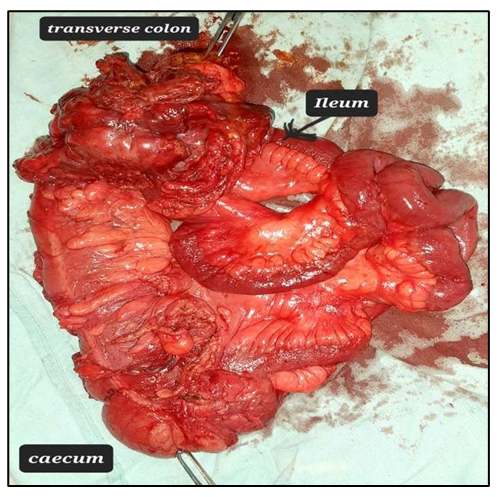
The localized omentum was inflamed and adhered to the site. In view of high suspicion of malignancy an extended Right Hemicolectomy with lymph node dissection was done (Fig 3).
End to end ileo-transverse anastomosis was done. Abdominal wall closure was done in layers, without placement of any mesh.
Postoperatively patient was started on full diet on day 4, which she tolerated well. Histopathology report revealed inflammatory myo-fibroblastic tumour (IMT) of ileum infiltrating transverse colon (Fig 4a & 4b), intermediate category with all resection margins free. On immunohistochemistry, spindle cells are diffusely immunopositive for smooth muscle actin (SMA)/muscle-specific actin(MSA) with focal immune-positivity for desmin.
A contrast enhanced computed tomography (CECT) scan of the abdomen and pelvis was done postoperatively, which showed no residual disease. Patient developed surgical site infection which was managed with daily cleaning and dressing. Secondary suturing was done. Patient was discharged with advice to follow up with positron emission tomography (PET) scan in oncology department, but was loss to follow up.
Discussion
Incisional hernia after open appendicectomy through a muscle-splitting (McBurney) incision is less common, occurring in less than 0.12% of operations for appendicitis.(3) The most common contents of the sac of incisional hernias are the small bowel, omentum or transverse colon because they are freely mobile in peritoneal cavity unlike ascending and descending colon.(4)
Neoplastic tumours found in hernias can be classified into three groups (5) - saccular, intra saccular, and extra saccular. Saccular tumours are tumours originating from the sac itself such as mesotheliomas. Extra saccular tumours are tumours present in proximity to the hernia sac, however, they are not contained within it. The last type, intra saccular tumours, are tumours originating from adjacent tissue and are contained within the sac itself.
The latter group most often originates from colo-rectal cancer(5) as was seen in the present case.
Inflammatory myo-fibroblastic tumor (IMT) is a rare tumour with borderline malignancy type. This tumour is very rare, but most commonly affects children, lung being the most common site. Intraabdominal IMT is rare. It affects mesenteric border of small bowel, anti-mesentric border being the rare site.(6)
Inflammatory Myofibroblastic Tumor (IMT) belongs to a class of rare spindle cell lesions showing a rather unpredictable biological behaviour with occasional tendency towards invasion to the surrounding tissue and local recurrence.
Symptoms are non-specific and depend on the location of the tumour. This lesion primarily described by Bahadori and Liebow in 1973.(7) Patients with intra-abdominal tumours most commonly present with intermittent abdominal pain due to the solid mass and the abdominal distention, as well as with weight loss, malaise, anorexia, and vomiting. Rarely, the presentation may be complicated by an intestinal obstruction, intussusception, or acute abdomen mimicking acute appendicitis, perforation.(2)
In hemodynamically stable patients with complicated hernia with hard lump, a CECT scan is advisable to rule out a malignancy pre-operatively. The mortality rates in patients with sepsis are high, if intervention not done early.
In present case, we suspected strangulated type incisional hernia, so an emergency surgical intervention without a confirmatory radiological diagnosis was done. Intraoperatively, after drainage of pus, we found that the palpable hard lump was tumour of ileum involving proximal part of transeverse colon, for which right hemicolectomy was performed. That tumour was histologically inflammatory myo-fibroblastic tumour of ileum of intermediate category.
IMTs generally have a benign course, and recurrence or distant metastasis is rare. The incidence of local recurrence has been reported to be 15-37 % when an IMT is located in the mesentery or the retroperitoneum. Nevertheless, to our knowledge, no precise recommendations exist in the literature on the necessity of any additional therapy. Complete surgical resection remains the treatment of choice for IMT.(2)
Conclusion
This is the first case of small bowel inflammatory myo-fibroblastic tumor infiltrating transverse colon, presenting as incarcerated incisional hernia with a contained perforation through open appendicectomy scar. A malignant tumour of bowel can be kept as differential for irreducible or incarcerated hernia with hard lump. Hemodynamically stable patient can be undergone contrast enhance computed tomography if feasible. However, unstable patients should be taken up for an emergency surgery. Resection according to malignant tumour resection protocols should be done, if intraoperative findings are suspicious of a malignancy.
References
- Secinti, Ilke E. Surprise in hernia sacs: Malignant tumor metastasis. Journal of Surgery and Medicine 6 (2022): 331-335.
- Salim, Maryam R. Inflammatory myofibroblastic tumor of the small bowel: a case report. Middle East Journal of Digestive Diseases 3 (2011): 134-137.
- Beltran, Marcelo A, Karina S. Cruces. Incisional hernia after McBurney incision: retrospective case-control study of risk factors and surgical treatment. World journal of surgery 32 (2008): 596-601.
- Choi, Pyong W. Incarcerated incisional hernia of the sigmoid colon after appendectomy: A case report. International journal of surgery case reports 31 (2017): 39-42.
- Hassan S, Mersad A, Etienne El-H. Perforated sigmoid colon cancer presenting as an incarcerated linguinal hernia: A case report. International Journal of Surgery Case Reports 72 (2020): 108-11.
- Oeconomopoulou, Alexandra, et al. Inflammatory myofibroblastic tumor of the small intestine mimicking acute appendicitis: a case report and review of the literature. Journal of Medical Case Reports 10 (2016): 1-4.
- Bahadori M. and Liebow A.A. Plasma Cell Granulomas of the Lung. Cancer ACS 31 (1973): 191-208.


