A Rare Case of Granular Cell Tumor of the Urethra Provoking Urinary Retention
Article Information
Karasavvidou Foteini1, Efstathiou Konstantinos2, Kakkas Grigorios2, Karathanasis Athanasios2, Kanti Stella2, Tzortzi de Paz Anna3, Tzortzis Vassilios3*
1Department of Pathology and Cytology, Faculty of Medicine-School of Health Sciences, University of Thessaly, University Hospital of Larissa, Larissa, Greece
2IASO Thessaly Clinic, Larissa, Greece
3Department of Urology, Faculty of Medicine, School of Health Sciences, University of Thessaly, University Hospital of Larissa, Larissa, Greece
*Corresponding Author: Dr. Vassilios Tzortzis, Department of Urology, Faculty of Medicine-School of Health Sciences-University of Thessaly, University Hospital of Larissa, Larissa, Greece
Received: 01 December 2020; Accepted: 28 December 2020; Published: 26 January 2021
Citation: Karasavvidou Foteini, Efstathiou Konstantinos, Kakkas Grigorios, Karathanasis Athanasios, Kanti Stella, Tzortzi de Paz Anna, Tzortzis Vassilios. A Rare Case of Granular Cell Tumor of the Urethra Provoking Urinary Retention. Archives of Nephrology and Urology 4 (2021): 018-022.
View / Download Pdf Share at FacebookAbstract
A 44-year-old woman was referred to urologist for urinary retention due to a urethral mass. Magnetic resonance imaging (MRI) showed a round mass that surrounded the urethra completely. The regional lymph nodes were normal and no other lesions were identified. Biopsy was performed revealing a tumor that was composed of large polygonal cells with eosinophilic and periodic acid-Schiff (PAS) - positive granular cytoplasm, indistinct cell margins and small round central nuclei. Immunohistochemical staining for S100 protein, vimentin, calretinin and inhibin Α was positive, indicating a granular cell tumor. Excision of the tumor including the urethra and an umbilical catheterized stoma using Mitrofanoff principle was performed.
Keywords
Female; Urethra; Granular tumor; Mitrofanoff
Female articles; Urethra articles; Granular tumor articles; Mitrofanoff articles
Female articles Female Research articles Female review articles Female PubMed articles Female PubMed Central articles Female 2023 articles Female 2024 articles Female Scopus articles Female impact factor journals Female Scopus journals Female PubMed journals Female medical journals Female free journals Female best journals Female top journals Female free medical journals Female famous journals Female Google Scholar indexed journals Urethra articles Urethra Research articles Urethra review articles Urethra PubMed articles Urethra PubMed Central articles Urethra 2023 articles Urethra 2024 articles Urethra Scopus articles Urethra impact factor journals Urethra Scopus journals Urethra PubMed journals Urethra medical journals Urethra free journals Urethra best journals Urethra top journals Urethra free medical journals Urethra famous journals Urethra Google Scholar indexed journals Granular tumor articles Granular tumor Research articles Granular tumor review articles Granular tumor PubMed articles Granular tumor PubMed Central articles Granular tumor 2023 articles Granular tumor 2024 articles Granular tumor Scopus articles Granular tumor impact factor journals Granular tumor Scopus journals Granular tumor PubMed journals Granular tumor medical journals Granular tumor free journals Granular tumor best journals Granular tumor top journals Granular tumor free medical journals Granular tumor famous journals Granular tumor Google Scholar indexed journals Mitrofanoff articles Mitrofanoff Research articles Mitrofanoff review articles Mitrofanoff PubMed articles Mitrofanoff PubMed Central articles Mitrofanoff 2023 articles Mitrofanoff 2024 articles Mitrofanoff Scopus articles Mitrofanoff impact factor journals Mitrofanoff Scopus journals Mitrofanoff PubMed journals Mitrofanoff medical journals Mitrofanoff free journals Mitrofanoff best journals Mitrofanoff top journals Mitrofanoff free medical journals Mitrofanoff famous journals Mitrofanoff Google Scholar indexed journals malignant peripheral nerve sheath tumours articles malignant peripheral nerve sheath tumours Research articles malignant peripheral nerve sheath tumours review articles malignant peripheral nerve sheath tumours PubMed articles malignant peripheral nerve sheath tumours PubMed Central articles malignant peripheral nerve sheath tumours 2023 articles malignant peripheral nerve sheath tumours 2024 articles malignant peripheral nerve sheath tumours Scopus articles malignant peripheral nerve sheath tumours impact factor journals malignant peripheral nerve sheath tumours Scopus journals malignant peripheral nerve sheath tumours PubMed journals malignant peripheral nerve sheath tumours medical journals malignant peripheral nerve sheath tumours free journals malignant peripheral nerve sheath tumours best journals malignant peripheral nerve sheath tumours top journals malignant peripheral nerve sheath tumours free medical journals malignant peripheral nerve sheath tumours famous journals malignant peripheral nerve sheath tumours Google Scholar indexed journals Urinary Retention articles Urinary Retention Research articles Urinary Retention review articles Urinary Retention PubMed articles Urinary Retention PubMed Central articles Urinary Retention 2023 articles Urinary Retention 2024 articles Urinary Retention Scopus articles Urinary Retention impact factor journals Urinary Retention Scopus journals Urinary Retention PubMed journals Urinary Retention medical journals Urinary Retention free journals Urinary Retention best journals Urinary Retention top journals Urinary Retention free medical journals Urinary Retention famous journals Urinary Retention Google Scholar indexed journals smooth muscle actin articles smooth muscle actin Research articles smooth muscle actin review articles smooth muscle actin PubMed articles smooth muscle actin PubMed Central articles smooth muscle actin 2023 articles smooth muscle actin 2024 articles smooth muscle actin Scopus articles smooth muscle actin impact factor journals smooth muscle actin Scopus journals smooth muscle actin PubMed journals smooth muscle actin medical journals smooth muscle actin free journals smooth muscle actin best journals smooth muscle actin top journals smooth muscle actin free medical journals smooth muscle actin famous journals smooth muscle actin Google Scholar indexed journals neuron?specific enolase articles neuron?specific enolase Research articles neuron?specific enolase review articles neuron?specific enolase PubMed articles neuron?specific enolase PubMed Central articles neuron?specific enolase 2023 articles neuron?specific enolase 2024 articles neuron?specific enolase Scopus articles neuron?specific enolase impact factor journals neuron?specific enolase Scopus journals neuron?specific enolase PubMed journals neuron?specific enolase medical journals neuron?specific enolase free journals neuron?specific enolase best journals neuron?specific enolase top journals neuron?specific enolase free medical journals neuron?specific enolase famous journals neuron?specific enolase Google Scholar indexed journals cluster of differentiation 68 articles cluster of differentiation 68 Research articles cluster of differentiation 68 review articles cluster of differentiation 68 PubMed articles cluster of differentiation 68 PubMed Central articles cluster of differentiation 68 2023 articles cluster of differentiation 68 2024 articles cluster of differentiation 68 Scopus articles cluster of differentiation 68 impact factor journals cluster of differentiation 68 Scopus journals cluster of differentiation 68 PubMed journals cluster of differentiation 68 medical journals cluster of differentiation 68 free journals cluster of differentiation 68 best journals cluster of differentiation 68 top journals cluster of differentiation 68 free medical journals cluster of differentiation 68 famous journals cluster of differentiation 68 Google Scholar indexed journals Granular cell tumors articles Granular cell tumors Research articles Granular cell tumors review articles Granular cell tumors PubMed articles Granular cell tumors PubMed Central articles Granular cell tumors 2023 articles Granular cell tumors 2024 articles Granular cell tumors Scopus articles Granular cell tumors impact factor journals Granular cell tumors Scopus journals Granular cell tumors PubMed journals Granular cell tumors medical journals Granular cell tumors free journals Granular cell tumors best journals Granular cell tumors top journals Granular cell tumors free medical journals Granular cell tumors famous journals Granular cell tumors Google Scholar indexed journals
Article Details
1. Introduction
Granular cell tumors have been reported to develop in soft tissues such as skin and subcutaneous tissue, bronchus, oesophagus, breast, tongue, larynx, pharynx, gingiva, trachea, right colon, vulva, and hypopharynx [1]. The incidence in the urinary tract is extremely rare with only few cases having been reported. Herein, we present a case of a granular cell tumor of a female urethra that in our knowledge is the first case of that size reported in the literature.
2. Case Report
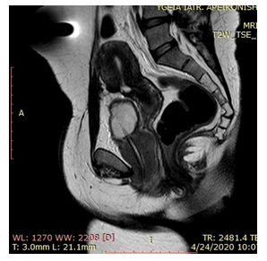
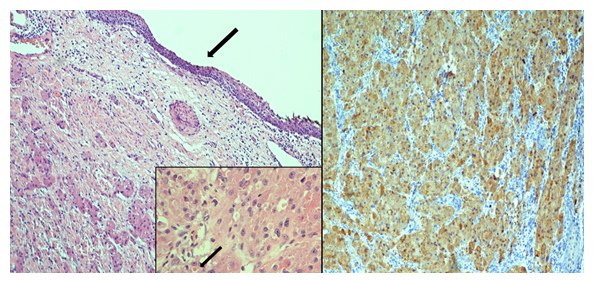
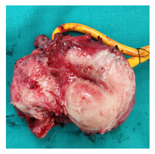
A 44-year-old woman was referred to the urologist by a gynecologist for urinary retention and a urethral solid mass that was found during a clinical examination. A Foley catheter was introduced and Magnetic Resonance Imaging (MRI) was performed. A round mass of 7.4 cm × 6.6 cm × 5.5 cm that completely surrounded the urethra was shown (Figure 1). Pelvic and inguinal lymph nodes were normal, and no other lesions were identified. Urinalysis was normal and urine cytology was negative. Biopsy of the tumor was performed. Histologically, the tumor was composed of large polygonal cells with abundant eosinophilic granular cytoplasms with coarse granules (representing phagolysosome aggregates). The nuclei showed mild to moderate nuclear atypia and small round central nuclei. Immunohistochemical staining for S?100 protein, neuron?specific enolase (NSE), smooth muscle actin (SMA), and cluster of differentiation 68 (CD68) was positive, indicating that it was compatible with granular cell tumor GCTs (Figure 2a, 2b). The staining was negative for cytokeratines ΑΕ1/ΑΕ3, caldesmon, GFAP, CD31, HMB45, MelanA, EMA, and SMA. Excision of the tumor, including the urethra, was performed after an informed consent of the patient (Figure 3).
The bladder neck was closed and urinary diversion was performed. Due to previous appendicectomy, an ileal segment of 15 cm using the Mitrofanoff principle was used as a neo-urethra. The ileal segment was remodeled around a Foley catheter of 14 Fr and implanted in the posterior wall of the native bladder. The length of the submucosal tunnel was 3-4 cm. The other side of the limb was brought out through the umbilicus. In addition, Martius flap was created in order to help bladder neck healing and vaginal reconstruction. Postoperative period was uneventful and clean intermittent catheterization (CIC) was initiated.

Figure 1: MRI indicating the mass around the urethra.

Figure 2: (a) Microscopic examination showed irregular polygonal cells with abundant eosinophilic cytoplasm with coarse granules. The urethral mucosa was intact (Large arrow shows the nonkeratinizing squamous epithelium of the urethra) (H/E, original magnification×100). (Inset) The nuclei showed mild to moderate nuclear atypia and small round central nuclei. Note the presence of large intracytoplasmasmic eosinophilic inclusions (small arrow) (H/E, original magnification ×400); (b) Tumor cells were positive for S?100 protein (S-100 IHC stain, original magnification ×100).

Figure 3: The mass with a Foley indicating the urethra
3. Discussion
Granular cell tumors (GCTs) are rare soft tissue neoplasms with neuroectodermal differentiation. They most often occur in the skin or subcutaneous tissue, of middle-aged adults with a slight female predominance. Virtually any anatomic site may be affected, but the head, neck regions and tongue are the most common [2]. GCTs of the female genitalia are extremely rare with less than 20 cases reported in the English literature [1]. Regarding the urethra, to the best of our knowledge, this is the third case reported and the second involving a female urethra [3, 4]. Granular cell tumours are also known as Abrikossoff tumors, first described by Abrikossof in 1926 [5]. In the beginning, such tumors were considered to originate from myogenic cells, and were called granular myoblastomas. Recent immunohistochemical studies revealed the most probable origin of these tumours to be Schwann cell derived [6]. However, Gurzu et al. proposed that the GCTs cannot be considered a purely neural tumor. An immunohistochemical profile study of 2250 soft tissue tumors suggested that the GCTs seemed to have an endomesenchymal origin [7]. Almost all granular cell tumors can be treated by simple excision. However, recurrences have been reported, which are considered to result from incomplete excision [8]. In rare cases, granular cell tumors may have a malignant course; they behave similarly to malignant peripheral nerve sheath tumours and have a 50% rate of metastasis. However, they constitute for less than 2% of all granular cell tumors [9]. Franbrug-Smith reported the largest series of malignant cases. They suggested that tumors with necrosis, spindling, vesicular nuclei with prominent nucleoli, increased mitotic activity (MIB-1 expression of more than 10%), high nucleocytoplasmic ratio and pleomorphism could be considered malignant. They also reported that the median tumor size in malignant cases was 4 cm (range, 1-12 cm) whereas in benign cases it was 2 cm [10]. Although in our case the tumor was considered to be benign microscopically, the size of it was relatively large which suggests that stricter follow-up should be required. Regarding the surgical technique, similarly to the literature reported it was impossible to separate the urethra from the tumor and urethrectomy was performed alongside with the Mitrofanoff procedure using an ileal segment due to an appendectomy.
References
- Mobarki M, Dumollard JM, Dal Col P, et al. Granular cell tumor a study of 42 cases and systemic review of the literature. Pathol Res Pract 216 (2020): 152865.
- Cardis MA, Ni J, Bhawan J. Granular cell differentiation: a review of the published work. J. Dermatol 44 (2017): 251-258.
- Chun-XiaoPu, Liang G, Yun-Jin B, et al. Granular cell tumor of the urethra: a case report and literature review. Asian J Androl 18 (2016): 946-948.
- Yokoyama H, Kontani K, Komiyama I, et al. Granular cell tumor of the urethra. Intern J Urol 14 (2007): 461-462.
- Abrikossoff A. Myomas originating from transversely atriated voluntary musculature. Virchow’s Arch. Pathol. Anat 260 (1926): 215-233.
- Mukai M. Immunohistochemical localization of S?100 protein and peripheral nerve myelin proteins (P2 protein, P0 protein) in granular cell tumors. Am J Pathol 112 (1983): 139-146.
- Gurzu S, Ciortea D, Tamasi A, et al. The immunohistochemical profile of granular cell (Abrikossoff) tumor suggests an endomesenchymal origin. Arch Dermatol Res 307 (2015): 151-157.
- Lack EE, Worsham GF, Callihan MD, et al. Granular cell tumor: a clinicopathologic study of 110 patients. J Surg Oncol 13 (1980): 301-316.
- Chelly I, Bellil K, Mekni A, et al. Malignant granular cell tumor of the abdominal wall. Pathologica 97 (2005): 130-132.
- Fanburg-Smith JC, Meis-Kindblom JM, Fante R, et al. Malignant granular cell tumor of soft tissue: diagnosis criteria and clinicopathologic correlation. Am. J. Surg. Pathol 22 (1998): 779-794.
