A New XXL Hysteroscope for Large Uterine Cavity and Obese Patients
Article Information
Margaux Jegaden1,2, Elodie Debras1,2, Déborah Couet1, Anne-Gaëlle Pourcelot1, Perrine Capmas1,2,3, Hervé Fernandez1,2,3
1AP-HP, GHU-Sud, Hospital Bicêtre, Department of Gynecology and Obstetrics, 78 rue du Général Leclerc, 94270 Le Kremlin Bicêtre, France
2Faculty of medicine, University Paris-Saclay, 63 rue Gabriel Péri, 94270 Le Kremlin, Bicêtre, France
3Université Paris-Saclay, UVSQ, Inserm, CESP, 94807, Villejuif, France
*Corresponding Author: Margaux Jegaden, Faculty of medicine, University Paris-Saclay, 63 rue Gabriel Péri, 94270 Le Kremlin, Bicêtre, France
Received: 18 June 2023; Accepted: 27 June 2023; Published: 04 July 2023
Citation:
Margaux Jegaden, Elodie Debras, Déborah Couet, Anne-Gaëlle Pourcelot, Perrine Capmas, Hervé Fernandez. A New XXL Hysteroscope for Large Uterine Cavity and Obese Patients. Journal of Surgery and Research. 6 (2023): 258-259.
View / Download Pdf Share at FacebookAbstract
Operative hysteroscopy is a safe surgical technique to treat intra-cavity uterine pathologies.
Keywords
XXL Hysteroscope, Uterine cavity, Obese patients
XXL Hysteroscope articles XXL Hysteroscope Research articles XXL Hysteroscope review articles XXL Hysteroscope PubMed articles XXL Hysteroscope PubMed Central articles XXL Hysteroscope 2023 articles XXL Hysteroscope 2024 articles XXL Hysteroscope Scopus articles XXL Hysteroscope impact factor journals XXL Hysteroscope Scopus journals XXL Hysteroscope PubMed journals XXL Hysteroscope medical journals XXL Hysteroscope free journals XXL Hysteroscope best journals XXL Hysteroscope top journals XXL Hysteroscope free medical journals XXL Hysteroscope famous journals XXL Hysteroscope Google Scholar indexed journals Uterine cavity articles Uterine cavity Research articles Uterine cavity review articles Uterine cavity PubMed articles Uterine cavity PubMed Central articles Uterine cavity 2023 articles Uterine cavity 2024 articles Uterine cavity Scopus articles Uterine cavity impact factor journals Uterine cavity Scopus journals Uterine cavity PubMed journals Uterine cavity medical journals Uterine cavity free journals Uterine cavity best journals Uterine cavity top journals Uterine cavity free medical journals Uterine cavity famous journals Uterine cavity Google Scholar indexed journals Obese patients articles Obese patients Research articles Obese patients review articles Obese patients PubMed articles Obese patients PubMed Central articles Obese patients 2023 articles Obese patients 2024 articles Obese patients Scopus articles Obese patients impact factor journals Obese patients Scopus journals Obese patients PubMed journals Obese patients medical journals Obese patients free journals Obese patients best journals Obese patients top journals Obese patients free medical journals Obese patients famous journals Obese patients Google Scholar indexed journals surgical technique articles surgical technique Research articles surgical technique review articles surgical technique PubMed articles surgical technique PubMed Central articles surgical technique 2023 articles surgical technique 2024 articles surgical technique Scopus articles surgical technique impact factor journals surgical technique Scopus journals surgical technique PubMed journals surgical technique medical journals surgical technique free journals surgical technique best journals surgical technique top journals surgical technique free medical journals surgical technique famous journals surgical technique Google Scholar indexed journals Operative hysteroscopy articles Operative hysteroscopy Research articles Operative hysteroscopy review articles Operative hysteroscopy PubMed articles Operative hysteroscopy PubMed Central articles Operative hysteroscopy 2023 articles Operative hysteroscopy 2024 articles Operative hysteroscopy Scopus articles Operative hysteroscopy impact factor journals Operative hysteroscopy Scopus journals Operative hysteroscopy PubMed journals Operative hysteroscopy medical journals Operative hysteroscopy free journals Operative hysteroscopy best journals Operative hysteroscopy top journals Operative hysteroscopy free medical journals Operative hysteroscopy famous journals Operative hysteroscopy Google Scholar indexed journals uterine pathologies articles uterine pathologies Research articles uterine pathologies review articles uterine pathologies PubMed articles uterine pathologies PubMed Central articles uterine pathologies 2023 articles uterine pathologies 2024 articles uterine pathologies Scopus articles uterine pathologies impact factor journals uterine pathologies Scopus journals uterine pathologies PubMed journals uterine pathologies medical journals uterine pathologies free journals uterine pathologies best journals uterine pathologies top journals uterine pathologies free medical journals uterine pathologies famous journals uterine pathologies Google Scholar indexed journals hysterometry articles hysterometry Research articles hysterometry review articles hysterometry PubMed articles hysterometry PubMed Central articles hysterometry 2023 articles hysterometry 2024 articles hysterometry Scopus articles hysterometry impact factor journals hysterometry Scopus journals hysterometry PubMed journals hysterometry medical journals hysterometry free journals hysterometry best journals hysterometry top journals hysterometry free medical journals hysterometry famous journals hysterometry Google Scholar indexed journals hysteroscope articles hysteroscope Research articles hysteroscope review articles hysteroscope PubMed articles hysteroscope PubMed Central articles hysteroscope 2023 articles hysteroscope 2024 articles hysteroscope Scopus articles hysteroscope impact factor journals hysteroscope Scopus journals hysteroscope PubMed journals hysteroscope medical journals hysteroscope free journals hysteroscope best journals hysteroscope top journals hysteroscope free medical journals hysteroscope famous journals hysteroscope Google Scholar indexed journals Delmont Imaging articles Delmont Imaging Research articles Delmont Imaging review articles Delmont Imaging PubMed articles Delmont Imaging PubMed Central articles Delmont Imaging 2023 articles Delmont Imaging 2024 articles Delmont Imaging Scopus articles Delmont Imaging impact factor journals Delmont Imaging Scopus journals Delmont Imaging PubMed journals Delmont Imaging medical journals Delmont Imaging free journals Delmont Imaging best journals Delmont Imaging top journals Delmont Imaging free medical journals Delmont Imaging famous journals Delmont Imaging Google Scholar indexed journals
Article Details
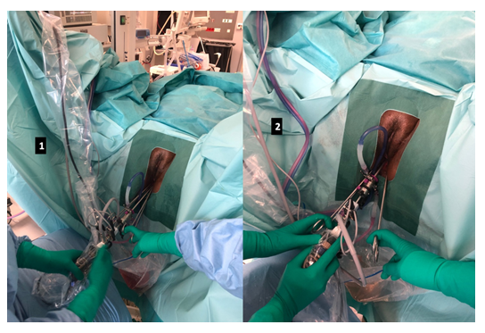
Operative hysteroscopy is a safe surgical technique to treat intra-cavity uterine pathologies [1-3]. The resectoscopes currently on the market are between 180 and 200 mm long. We are more and more confronted with technical difficulties and incomplete surgeries due to a too short length of resectoscopes. Indeed, due to the increase of the average BMI in the population, we are regularly confronted with patients with a BMI > 35 [4,5]. Their peri-perineal excess fat does not allow us to work easily, with a surgical handle in contact with the patient, limiting the procedure and access to the fundic wall and the anterior part of the uterine cavity.
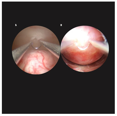
Also, in the context of polyfibroid uterus, patients could have a large cavity with a hysterometry more than 12cm. Actual resectoscopes does not allow to reach the pathologies of the uterine fundus. Fibroids affect 20 to 50% of women of childbearing age and their incidence increases with age, affecting more than one woman in two from the age of 35 years old [6]. With the advancement of the age of first pregnancy and the possibility of gamete donation , we are faced with an increase in the management of these polyfibroid uteri with large cavities and the presence of intra-cavity myomas [7,8].
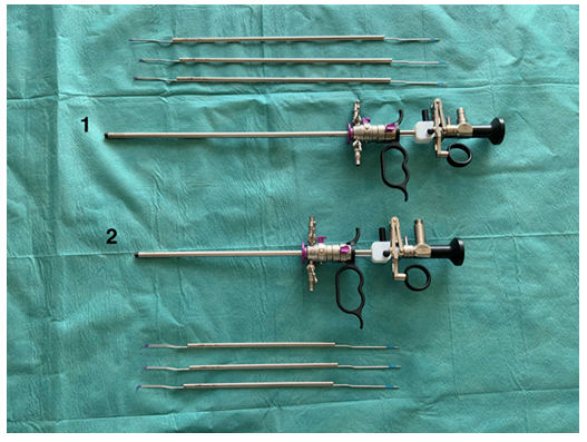
In order to remedy these technical difficulties, Delmont Imaging (Avenue du Mistral 13 600 La Ciotat / France) has developed a new surgical hysteroscope "XXL Resecare" to respond to these more frequent difficulties (Figure 1). They have developed a hysteroscope with a diameter of 6.9mm (18 French) and a length of 287mm with bipolar electrodes adapted to the size of the hysteroscope: curved loop, roller ball and knife. This resectoscope allows to reach the pathology of the uterine fundus and an optimal position of the surgeon (Figures 2 and 3). With an experience of 42 patients treated, we were able to perform a complete hysteroscopic treatment in those patients who could not be treated entirely with a standard-size hysteroscope.

Figure 1: Comparison of the size of the two resectoscopes
- XXL Resecare, diameter of 6.9mm (18 French) and a length of 287mm, with electrods, Delmont Imaging (La Ciotat/ France)
- Resecare, diameter of 6.9mm (18 French) and a length of 187mm, with electrods Delmont Imaging (La Ciotat/ France)

Figure 2: Comparison of the surgeon's position between the two hysteroscopes on a patient with BMI > 35
- XXL Resecare, length of 287mm (18 French) Delmont Imaging (La Ciotat/ France)
- Resecare, length of 187mm (18 French), Delmont Imaging (La Ciotat/ France)

Figure 3: Comparison of accessibility to fundic pathology, loop maximally extended, between the two hysteroscopes
- XXL Resecare, length of 287mm (18 French), Delmont Imaging (La Ciotat/ France)
- Resecare, length of 187mm (18 French), Delmont Imaging (La Ciotat/ France)
References
- American Association of Gynecologic Laparoscopists (AAGL): Advancing Minimally Invasive Gynecology Worldwide. AAGL practice report: practice guidelines for the diagnosis and management of submucous leiomyomas. J Minim Invasive Gynecol 19 (2012): 152-171.
- American Association of Gynecologic Laparoscopists. AAGL practice report: practice guidelines for the diagnosis and management of endometrial polyps. J Minim Invasive Gynecol 19 (2012): 3-10.
- AAGL Elevating Gynecologic Surgery. AAGL practice report: practice guidelines on intrauterine adhesions developed in collaboration with the European Society of Gynaecological Endoscopy (ESGE). Gynecol Surg 14 (2017): 6.
- De Saint Pol T. Evolution of obesity by social status in France, 1981-2003. Economics & Human Biology 7 (2009): 398-404.
- Organization WH. Obesity: Preventing and Managing the Global Epidemic: Report of a WHO Consultation. World Health Organization (2000).
- Parker WH. Etiology, symptomatology, and diagnosis of uterine myomas. Fertil Steril 87 (2007): 725-736.
- Kohler HP, Billari FC, Ortega JA. The Emergence of Lowest-Low Fertility in Europe During the 1990s. Population and Development Review 28 (2002): 641-680.
- Sobotka T. Is Lowest-Low Fertility in Europe Explained by the Postponement of Childbearing? Population and Development Review 30 (2004): 195-220.
