A Giant Aneurysm of the Right Coronary Artery
Article Information
Jeanne Combis*, Sarah Badiche, Samantha Guimaron, Aurelien Hostalrich, Bertrand Marcheix
Department of Thoracic and Cardio vascular Surgery, Toulouse University Hospital, Toulouse, France
*Corresponding Author: Jeanne Combis, Department of Thoracic and Cardio vascular Surgery, Toulouse University Hospital, Toulouse, France
Received: 09 October 2023; Accepted: 09 November 2023; Published: 05 January 2024
Citation: Jeanne Combis, Sarah Badiche, Samantha Guimaron, Aurelien Hostalrich, Bertrand Marcheix. A giant aneurysm of the right coronary artery. Journal of Surgery and Research. 7 (2024): 03-05.
View / Download Pdf Share at FacebookAbstract
The management of giant coronary artery aneurysms is poorly established due to their rarity. They are found in 0.02% of the population. Treatment includes either angioplasty or surgery, therefore involving multidisciplinary management by interventional cardiologists and cardiac surgeons. In this case, we present a case of a giant aneurysm of the right coronary artery in a 67-year-old patient, revealed by an acute coronary syndrome with ST-segment elevation, treated by coronary arteries bypass grafting.
Keywords
Giant coronary aneurysm, Acute coronary syndrome, Coronary artery bypass
Article Details
Introduction
Acute coronary syndrome with ST-segment elevation (STEMI) is an emergency. The standard course of treatment is angioplasty with treatment of the culprit lesion. In some cases, angioplasty may be difficult to perform due to the severity of lesions, calcifications, tortuosities and diffuse infiltrations of the arteries. Treating coronary aneurysms is challenging as they are rare and percutaneous treatment can be tricky. We hereby present the case of a giant aneurysm of the right coronary artery (RCA) revealed by a STEMI and treated by surgery.
Case Report
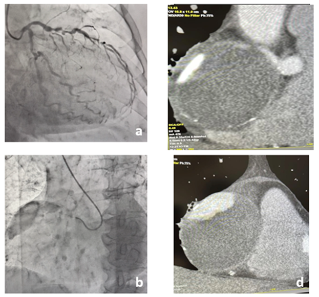
A 67-year-old female patient with no medical history nor cardiovascular risk factors presented herself with persistent constrictive chest pain. She was admitted to the emergency department of a local hospital with suspicion of a sub-acute inferior myocardial infarction (MI). Her electrocardiogram (EKG) showed a sinus rhythm with thin QRS waves, a 2 mm ST-segment elevation in the inferior derivations, thin Q waves and a discrete ST-segment depression in V1 and V2 derivations. She then received emergency coronary angiography. Catheterization of the RCA showed a healthy proximal segment followed by a 7cm-large aneurysm without opacification of its downstream bed. However, catheterization of the left main coronary artery revealed retrograde opacification of the distal RCA downstream branches (Figure 1a and 1b). Multivessel disease was also visualized with a 60% stenosis of the ostial left anterior descending (LAD) artery associated to multiples stenoses of its proximal and middle segments and a 70% stenosis of the ostial circumflex artery.
Pre-operative assessment included a coronary angiographic CT scan which revealed an oval, 76mm-long, partially thrombosed aneurysm of the RCA with calcified walls (Figures 1c and 1d). Trans-thoracic echocardiography (TTE) showed preserved left and right ventricular ejection fractions despite an inferior hypokinesia. There were no signs of valvular diseases.
Physicians concluded on a STEMI due to multivessel coronary disease associated to a RCA aneurysm. The patient was then transferred to our center for a triple CABG procedure and treatment of the coronary aneurysm.
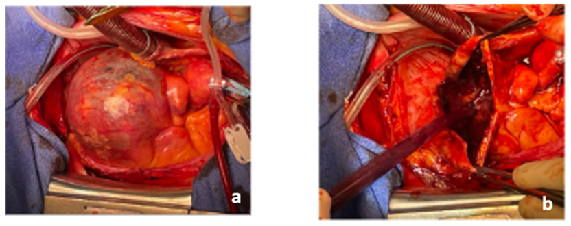
Surgery began with harvesting both mammary arteries and a left radial arterial graft. The patient was then put under cardiopulmonary bypass and the aorta clamped. We first located the proximal neck of the giant aneurysm (Figure 2a). Then, we opened the aneurismal sac which was completely thrombosed. We did not visualize any downstream arteries through the inside of the aneurysm. We then performed a partial resection of the aneurysm (Figure 2b).
Once the resection completed, we exposed a posterior descending artery (PDA) which had been visible through the left main coronary artery catheterization but not through the right one. As this artery was of good caliber, we grafted it with the radial conduit. Proximal anastomosis was done on the aorta. The right mammary artery was grafted on the lateral branch of the circumflex artery and the left mammary artery was grafted on the LAD artery. Careful de-airing was conducted, and bypass weaning was done without difficulty. Aortic clamping and cardiopulmonary bypass durations were respectively 58 and 68 minutes.
Pathology analyses of the aneurysmal sac revealed a heavily fibro-calcified wall associated with fibrino-cruoric thrombosis of the aneurism. There were also myxoid deposits associated with fibrosis and inflammation of the medial layer.
Post-operative stay was uneventful. The patient was discharged on day 13 with no abnormalities on follow-up TTE and EKG. So far, the patient remains asymptomatic and well after one month of surgery.
Discussion
A coronary aneurysm is defined by the dilation of one of its segments with a diameter greater than 1.5 times the diameter of the adjacent healthy segments. In rare cases, aneurysms are called giant coronary aneurysms when their sizes are greater than 2 cm. They occur in 0.02% of the population. Among coronary aneurysms, RCA aneurysms are the most frequently encountered (40%).
Giant coronary aneurysms are diagnostic and therapeutic emergencies because of their risk of rupture and embolization in distal territories.
Given the rarity of giant coronary aneurysms, there is no real consensus regarding their management option. The physiopathology of their formation is unclear, although various causes have been suggested such as atherosclerosis, connective tissue diseases, or inflammatory diseases in the case of pediatric patients (i.e.Takayasu and/or Kawasaki diseases).
In this case, surgery consisted of resection and exclusion of the aneurysmal sac and ligation of its collateral arteries. We concomitantly treated the associated coronary lesions, since the patient was admitted for a STEMI.
Conclusions
The management of coronary aneurysms is poorly codified due to their rarity. It requires a multidisciplinary approach involving interventional cardiologists and cardiac surgeons. The goal is to provide an optimal solution for patients adapted to the anatomical variant of their coronary arteries.
References
- Pham V, Hemptinne Q, Grinda JM, et al. Giant coronary aneurysms, from diagnosis to treatment: A literature review. Arch Cardiovasc Dis 113 (2020): 59-69.
- Kawsara A, Núñez Gil IJ, Alqahtani F, et al. Management of Coronary Artery Aneurysms. JACC Cardiovasc Interv 11 (2018): 1211-1223
- Li D, Wu Q, Sun L, et al. Surgical treatment of giant coronary artery aneurysm. J Thorac Cardiovasc Surg. 130 (2005): 817-821.
- Kurnauth V, Morey M, Jarvis R. Giant right coronary artery aneurysm - A rare presentation. J Med Imaging Radiat Oncol 64 (2020): 252-254.
- Altarabsheh SE, Deo SV, Rababa'h A, et al. Giant right coronary artery aneurysm in the setting of the acute coronary syndrome: A case report. J Card Surg 35 (2020): 2379-2381.


