Pneumatocele in a 33-Year-Old Ghanaian Woman: A Case Report
Article Information
Emmanuel Kobina Mesi Edzie1*, Klenam Dzefi-Tettey2, Philip Narteh Gorleku1, Henry Kusodzi1, Abdul Raman Asemah1
1Department of Medical Imaging, School of Medical Sciences, College of Health and Allied Sciences, University of Cape Coast, Cape Coast, Ghana
2Department of Radiology, Korle Bu Teaching Hospital. 1 Guggisberg Avenue, Accra, Ghana
*Corresponding Author: Dr. Emmanuel Kobina Mesi Edzie, Department of Medical Imaging. School of Medical Sciences, College of Health and Allied Sciences, University of Cape Coast, PMB, Cape Coast, Ghana
Received: 06 July 2020; Accepted: 17 July 2020; Published: 29 July 2020
Citation: Emmanuel Kobina Mesi Edzie, Klenam Dzefi-Tettey, Philip Narteh Gorleku, Henry Kusodzi, Abdul Raman Asemah. Pneumatocele in a 33-year-old Ghanaian Woman: A Case Report. Journal of Radiology and Clinical Imaging 3 (2020): 080-084.
View / Download Pdf Share at FacebookAbstract
A 33-year-old female baker presented to our facility with a chronic cough of 7-month duration, difficulty in breathing, and tachypnea and had been on anti-tuberculous treatment for the past six months prior to her referral. A chest radiograph revealed huge thin-walled rounded hyper luscencies with smooth inner margins and without vascular markings and fluid in both upper zones subsequently diagnosed as bilateral pneumatoceles with mild compressive effects towards the left. Her condition was stable and subsequently improved after passage of chest tubes. The ability to differentiate pneumatoceles from other conditions like pneumothorax, giant bullae, and cavities is crucial since an error in diagnosis may affect the management ultimately.
Keywords
Pneumatocele; Lung; Chest radiograph; Adult
Pneumatocele articles, Lung articles, Chest radiograph articles, Adult articles
Pneumatocele articles Pneumatocele Research articles Pneumatocele review articles Pneumatocele PubMed articles Pneumatocele PubMed Central articles Pneumatocele 2023 articles Pneumatocele 2024 articles Pneumatocele Scopus articles Pneumatocele impact factor journals Pneumatocele Scopus journals Pneumatocele PubMed journals Pneumatocele medical journals Pneumatocele free journals Pneumatocele best journals Pneumatocele top journals Pneumatocele free medical journals Pneumatocele famous journals Pneumatocele Google Scholar indexed journals Lung articles Lung Research articles Lung review articles Lung PubMed articles Lung PubMed Central articles Lung 2023 articles Lung 2024 articles Lung Scopus articles Lung impact factor journals Lung Scopus journals Lung PubMed journals Lung medical journals Lung free journals Lung best journals Lung top journals Lung free medical journals Lung famous journals Lung Google Scholar indexed journals Chest radiograph articles Chest radiograph Research articles Chest radiograph review articles Chest radiograph PubMed articles Chest radiograph PubMed Central articles Chest radiograph 2023 articles Chest radiograph 2024 articles Chest radiograph Scopus articles Chest radiograph impact factor journals Chest radiograph Scopus journals Chest radiograph PubMed journals Chest radiograph medical journals Chest radiograph free journals Chest radiograph best journals Chest radiograph top journals Chest radiograph free medical journals Chest radiograph famous journals Chest radiograph Google Scholar indexed journals Adult articles Adult Research articles Adult review articles Adult PubMed articles Adult PubMed Central articles Adult 2023 articles Adult 2024 articles Adult Scopus articles Adult impact factor journals Adult Scopus journals Adult PubMed journals Adult medical journals Adult free journals Adult best journals Adult top journals Adult free medical journals Adult famous journals Adult Google Scholar indexed journals hemophilus influenza articles hemophilus influenza Research articles hemophilus influenza review articles hemophilus influenza PubMed articles hemophilus influenza PubMed Central articles hemophilus influenza 2023 articles hemophilus influenza 2024 articles hemophilus influenza Scopus articles hemophilus influenza impact factor journals hemophilus influenza Scopus journals hemophilus influenza PubMed journals hemophilus influenza medical journals hemophilus influenza free journals hemophilus influenza best journals hemophilus influenza top journals hemophilus influenza free medical journals hemophilus influenza famous journals hemophilus influenza Google Scholar indexed journals streptococcus pneumonia articles streptococcus pneumonia Research articles streptococcus pneumonia review articles streptococcus pneumonia PubMed articles streptococcus pneumonia PubMed Central articles streptococcus pneumonia 2023 articles streptococcus pneumonia 2024 articles streptococcus pneumonia Scopus articles streptococcus pneumonia impact factor journals streptococcus pneumonia Scopus journals streptococcus pneumonia PubMed journals streptococcus pneumonia medical journals streptococcus pneumonia free journals streptococcus pneumonia best journals streptococcus pneumonia top journals streptococcus pneumonia free medical journals streptococcus pneumonia famous journals streptococcus pneumonia Google Scholar indexed journals lung parenchyma articles lung parenchyma Research articles lung parenchyma review articles lung parenchyma PubMed articles lung parenchyma PubMed Central articles lung parenchyma 2023 articles lung parenchyma 2024 articles lung parenchyma Scopus articles lung parenchyma impact factor journals lung parenchyma Scopus journals lung parenchyma PubMed journals lung parenchyma medical journals lung parenchyma free journals lung parenchyma best journals lung parenchyma top journals lung parenchyma free medical journals lung parenchyma famous journals lung parenchyma Google Scholar indexed journals Pneumatoceles articles Pneumatoceles Research articles Pneumatoceles review articles Pneumatoceles PubMed articles Pneumatoceles PubMed Central articles Pneumatoceles 2023 articles Pneumatoceles 2024 articles Pneumatoceles Scopus articles Pneumatoceles impact factor journals Pneumatoceles Scopus journals Pneumatoceles PubMed journals Pneumatoceles medical journals Pneumatoceles free journals Pneumatoceles best journals Pneumatoceles top journals Pneumatoceles free medical journals Pneumatoceles famous journals Pneumatoceles Google Scholar indexed journals erythrocyte sedimentation rate articles erythrocyte sedimentation rate Research articles erythrocyte sedimentation rate review articles erythrocyte sedimentation rate PubMed articles erythrocyte sedimentation rate PubMed Central articles erythrocyte sedimentation rate 2023 articles erythrocyte sedimentation rate 2024 articles erythrocyte sedimentation rate Scopus articles erythrocyte sedimentation rate impact factor journals erythrocyte sedimentation rate Scopus journals erythrocyte sedimentation rate PubMed journals erythrocyte sedimentation rate medical journals erythrocyte sedimentation rate free journals erythrocyte sedimentation rate best journals erythrocyte sedimentation rate top journals erythrocyte sedimentation rate free medical journals erythrocyte sedimentation rate famous journals erythrocyte sedimentation rate Google Scholar indexed journals full blood count articles full blood count Research articles full blood count review articles full blood count PubMed articles full blood count PubMed Central articles full blood count 2023 articles full blood count 2024 articles full blood count Scopus articles full blood count impact factor journals full blood count Scopus journals full blood count PubMed journals full blood count medical journals full blood count free journals full blood count best journals full blood count top journals full blood count free medical journals full blood count famous journals full blood count Google Scholar indexed journals
Article Details
1. Introduction
Pneumatoceles are thin-walled air filled cystic lesions that develop within the lung parenchyma [1]. Most often, they occur as a sequela to acute pneumonia, commonly caused by staphylococcus aureus and other infectious bacteria such as streptococcus pneumonia and hemophilus influenza [2]. Pneumatoceles also occur from other noninfectious agents such as hydrocarbon ingestion, trauma and positive pressure ventilation [3]. Typically asymptomatic, pneumatoceles are generally observed soon after the development of pneumonia and may not require surgical intervention [4]. Because the lesion generally resolves spontaneously and patients rarely die during the acute illness, the precise pathogenesis and pathologic anatomy are in most instances uncertain [5]. In adults, this condition can be misdiagnosed as cavitating lung mass. Pneumatoceles are seen in all age groups but are often common in children and rare in adults [6].
2. Case Report
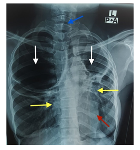
We report a case of a 33-year-old female baker who presented at the chest clinic of the Cape Coast Teaching Hospital (CCTH) with a history of chronic cough of 7-month duration. The cough was unproductive but persistent and associated with chest pains and dyspnea. She did not have fever on examination. She was tachypneic with a respiratory rate of 24 breaths per minute with reduction in chest movements bilaterally. The trachea was shifted to the left. The percussion notes were hyper resonant worse on the right upper zone. Breath sounds were reduced bilaterally with lower zone crackles. Her temperature was 36.5 degrees Celsius, pulse-92beats per minute (bpm) and a blood pressure of 120/76 mmHg was recorded. Heart sounds were present and normal. The gastrointestinal system and musculoskeletal system were unremarkable. The patient had been on anti-tuberculous treatment for the past six months prior to her referral to CCTH. A provisional diagnosis of atypical pneumonia with pneumothorax was made for further investigation. The patient was asked to do a chest X-ray (CXR), erythrocyte sedimentation rate (ESR), sputum culture and full blood count (FBC). The ESR was markedly raised (204 mm per hour), sputum culture was negative and FBC showed leukocytosis with predominant lymphocytosis. The CXR revealed huge thin-walled rounded hyper luscencies with smooth inner margins and without vascular markings and fluid in both upper zones. The lung fields visualized in both middle zones showed diffuse reticulonodular shadows with a reduction in the lung volume on the left. The trachea was shifted to left. A radiological diagnosis of bilateral pneumatoceles with mild mediastinal shift to the left side secondary to tuberculosis was made (Figure 1). The patient was managed with continuation of the anti-tuberculous treatment and thoracotomy was done after admission at the chest ward. She was followed up till the respiratory rate became 13 breaths per minute. A repeat chest radiograph (CXR) showed significant improvement four weeks after admission. The cough and dyspnea improved markedly. She was discharged and enrolled into the chest clinic. She is currently well.

Figure 1: A PA chest radiograph of a female showing huge thin-walled rounded hyper luscencies with smooth inner margins and without vascular markings and fluid in both upper zones (Pneumatoceles) as shown by the white arrows. The trachea (blue arrow) and heart (red arrow) are displaced to the left. Both middle zones show diffuse reticulonodular shadows (yellow arrows) with a reduction in the lung volume on the left.
3. Discussion
The formation of pneumatoceles has been advanced from three theories to better understand its mechanism but till date the exact mechanism in the formation of pneumatoceles remain unknown. One theory postulates that, the respiratory valve is obstructed and it caused by inflammatory exudate within the airway lumen leading to the distal dilatation of the bronchi and alveoli [7]. Another theory suggests that, the focal collection of air in the pleura dissects down the corridors to the pleura and forms pneumatoceles [8]. Finally, a third theory suggests that, after the initial drainage of the necrotic lung parenchyma, subsequent enlargement of the pneumatocele is caused by check-valve bronchiolar obstruction, resulting from a pressure from the adjacent pneumatocele or intraluminal inflammatory exudate [9]. The majority of pneumatoceles resolve spontaneously. In children, the development of tension pneumatocele commonly occurs as a result of severe complications of pneumococcal infection and bacterial pathogens like streptococcus pneumonia and staphylococcus aureus. Development of tension pneumatocele is rare in adults. Tension pneumatoceles can rupture and cause pneumothorax or bronchopleural fistula [10]. The clinical presentation may vary depending on the stage of the pneumonia. The symptoms are often initially subtle and nonspecific. The patient may be asymptomatic or present with some additional underlying pathology [11]. Pneumatoceles can be single but are more often multiple and size can vary from greater than 1 cm in diameter to occupying entire hemithorax, with walls less than 4 mm and are of uniform thickness. In a report by McGarry et al on pneumatocele formation in adult pneumonia, all three adults were severely ill and two expired [11]. Tension pneumatocele enlarges significantly compressing adjacent lung and mediastinum resulting in cardiovascular collapse. Our patient had multiple pneumatoceles and were confined at the upper zone and presented with chronic cough associated with chest pains and dyspnea. The diagnosis of pneumatoceles are usually done with the aid of CT scan and X ray examination but are rarely observed on initial chest radiographs. Between these two modalities, CT scan has a relatively high sensitivity in terms of early detection and differential diagnosis. Despite the low sensitivity of X-rays, they are still used as the first imaging modality [12]. Different forms of imaging guided treatment modalities such as the percutaneous drainage, tube drainage, compression, and catheter drainage have been described as the effective treatment modalities for single tension pneumatocele [13]. In our case, the patient was managed on her anti-tuberclous treatment and chest tube drainage was done at the chest ward successfully.
4. Conclusion
Different forms of pulmonary infections are encountered in everyday clinical practice and deciding on the most likely diagnosis may be challenging. Pneumatoceles can be confused with giant bullae, cavities, and pneumothorax and treatment of these conditions may vary, hence the ability to identify pneumatoceles which is rare in adults will prevent an error in diagnosis and treatment.
Ethical Consideration
A written informed consent was obtained from the patient. The patient was assured of confidentiality and anonymity. Ethical clearance was not required for this study.
Acknowledgement
We are grateful to the 33-year-old woman for allowing us to report this case.
Conflict of Interest
The authors have no conflict of interest to declare.
Authors’ Contributions
All authors contributed equally to the concept, design, data collection, drafting and revision of the manuscript before submission.
Funding
No funding secured for this study.
References
- Aftabuddin M, Rahman MM, Khan OS, et al. Tension Pneumatocele a Rare Presentation. Medicine Today 27 (2015): 37-39.
- Hussain N, Noce T, Sharma P, et al. Pneumatoceles in preterm infants—incidence and outcome in the post-surfactant era. Journal of Perinatology 30 (2010): 330-336.
- Splittgerber M, Pancholy B. Pneumatocele in an Adult: Case Report and a Review of the Literature. InB45. OBSTRUCTIVE LUNG DISEASE: INTERESTING CASES (2016): A3608-A3608.
- Imamoglu M, Çay A, Kosucu P, et al. Pneumatoceles in postpneumonic empyema: an algorithmic approach. Journal of pediatric surgery 40 (2005): 1111-1117.
- Quigley MJ, Fraser RS. Pulmonary pneumatocele: pathology and pathogenesis. American Journal of Roentgenology 150 (1988): 1275-1277.
- Wan-Hsiu L, Sheng-Hsiang L, Tsu-Tuan W. Pneumatocele formation in adult pulmonary tuberculosis during antituberculous chemotherapy: a case report. Cases journal 2 (2009): 8570.
- Conway DJ. The origin of lung cysts in childhood. Archives of disease in childhood 26 (1951): 504.
- Boisset GF. Subpleural emphysema complicating staphylococcal and other pneumoniae. J Pediatr 81 (1972): 259-266.
- Flaherty RA, Keegan JM, Sturtevant HN. Post-pneumonic pulmonary pneumatoceles. Radiology 74 (1960): 50-53.
- Zuhdi MK, Spear RM, Worthen HM, et al. Percutaneous catheter drainage of tension pneumatocele, secondarily infected pneumatocele, and lung abscess in children. Critical care medicine 24 (1996): 330-333.
- McGarry T, Giosa R, Rohman M, et al. Pneumatocele formation in adult pneumonia. Chest 92 (1987): 717-720.
- Jamil A, Kasi A. Pneumatocele. [Updated 2020 Mar 21]. In: StatPearls [Internet]. Treasure Island (FL): StatPearls Publishing; (2020).
- Wu ET, Chen JS. Management of multiple tension pneumatoceles refractory to tube thoracostomy decompression. The Annals of thoracic surgery 81 (2006): 1482-1484.
